
Abstract
Background:
After open or thoracoscopic lung biopsy, it is common to leave a chest tube as a postoperative drain that is typically removed on the first or second postoperative day. Standard technique is to apply an occlusive dressing at the site of chest tube removal using gauze and some form of tape.
Methods:
We reviewed the charts of children who underwent thoracoscopic lung biopsy at our institution for the past 9 years, many of whom left the operating room with a chest tube. When the tube was removed, the site was dressed, based on attending surgeon preference, with either cyanoacrylate tissue adhesive (Dermabond®; Ethicon, Cincinnati, OH) or a standard dressing with gauze and transparent occlusive adhesive dressing. Endpoints included wound complications and need for a secondary dressing.
Results:
Of 134 children who underwent thoracoscopic biopsy, 71 (53%) were given a chest tube. Chest tubes were removed at bedside in standard manner after a mean of 2.5 days. In 36 (50.7%) cyanoacrylate was used and in 35 (49.3%) a standard occlusive gauze dressing was used. No patient in either group suffered a wound dehiscence or needed a rescue dressing. There were no wound-related complications or surgical site infections in either group.
Conclusion:
Cyanoacrylate dressings are effective for closure of chest tube drain sites and appear to be safe. They might also save patients from having to deal with a bulky bandage and the discomfort of having a strong adhesive removed from their surgical site.
Introduction
Chest tube placement (thoracostomy) remains a common practice among pediatric surgeons after thoracotomy and thoracoscopic lung biopsy. The purpose of these drains is to allow egress of blood, fluid, and air that might otherwise accumulate within the chest in the immediate postoperative period. These drains are traditionally removed at the bedside when the amount of fluid draining is below a certain threshold and there is no evidence of an air leak, most often on the first or second postoperative day. A standard practice includes removing the large adhesive dressing, cutting the stitch holding the tube in place, asking the patient to take and hold a big breath, pulling the tube swiftly but deftly, and immediately placing an occlusive dressing over the site.
This occlusive dressing in the past might have included Vaseline gauze, a large gauze pressure dressing, and a great deal of adhesive tape. Today, most surgeons use a smaller dressing and perhaps an air-tight occlusive plastic film (Tegaderm™; 3M Corporation, St. Paul, MN), but it still involves placing an adhesive dressing that is left in place for several days to a week and the wound itself is essentially left open to heal by secondary intention.
For the past two decades, 2-octyl cyanoacrylate tissue adhesive (Dermabond®; Ethicon, Cincinnati, OH) became standard in many centers to close surgical incisions 1 and repair lacerations.2,3 Although we generally use it more as a dressing than the sole means of skin closure (we almost always use absorbable subcuticular sutures before applying the adhesive), our experience has convinced us, and the literature appears to support, that it is safe, effective, and well tolerated by patients and their families. Furthermore, the risk of wound infection appears to be lower,4–10 the cosmetic appearance is the same or better,2,11 and the ability to let the children bathe and swim sooner after surgery substantially improves their quality of life. 12 This made us wonder if it might be just as useful for other types of wound closure, including after removal of tunneled central venous catheters and chest tubes.
Methods
After obtaining approval from the Institutional Review Board of our hospital, we reviewed the records of all children and adolescents who underwent thoracoscopic lung or pleural biopsy at our institution between 2013 and 2021 inclusive. Charts were reviewed with special attention paid to management of the chest tube and how the chest tube incision was closed upon removal of the drain. Calls to the office and visits to the clinic for wound issues were reviewed with attention to the need for rescue dressings and infection.
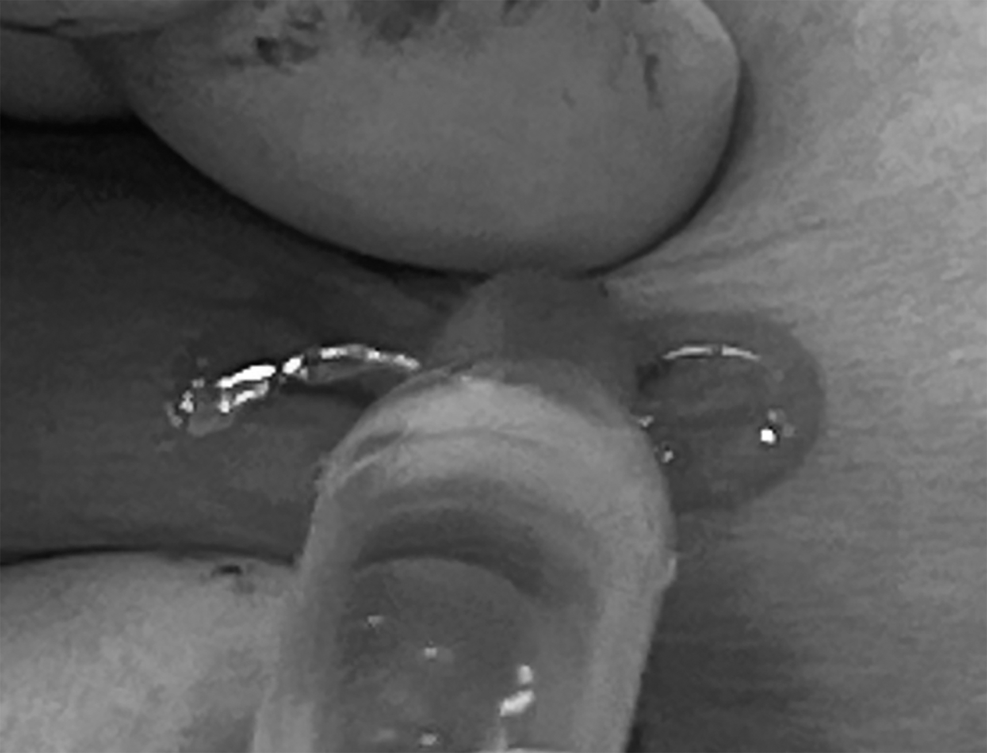
In patients receiving a standard dressing, sterile gauze and, most commonly, an adhesive plastic film dressing were applied. Vaseline-impregnated gauze was never used. Parents were instructed to leave the gauze dressing on for a minimum of 3 days and then apply antibiotic ointment or Vaseline and a band-aid on the incision until it was dry. In other patients, the skin edges were gently pinched together with our gloved fingers (Fig. 1) and cyanoacrylate tissue adhesive was used to close the wound (Fig. 2); no other dressing was applied (Fig. 3). Patients were instructed to leave the glue uncovered without any ointment and to let it fall off or flake off over the course of the next few weeks. Meanwhile, they were allowed to bathe right away and swim the next day.

After removal of the chest tube, the skin is gently pinched to appose the skin edges. We generally tunnel the tube for a short distance, allowing the practitioner to apply gentle pressure on the intercostal site of entry if escape or entrainment of air is a concern. This has not been an issue in our experience.
With the skin edges together, the cyanoacrylate tissue adhesive is applied, being careful not to let it get directly into the wound as this can cause pain and potentially a foreign-body reaction. At first, this may require 2 people to perform properly.
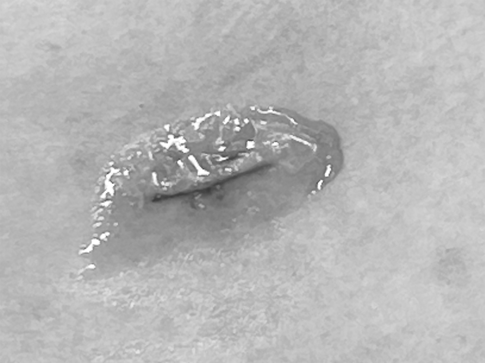
The final appearance of the adhesive having been applied. No other dressing is recommended or necessary.
Results
A total of 153 patients were identified and charts reviewed (mean age 11.5 years, range 0.5–21). Diagnoses included tumor in 97 (63%), infection in 41 (27%), and spontaneous pneumothorax in 15 (10%). We found that 71 (53%) had a chest tube placed before leaving the operating room. Chest tubes were removed at bedside after a mean of 2.5 days (range 1–8); none required a second or replacement tube. In 36 patients (50.7%), the chest tube site was closed with cyanoacrylate tissue adhesive. In 35 (49.3%), the wound was left open and a standard dressing applied. No patient returned to the ED to address issues related to the chest tube incision and there were no surgical site infections. In the cyanoacrylate group, no patient called the office or was seen in the clinic due to the glue coming off prematurely (<3 days) and none required a rescue dressing.
Discussion
Cyanoacrylate tissue adhesive for closure of skin incisions and lacerations was available in Europe and elsewhere in the 1990s and eventually approved by the Federal Drug Administration for use in the United States in 1998. 13 Since then, it has become a mainstay of wound closure in adults and in children. It appears to be safe and highly effective. Initial admonitions expressed by surgeons of a prior generation that using an occlusive dressing on a surgical wound would increase the risk of wound infection have not only proved to be unfounded, but the risk of infection appears to be significantly less when cyanoacrylate is used.
Whether this is due to the occlusive nature of the dressing or some inherent antimicrobial properties is unclear. For years we have routinely used it, for example, to close wounds in children with contaminated wounds (perforated appendicitis, enterostomy closure, and bowel resections with stool contamination) and our already low wound infection rates have continued to fall. This has made us believe that it would be safe to use it as a closure for wounds traditionally considered colonized, such as after Hickman catheter removal and, as reported in this study, after discontinuation of a chest tube. It has also been described for the control of fluid leakage after paracentesis for ascites. 14
Although their routine use after uncomplicated thoracoscopic procedures is increasingly questioned, in many centers chest tubes are de rigueur after any thoracic operation. And although we have learned that smaller single tubes are adequate for most situations, children with a chest tube still often suffer from discomfort and anxiety, which increases dramatically when they anticipate removal of the tube, typically performed at the bedside without sedation or local anesthetic. When asked, many will admit that the worst part of the process was removal of the large amount of adhesive tape many surgeons still use. We, therefore, have endeavored to always use the minimum amount of adhesive necessary and started to use cyanoacrylate glue >10 years ago, which we hypothesized would be a low-maintenance, well-tolerated, and effective skin closure and dressing in one.
The technique takes practice and at first is usually a 2-person job—one to gently pinch the skin and another to apply the glue—especially in a frightened and squirming young child. Nevertheless, most of our residents and nurse practitioners eventually learn to do it by themselves. The closure is quite durable. Although we anticipated that as many as half would fall off prematurely and require a rescue dressing, in the past 9 years of this study we have found no such case documented in the office medical record. If it were to occur, we expect that a simple dressing with ointment and a band-aid—essentially what would have been done had a standard dressing been applied—should be adequate. In fact, many patients arrive for their routine postoperative visit 3 weeks later and ask what they can use to help remove the glue that persists on the site.
Cyanoacrylate tissue adhesive remains the default dressing after chest tube removal in our practice, including patients who are recovering from thoracotomy and regardless how long then tube has been in place. Although anecdotally some report a significant incidence of local skin reactions to cyanoacrylate, this has not been our experience.
Our standard approach for thoracoscopic lung biopsy is to use an endo-stapler with a vascular load fired intracorporeally if there is enough space for this to be performed safely. In infants or toddlers, we have instead felt the need to enlarge one of the port sites enough to deliver the specimen and fired the stapler externally. We use a standard three-port approach in almost every instance, a 30° angled lens for visualization, and atraumatic graspers to manipulate the lung, attempting always to provide a minimum of 1 cm margin when feasible. In smaller children we use mainstem intubation or low-pressure insufflation only, whereas in children >30 kg we prefer to use a double-lumen endotracheal tube to isolate the operative side. We only leave a chest tube in roughly half of patients who undergo thoracoscopic biopsy and, when we do, it is almost always a standard 16- or 20-French straight polyvinyl chloride tube, depending on the size and age of the patient.
Our study has certain limitations. It was a retrospective study and thus, although we doubt patients would have felt the need to manage a wound infection or minor dehiscence on their own without having notified our team, this remains a possibility. In addition, as we did not track the day-to-day appearance of the dressing, it is possible that some fell off early and the subsequent eschar that formed was mistaken by the patient to be glue. Again, we expect this not to be an issue as we know that small incisions generally heal quite nicely by secondary intention.
Finally, without having been randomized, it is difficult to know whether patients prefer one type of dressing over the other; however, we have very few specific complaints about the use of the glue except, perhaps, that some find it difficult to remove when it has outlived its usefulness several weeks after the operation. There is also the possibility that in a larger cohort of patients we might discover that some develop a foreign body reaction 15 or contact dermatitis 16 to the glue, but there were no such reactions in our patients. Finally, almost all of our chest tubes are removed in the first 3 days after surgery. It is conceivable that the risk of infection or superficial wound dehiscence might be higher when the glue is applied to chest tube sites that are more than a week or two old.
Conclusion
Cyanoacrylate skin adhesive appears to be safe, effective, and well tolerated for the closure of thoracostomy incisions in children.
Footnotes
Acknowledgment
The author would like to thank Rosa Hwang, clinical research coordinator, for help in identifying patients and generating the data set.
Authors' Contributions
Conceptualization, methodology, validation, formal analysis, investigation, resources, data curation, writing, and visualization by P.M.
Disclosure Statement
No competing financial interests exist.
Funding Information
The author (P.M.) is funded partially by the Morgan Family Endowed Chair for Pediatric Surgical Research.