
Abstract
Background:
General Surgery course is a mandatory in medical schools and continuing surgery training is important even to experienced surgeons which they need to maintain and/or improve their surgical skills. Additionally, the models used for that practice are human cadavers, anesthetized porcine, or simulators and are not accessible for medicine schools or physicians in many countries. Therefore, we present a new technical procedure for preparation of frozen experimental animal's cadavers for medical surgical training.
Materials and Methods:
To perform the study, one porcine slaughtered and frozen at −20°C was used. The porcine cadaver was thawed at room temperature (25°C) and then the pneumoperitoneum test was performed and viscera inspection carried out.
Results:
The porcine cadaver took 20 hours to completely thaw. The pneumoperitoneum was successfully performed with total distention of the abdominal cavity. All viscera were well preserved maintaining important in vivo characteristics for consistency.
Conclusion:
The use of thawed porcine cadaver as a model to train many surgical procedures including videolaparoscopy is feasible. The tissues were well preserved by this method and was financially accessible and could be used for different techniques, equipment, and material tests.
Introduction
The videosurgery and general surgery teaching demands time training and dedication, beyond that also had the necessity of senseless surgical models such as experimental animals, simulators, or human cadavers. 1 All of these models have their applications, costs, and limitations. The use of tissue models with experimental animals (porcine represents 70%) 1 or fresh cadavers is the one that is closest to the surgical reality and provides training as the surgical act itself. 2 On the other hand, these methods have high-cost inherent to the preparation, anesthesia, and maintenance whether of animals or fresh cadavers, the latter being rarer and less available.1,3
Many countries have difficulties in using fresh human cadavers, whether by public policy, ethics, or religion. As an example in Brazil, there is no legislation that allows the use of fresh human cadavers; for this reason it must be imported for teaching/training proposes (e.g., in United States, in 2014, a cadaver could cost US$ 8385.00 per training program, 4 then one can speculate on the even higher cost plus the cost of importation to be used in Brazil).
To make these procedures economically viable and therefore make them more accessible, we idealized a new tissue preparation that will allow a greater number of doctors to have easy access to high-quality surgical training. The aim of our study was to present a new technical procedure for preparation of frozen experimental animal's cadavers for medical surgical training.
Materials and Methods
The test proposal was approved by the Institution Animal Ethics Committee protocol 2023/17.
Study design
To perform the test, the slaughterhouse was requested that a female swine, TopigNorsvin TN70 hybrid breed, 25 kg, 90 days old, slaughtered according to the 03-Normative Instruction by the Brazilian Ministry of Agriculture, Livestock, and Food Supply be made available. Also, for the study proposal, it was requested that the animal had 24 hours fast, all viscera be maintained after being slaughtered, and the head and the distal portion of four limbs be resected. The cadaver was frozen and kept in −20°C for 2 days for further thawing assessments (Fig. 1).
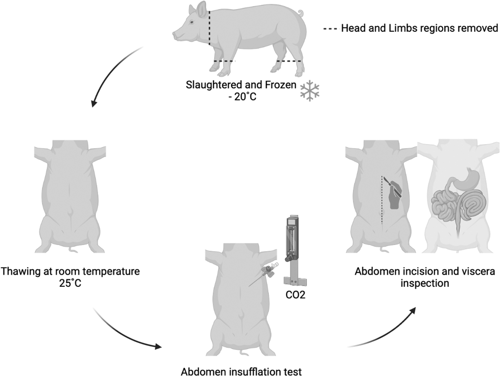
Study design, experimental test phases.
Thawing process
The porcine cadaver was removed from the freezer and was placed on an inox V-type small animal operation table in dorsal recumbency at room temperature, 25°C. The thaw process was verified hourly until the cadaver was completely thawed.
Cadaver assessment
Once the cadaver was thawed, the abdominal insufflation (pneumoperitoneum) was tested, using a 16G catheter 45° inserted caudal from the region below the costal arch laterally to the midline with CO2 insufflation. After that, the abdominal cavity was inspected; a large incision was performed on the middle line (30 cm) using a 23 scalpel blade. After viscera inspection, the cadaver was discarded.
Results
Thawing evaluation
The thawing process was verified by cadaver palpation, and it took 20 hours at room temperature, 25°C, until the porcine cadaver was completely thawed. In the first 6 hours, the skin and abdomen region became flaccid, but viscera, thoracic region, and limbs were still frozen. After 14 hours, it was observed that the abdomen was free, flaccid, and mobile. It was also noted that the viscera remained frozen, mainly small intestine. The limbs were with joint mobility; however, the dorsal region was still rigid. For this reason, the cadaver was repositioned in sternal recumbency for 6 hours. After 20 hours of thawing process, the porcine cadaver was completely thawed.
Cadaver evaluation
Tests were performed 3 hours after the cadaver was detected thawed. Pneumoperitoneum was performed with gradual insufflation until there was no further abdomen distention (Fig. 2), then the gas was removed from the abdominal cavity to allow the abdominal inspection by laparotomy. When the cavity was incised, a fetid odor was noted, characteristic of bacterial fermentation, possibly intestine bacterial proliferation as there were remnants of feces in the colon and sigmoid region. In addition, the stomach and parts of small intestine/colon had mild-to-moderate gas distention. All viscera were considered well preserved similar to in vivo characteristics of color, consistency, and anatomical position (Fig. 3). The animal has a cyst in the umbilical region, without relevance to this study.
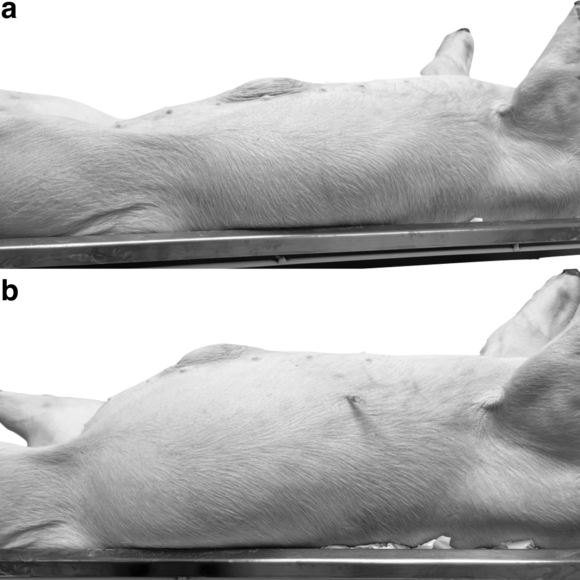
Pneumoperitoneum test. In

Porcine abdominal viscera inspection: stomach, delimited by grey punctuate line; ascending colon, black arrow; spleen, delimited by white punctuate line; cecum, black arrow head; small intestine, white arrows; umbilical cyst, white arrow.
The experimental cost in general was around US$ 250.00, considering the slaughter, transport, freezing maintenance, test materials, and animal discard.
Discussion
This conservative model seemed to be feasible and generated a great impact in the surgical training/teaching field.
The thawing process demonstrates that previous logistic organization is necessary to prepare the cadaver for the training/teaching day. It was observed that at least 20 hours was needed for porcine cadaver to completely defrost at 25°C, and it is similar to the human lower extremities thawing process at 15°C that was also frozen at −20°C. 5 For human torsos, the thawing process needed ∼3 days at 15°C, 5 and for porcine specimens frozen at −70°C also 3 days of thawing are necessary. 6 The method used for thawing in this study allowed the performance of the abdominal insufflation test in a satisfactory manner.
Pneumoperitoneum test in porcine cadaver was successfully performed in this study and could benefit a lot of hands-on videolaparoscopy training. The abdominal skin after thawing was still preserved and kept the elasticity, promoting a good distention during the insufflation similar to in vivo experiments. Other studies using in vivo porcine models, reported a good distention of pneumoperitoneum according to their study goals. The evaluation of histopathological changes of porcine induced-pneumoperitoneum intra-abdominal hypertension 7 and the laparoscopic transplantation of cell sheets in liver, colon, small intestine, and stomach by pneumoperitoneum. 8
The viscera inspection was carried out to verify whether it was possible to perform surgical procedures after thawing. Viscera observation was a normal aspect by visualization and palpation, which allows surgical training by laparoscopic or laparotomy approaches as well as studies which used in vivo models. 8
Some considerations have to be taken for the next steps. A new study is already in progression with a major experimental-N, also comparing different types of thawing at room temperature versus refrigerator and performing the tests immediately after thawing to avoid the fetid odor and bacterial proliferation.
Conclusion
The use of thawed porcine cadaver as a model for doctors and medical students training for many surgical procedures including videolaparoscopy is feasible and reasonable. This method maintains the tissues well for miscellaneous techniques, equipment, and material tests. Also, the use of thawed porcine cadaver reduces waste materials, teaching costs, and even the use of volatile anesthetics, allowing pregnant doctors/students to stay in an operation room during training labs.
Footnotes
Acknowledgment
We thank the Brazilian Coordination for the Improvement of Higher Education Personnel (CAPES).
Authors' Contributions
B.Z.: Conceptualization, Methodology, Investigation, and Supervision. B.B.: Methodology, Investigation. D.D.M.: Writing—review. R.B.: Investigation. J.P.H.P.: Methodology, Investigation, Writing—original draft and Project Administration. B.Z. purchased the animal for the purposes of the study.
Disclosure Statement
No competing financial interests exist.
Funding Information
Funded by the author's own resources.