
Abstract
Background:
Diastasis recti (DR) is a common condition, especially in women after pregnancy, often associated with concomitant hernia defects and defined as a rupture of the midline and a separation of the rectus muscle more than 2 cm. Symptoms related to this are low back pain, urinary incontinence and pelvic prolapse, as well as abdominal bulging and core instability. We analyzed clinical and functional outcomes after treatment of DR alone or associated with midline hernias in 219 patients who underwent a midline reconstruction using miSAR® technique (minimally invasive stapled abdominal wall reconstruction).
Methods:
Between April 2019 and April 2022, 219 patients were treated with miSAR®. All patients were requested to quantify preoperative and postoperative functional symptoms (urinary incontinence, low back pain, abdominal swelling, and respiratory distress).
Results:
Twenty-seven men and 192 women underwent the miSAR® technique. The mean body mass index was 23.9 kg/m2. We performed the miSAR® technique in patients affected by incisional midline hernia and umbilical hernia alone or associated with DR. Composite mesh was used in 91.8% of cases. The average operating time was 90 minutes. Seven percent of the patients had postoperative complications, including two retromuscular hematomas, two retromuscular seromas, and one postoperative bleeding event. Two patients were readmitted for bowel obstruction. After surgery, there was symptomatic improvement in urinary incontinence, low back pain, respiratory symptoms, and abdominal swelling; this improvement was confirmed at 6 months and at 1- and 2-year follow-up. At the 1-year follow-up, the overall recurrence rate was 2.83%.
Conclusion:
miSAR® is a feasible and effective technique and shows promising results in the treatment of DR and ventral hernia. Possible enhancements include use of preoperative Botox to treat defects larger than 6 cm. Multicentric analysis is needed to validate the technique, and longer follow-up is required to assess the recurrence rate.
Introduction
Diastasis recti (DR) is a very common condition, especially in women after pregnancy, and it is often associated with concomitant hernia defects. According to International Endohernia Society, it is defined as a separation of the recti muscles more than 2 cm, identifying three categories: < 3 cm, 3–5 cm, and > 5 cm. 1 The prevalence is probably underestimated, as demonstrated by a recent study published by Kaufman et al., who found a prevalence of 57% of DR in an asymptomatic population, aged between 18 and 90 years, involving both sexes. 2 Commonly related symptoms are low back pain (LBP), urinary incontinence (UI), and pelvic prolapse, as well as abdominal deformations, such as bulging and core instability. Risk factors include obesity and multiple pregnancies.2,3Minimally invasive stapled abdominal wall reconstruction (miSAR®) technique was presented in a previous article. 4 This procedure restores the integrity of the linea alba, using a reinforced stapled suture. It has all the advantages of a minimally invasive approach and achieves sublay mesh positioning through the creation of a retromuscular virtual cavity.
In this article, we present a case series of 219 patients treated consecutively using the miSAR® technique, consisting of laparoscopic reconstruction of the midline using a linear stapler, with an analysis of clinical and functional outcomes after 6, 12, and 24 months.
Methods
Between April 2019 and April 2022, 219 patients (27 men and 192 women) were treated using the miSAR® technique at San Giovanni Hospital in Rome. The procedure was conducted in accordance with ethical standards, and all patients signed a written informed consent before undergoing surgery.
Patients between 22 and 83 years of age were included in the study (mean age 46.1 years old). The mean body mass index (BMI) was 23.9 kg/m2, and only 6 patients with a BMI >30 kg/m2 were included.
Patients with a BMI >24.9 kg/m2 were advised to go on a compulsory diet before surgery. All patients were requested to quantify preoperative functional symptoms (UI, lower back pain, abdominal swelling [AS], and respiratory distress [RD]) by assigning a value from 0 to 10; the same procedure was repeated after 6 months, 1 year, and 2 years.
Patients who asked for associated abdominoplasty surgery were visited by a plastic surgeon in order to decide if it was needed.
On the first postoperative day, patients were questioned about their pain from 0 to 10, according to the Numeric pain scale (NRS).
The miSAR® technique was used for reconstruction of the linea alba in DR, midline ventral hernia, and midline incisional hernia. The technique was associated with other surgical procedures, such as cholecystectomy, inguinal hernia repair, abdominoplasty, appendectomy, and hysterectomy.
Transversus abdominis plane block (TAP block) was performed after the procedure.
During hospitalization, paracetamol and ketorolac were administered for pain control. After surgery, a postoperative band was suggested for 2 months.
Surgical technique
The patient was placed in a supine/combined lithotomy position with thighs extended (120°). Preoperative antibiotics were administered, and general endotracheal anesthesia was introduced. A Foley catheter was placed and then removed at the end of the procedure. The abdomen was prepped and draped in the usual sterile manner.
Pneumoperitoneum was induced using an open technique, placing the first 12 mm trocar 2 cm into the left iliac fossa. The abdomen was insufflated with carbon dioxide up to a pressure of 6–8 mmHg.
A 30° laparoscope was inserted, and the abdomen and abdominal wall were inspected.
Additional trocars were inserted under direct vision in the following locations: a 12 mm trocar in the sovrapubic space and a 5 mm trocar in the right iliac fossa. If a 5 mm laparoscope was available, two 5 mm trocars and one 12 mm trocar were used. When necessary, lysis of adhesions to the abdominal wall was performed. Redundant adipose tissue was dissected from the posterior layer of the anterior abdominal wall until the xiphoid process, and the falciform ligament was dissected. The adipose tissue was removed using an endobag. The diastasis and midline hernias were exposed.
The peritoneum and the posterior rectus sheaths were incised bilaterally on the arcuate line, and the retromuscular space was exposed.
At this point, the laparoscope was moved to the trocar in the left iliac fossa. The rectus sheath was dissected from the muscle using a blunt dissector introduced into the space previously created. The abdominal pressure was lowered to 6 mmHg. A 60 mm endoscopic reinforced stapler (purple load) was introduced through the suprapubic trocar; its jaws were opened and introduced into the two retromuscular pockets previously created (Fig.1). The stapler was fired, binding the rectus sheath in the lower and upper lines. After this procedure, a cavity was created between the muscle at the top and the posterior sheath at the bottom (Fig.2). Dissection of the muscle from the sheath was carried out cranially and laterally to reduce lateral tension and to reach the xiphoid process, the transverse muscle, and the lower margin of the ribcage cranially. The firing process was repeated in a cranial direction until the xiphoid process was reached; a mean of five loads was required. A polypropylene or composite mesh was placed in the space between the rectus muscles and the posterior sheaths, without using any fixation device. If the rectus muscles were distant along the midline, oversewing with a small-bite barbed absorbable suture was implemented along the superior stapled suture (Fig. 3).
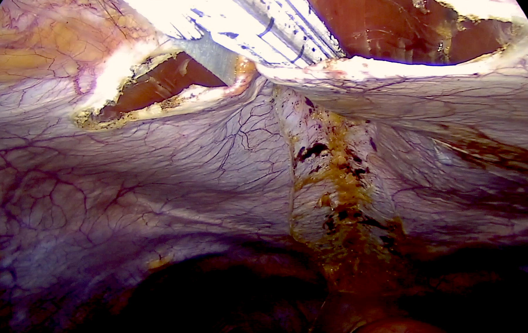
Start of midline stapled reconstruction introducing stapler in posterior fascia’s incisions.

Retromuscular pocket; stapled sutures (anterior and posterior).

Handsewn reinforcement of anterior suture and sublay mesh.
Subsequently, the laparoscope was moved back to the suprapubic trocar. A barbed absorbable suture was used to close the initial opening in the posterior sheath. TAP block was performed using 7.5 mg/mL Naropin diluted in 20 mL of saline solution. The trocars were removed under direct vision. Access for all trocars >5 mm was closed with 2.0 polyglactin suture at the fascial layer.
Defects >6 cm can be treated with Botox® infiltration 40 days before surgery to ensure greater laxity of the muscle structures. Botox infiltration was conducted using 200 units of botulinum toxin A (Botox) in a 2:1 dilution with saline injected under ultrasonographic guidance into the external and internal oblique muscles at three separate sites bilaterally for a total of six injections.
Neuraxial anesthesia was used in two patients.
Statistical analysis
All data were initially entered into an Excel database (Microsoft, Redmond, Washington, USA), and the analysis was performed using SAS software (Version 9.3). The statistical significance of the categorical variables was evaluated using the Wilcoxon two-sample signed-rank test; Student’s t-test was used for analysis of continuous variables. A P value <.05 was considered statistically significant. Descriptive statistics consisted of the mean ± standard deviation for parameters with normal distributions (after confirmation with histograms and the Kolmogorov–Smirnov test) and the median and range (min. and max.) for variables with no normal distribution.
Guidelines do not require the institutional board’s approval.
Results
The mean age was 46.1 ± 11.7 years. There were 27 men and 192 women. The mean BMI was 23.9 ± 3.7 kg/m2.
We performed the miSAR® technique in 24 patients affected by incisional midline hernia, 23 with diastasis and midline ventral hernia (no umbilical hernia), 3 with recurrent recti diastasis, 118 with recti diastasis and umbilical hernia, and 51 with recti diastasis alone. Abdominoplasty was performed after laparoscopy in 42 patients. TAPP repair was performed in 3, cholecystectomy in 9, appendectomy in 1, and hysterectomy in 2 patients. The characteristics of the patient population are shown in Table 1 and Table 2.
Conditions Treated with the miSAR Technique
miSAR, minimally invasive stapled abdominal wall reconstruction.
miSAR® Associated Procedures
miSAR, minimally invasive stapled abdominal wall reconstruction.
All procedures were performed using the same technique, and open conversion was not needed in any case.
Polypropylene mesh was used in 18 patients, and no mesh was used in 2 patients. Composite mesh was used in 199 patients. In the first 50 patients, no reinforcement of the mechanical suture was applied. The average operating time was 90 minutes.
Hospitalization was for 2 days after surgery using the miSAR procedure and 3 days on average if abdominoplasty was performed.
Fifteen patients (6.8%) had postoperative complications. Four patients had experienced retromuscular hematoma, three of whom were treated conservatively; one hematoma was drained. Two patients had retromuscular seroma, which was treated conservatively. One case of postoperative bleeding occurred when abdominoplasty was performed at the same time as the miSAR procedure. Two patients experienced a ventral hernia on the epigastric trocar incision, and 2 patients had postoperative wound complications. Two patients were readmitted for bowel obstruction: one on the 10th postoperative day and the other on the 4th postoperative day. Both patients vomited after discharge, and the parietal recess between the rectus muscle and the posterior sheath was torn. One patient developed deep vein thrombosis, and 1 patient had a vesical fistula (Table 3).
Postoperative Complications
The median pain score on the first postoperative day according to the NRS scale was 5.6 ± 2.
A total of 141 patients completed the preoperative and postoperative questionnaire at the 6-month follow-up, giving a value from 0 to 10 for functional symptoms (UI, LBP, respiratory symptoms [RS], and AS); the value did not change after 1-year and 2-year follow-up.
Among the patients, 75 (53%) had UI before the surgery, with a mean quantitative value of 3.37; LBP was reported before the surgery by 111 patients (79%), and the mean quantitative value was 5.76. In total, 59 patients (42%) had RS, mostly shortness of breath, with a mean quantitative value of 2.65. AS and feelings of distress were the most common symptoms in 131 patients (93%), with a mean quantitative value of 8.31.
The 6-month follow-up questionnaire showed significant improvement in all these symptoms (Table 4 and Table 5). Fifty-three patients (70%) had full resolution of UI, 18 patients (24%) had improvement of UI, and only 4 patients (5.4%) did not benefit. The postoperative mean value was 0.54. LBP disappeared in 65 patients (58%), and it was improved in 42 patients (38%); 3 patients (2.7%) did not benefit. The postoperative mean value was 1.16. Forty-five patients (76.3%) had resolution of RS; RS was improved in 82 patients (62.6%), although 4 patients did not benefit. The mean postoperative value was 0.38. AS disappeared in 45 patients (34.3%) and was improved in 82 (62.6%); 4 patients (3%) did not benefit. The mean postoperative value was 2.64.
Preoperative and Postoperative Values of Functional Symptoms
AS, abdominal swelling; LBP, lower back pain; RD, respiratory distress; UI, urinary incontinence.
Postoperative Symptoms
AS, abdominal swelling; LBP, lower back pain; RD, respiratory distress; UI, urinary incontinence.
During follow-up, 3 (6%) of the patients treated using no reinforced load had a recurrence, with 1 having associated ventral hernia. Reinforced suture was used in 91 patients, and only 1 had recurrence (1.1%); 7 patients were lost during follow-up. No statistical significance was found between the two groups (P = .09) (Table 6).
Recurrence
Discussion
Abdominal wall defects represent one of the most common surgical pathologies; therefore, several techniques have been developed in the last decade. Over the years, the superiority of mesh repair over direct suture has been proven. The sublay mesh position seems to be the most favorable placement; however, widespread laparoscopic repairs with onlay mesh have become increasingly frequent and have shown good results. 5 Nevertheless, in some cases, the presence of a foreign body in the peritoneal cavity has led to serious complications such as infection, fistula, bowel injury, and bowel adhesions. Despite remarkable development in mesh composition, the risk associated with intraperitoneal positioning has not been removed. For these reasons, open Rives–Stoppa still holds a central role in abdominal wall surgery.6–8
For RD, recent guidelines define the condition as a separation between the rectus muscles wider than 2 cm, and a new classification system has been introduced. Plication of the linea alba is suggested, and mesh-based repair is recommended when concomitant midline hernias are present. However, no indication for the surgical technique has been made.1,9
Restoration of the linea alba is an extremely important topic in hernia surgery; partially abandoned with widespread IPOM, it has regained importance with the introduction of IPOM plus. This technique combines intraperitoneal mesh placement with direct fascia closure of the defect, overcoming some of the most common complications of conventional IPOM, such as seromas and bulging; moreover, restoration of normal anatomy of the abdominal wall favors better functional outcomes.10–13
The miSAR® technique, first proposed by our group in 2021 to correct DR, has the objective of achieving sublay mesh positioning through a complete laparoscopic approach, and it has proven to be safe and feasible in the treatment of all midline hernias. 4 At first, the technique was used only in patients with DR, and the stapler was employed to achieve plication of the linea alba while maintaining even tension on the suture. With experience, we extended the surgical indication to patients with midline abdominal wall defects, because it allowed us to restore the anatomy of the abdominal wall, to place the mesh in an ideal space, and to attain optimal overlap. One hundred sixty-five patients with ventral hernia were treated successfully. Among them, only 1 patient (0.6%) had recurrence at the 1-year follow-up. Further studies are needed to assess the long-term results of the technique in the setting of hernia repair. Compared with traditional IPOM, miSAR® has a very low rate of intra-abdominal complications. Indeed, only 2 patients (1.1%) had bowel obstruction due to herniation of intestinal loops due to tearing of the manual suture of the opening in the posterior sheath; in neither case was intestinal resection needed, and the surgical stapled suture of the midline was intact. No adhesion was present due to the beneficial effect of composite mesh.
Moreover, the retromuscular prosthesis position avoids displacement without the need for fixation devices, which may contribute to a reduction in postoperative pain. 14 According to the NRS scale, postoperative pain in our patients was low and manageable in most cases with paracetamol alone. As shown by Cavallaro et al., 15 TAP block is effective to reduce postoperative pain.
An interesting randomized control trial by Bernardi et al. analyzed the effect on quality of life in patients undergoing hernia surgery: one group receiving primary fascia closure before mesh placement and another treated only with intraperitoneal mesh with a bridging technique. IPOM resulted in a nonfunctional portion of the abdomen, whereas fascia closure and restoration of the integrity of the abdominal wall produced statistically significant improvement in the quality of life of patients over time. This was a key outcome because the indication for surgery in ventral hernias is often based on symptoms. 16
For patients treated with miSAR®, there was a significant improvement in associated symptoms after surgery, as based on a questionnaire at 6-month, 1-year, and 2-year follow-up; a greater benefit regarding urinary incontinence and abdominal swelling was found, with improvement reported in up to 90% of patients. Reconstruction of the midline restores the abdominal core and balances abdominal forces, reaching the physiological anatomy of the linea alba and resolving or improving functional symptoms.
When considering DR, in the last decade, many functional disabilities have been associated with the condition; therefore, the frequency of surgical reconstruction has increased. Common related symptoms are low back pain, abdominal pain, posture, urinary incontinence, abdominal muscle strength, and reduced quality of life. However, there is no standard method to evaluate symptoms, and it is difficult to compare surgical outcomes among techniques. Most studies, including ours, refer to a patient’s self-evaluation and improvement in the perception of the condition. There is consistent evidence that surgical repair improves physical function, decreases urinary incontinence, and enhances quality of life. 17
Overall, stapled reconstruction of the midline distributes the tension evenly along the suture, preventing fascial tearing and recurrence.
Stapled reconstruction is supported in a recent study by Lauro et al., 18 who compared forces and resistance of stapled sutures, oversewn stapled sutures, and simple handsewn sutures using ex vivo models of the human fascia lata. Four items were evaluated: maximal stress, deformability, rigidity, and ability to absorb mechanical energy up to the breaking point. The study showed that stapled and hybrid sutures have better strength performance than handsewn sutures, suggesting that stapled sutures can resist higher tensile forces but are less deformable. Compared with handsewn sutures, stapled sutures exhibit uniform distribution of forces along the closure because knots represent an area of weakness, although the behavior at the breaking point is “all or nothing.” Hybrid sutures have an intermediate failure behavior. Handsewn suture presented a strength of 0.83 MPa, stapled 2.10 MPa, and hybrid 2.68 MPa. These data should be considered in the surgical application of staplers in the reconstruction of the midline for DR and ventral hernia and may guide surgeons in tailoring the surgical technique.
A limit of the technique is represented by the width of the defect; in fact, a large loss of substance does not allow correct use of the stapler. Application of preoperative Botox injection might overcome this limit and extend indications, as shown in the literature.19,20 In our center, we have tested Botox injections in five cases of diastasis wider than 10 cm and obtained good results, yet our data are preliminary, and a larger series is needed to standardize the technique.
A final highlight of miSAR® is that it is completely intraperitoneal, so it is possible to associate other procedures, such as laparoscopic cholecystectomy, appendectomy, or groin hernia repair, as shown by our series. Moreover, as the stapled suture of the posterior sheath does not compromise umbilical vascularization, even abdominoplasty is practicable and safe.
Although multicentric analysis and randomized trials are needed to validate the technique, as well as a longer follow-up, miSAR® shows promising results in treating DR and ventral hernias. Further development may be represented by its use in exclusive hernia defect.
Footnotes
Acknowledgment
The authors would like to thank their medical colleagues and operating room staff.
Authors’ Contributions
G.M.: Conceptualization, methodology, validation, investigation, supervision, and project administration. M.G.L.: Methodology, validation, investigation, resources, data curation, writing, and visualization. E.B.: Investigation, formal analysis, and writing. G.N.: Supervision and project administration.
Disclosure Statement
The authors have no conflict of interests or financial ties to disclose.
Funding Information
The authors received no financial support for the research, authorship, and publication of this article.