
Abstract
Abstract
Background:
Lipopolysaccharide (LPS) is a structural component of the outer membrane of gram-negative bacteria. LPS activates the host cells, leading to the production and release of proinflammatory cytokines. Given the induction duration for the release of cytokines, the initial mechanisms that produce LPS action on a timescale of minutes are not fully understood. We studied the effect of initial LPS-induced action on the lymphatic system by measuring the time-dependent changes in mesenteric lymph flow in guinea pigs in vivo. In addition, we determined the leakage of plasma protein into the lymphatic system using Evans blue dye.
Methods and Results:
The mesenteric lymphatic vessel was cannulated with a polyethylene catheter. We administered drugs via a catheter in jugular vein. The control animals received vehicle intravenously (i.v.). The experimental group received 1 mg/kg or 10 mg/kg LPS i.v. Twenty minutes before injection of the vehicle or LPS, Evans blue dye (5 mg/kg i.v.) was administered. Lymph output was measured every 20 min. The amount of Evans blue in the lymph was determined by spectrophotometry. The mesenteric lymph showed a steady flow rate of approximately 290 μL/kg/20 min. The lymph flow immediately increased after the administration of LPS and reached 3.4-fold and 7.4-fold after 1 h of 1 mg/kg and 10 mg/kg LPS injection, respectively. The albumin content in lymph significantly increased in proportion to the increased lymph volume.
Conclusions:
These results suggest that the early increase in mesenteric lymph flow rate in guinea pigs produced by LPS is mediated by vascular hyperpermeability and plasma albumin leakage.
Introduction
Previous reports in a rabbit LPS-induced pleurisy model showed that LPS-induced acute endothelial hyperpermeability occurred not only in skin with acute peripheral inflammation, 4 but also during systemic inflammation. 5 Administration of LPS results in structural changes in the capillary endothelial cells, which increases permeability and results in a loss of barrier function. Extravascular fluid is reabsorbed by postcapillary venules, but the lymphatic system also contributes to drainage. It can be hypothesized that lymphatic drainage of the extravascular fluid increases under conditions of altered vascular permeability. In fact, spleen lymph flow increased 8-fold after LPS exposure in the rat. 6
In the present study, we examined the effect of LPS on the lymphatic flow using mesenteric lymphatic vessels, because the mesenteric lymph accounted for the highest percentage of lymph in the whole lymph flow. 7 To estimate the volume of leaked serum albumin, we determined the amount of Evans blue dye leakage. When Evans blue dye is administered intravenously, it binds to plasma proteins, particularly albumin, and can be used as a marker of plasma extravasation in vascular permeability studies. 8 The tissue distribution of I131-labeled serum albumin has been shown to correspond to the distribution of Evans blue dye.9,10
In guinea pigs, the mesenteric lymph flow in endotoxemia has never been determined. We chose to perform our study in guinea pigs because, like humans, they are highly sensitive to endotoxins.11,12 Additionally, the lipid metabolism pathway in guinea pigs is similar to the human pathway, providing a good model to study lipid metabolism. 13 Given that LPS is a glycolipid and lymph is involved in transferring lipids, the guinea pig is an appropriate model for our study.
Methods
Animals
Adult male, Hartley guinea pigs (weighing 350–450 g, Sankyo-labo Co. Tokyo, Japan) were used in the study. The guinea pigs were provided free access to water and food, and were exposed to light on a 12:12 h cycle in a humidity- and temperature-controlled environment. The experiments were approved by the Graduated University of Japan Traditional Medicine and Science Animal Care Committee.
Cannulation of blood vessels
The guinea pigs were anesthetized with an intraperitoneal (i.p.) injection of pentobarbital sodium (30 mg/kg body weight). While anesthetized, the body temperature was maintained using a heating pad. The carotid artery was dissected out and cannulated with polyethylene tubing containing heparinized (heparin sodium, 25 U/mL) saline to monitor blood pressure. Next, the cannulation of the internal jugular vein was performed with a 3-Fr silicone catheter. Chemicals were administered via the catheter. Blood pressure was measured from 1 h before administration of LPS to 3 h after, with readings taken every 20 min.
Cannulation of the efferent mesenteric lymph duct
A midline laparotomy was performed on the abdominal wall to access the peritoneal cavity. A portion of the jejunum was hung on two fishhooks to visualize the base of the mesentery (Fig. 1).The mesenteric lymph duct was located superior and parallel to the mesenteric artery. Under a dissection microscope, the efferent mesenteric lymph duct was cannulated with a polyethylene catheter (internal diameter of 0.3 mm). The lymph duct was then doubly ligated with a 10-0 silk nylon thread. The catheter was brought out through an incision in the left flank. A constant infusion of Ringer's lactate (Lactec injection, Otsuka Pharmaceutical Co. Tokyo, Japan) was initiated through the internal jugular catheter at a rate of 1.5 mL/h using a constant infusion pump (Terufusion syringe pump TE-311, Terumo Co. Tokyo, Japan). Lymph samples were collected every 20 min in sterile tubes and measured by weight.
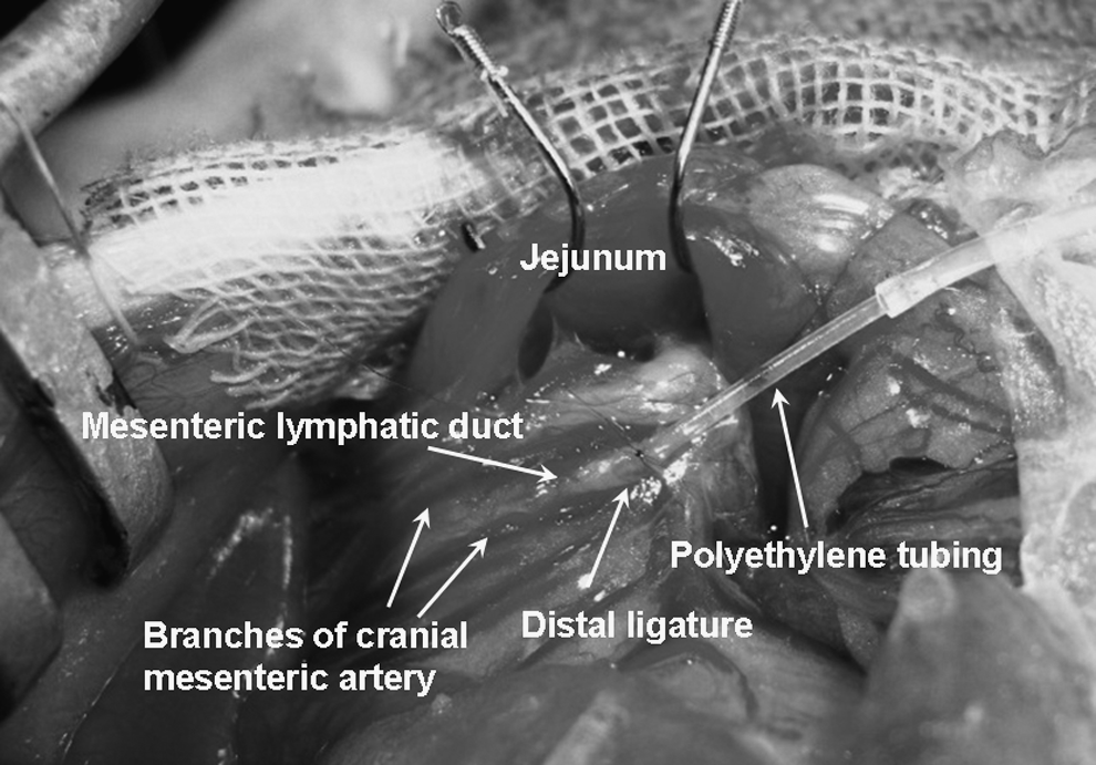
Cannulated mesenteric lymphatic duct.
Experimental design
The guinea pigs were divided into three groups as follows. Group 1: intravenous (i.v.) injection of Evans blue and saline (vehicle). Group 2: i.v. injection of Evans blue and 1 mg/kg LPS. Group 3: i.v. injection of Evans blue and 10 mg/kg LPS.
Permeability assay
Microvascular permeability was determined using a modified method based on a procedure reported by Lin et al. 14 The animals were injected with 5 mg/kg of Evans blue dye through the internal jugular catheter. Twenty minutes after injection of the dye, the animals were injected with 500 μL of saline or LPS. Stained lymph samples (50 μL) were infiltrated with 300 μL of sodium sulfate and 700 μL of acetone overnight to extract the Evans blue extravasations. The infiltrated solutions were centrifuged at 2000 g for 10 min. Then the supernatant was collected, and the concentration of Evans blue extracted in acetone was determined using spectrophotometry at a wavelength of 620 nm with a spectrophotometer (Tech-jam AC-114, Optima Inc. Tokyo, Japan). The results were plotted on a standard curve for Evans blue (6.25–100 μg/mL).
Chemicals
Lipopolysaccharide (Escherichia coli serotype O111:B4) and Evans blue were purchased from Sigma Chemical Co. (St. Louis, MO) and diluted in sterile saline. Pentobarbital sodium was purchased from Schering Plough (Kenilworth, NJ), and heparin sodium was purchased from Wako Pure Chemicals Co. (Osaka, Japan).
Statistical analysis
All experimental results are presented as the mean±standard error of the mean (SEM). Comparisons of the groups as a function of time were performed using repeated measures analysis of variance (ANOVA), followed by post hoc tests with the Bonferroni correction. P values less than 0.05 were considered statistically significant.
Results
Effect of LPS on mesenteric lymph flow
Under anesthesia, the mesenteric lymph flow during Ringer's lactate infusion showed a steady state of approximately 290 μL/kg/20 min (Fig. 2). Stable lymph flow was observed over 5 h. Vehicle injection did not alter the mesenteric lymph flow. However, injection of LPS immediately increased the mesenteric lymph flow dose-dependently and significantly. The lymph flow rate 20 min after the 1 mg/kg LPS injection was 574.9±43.3 μL/kg/20 min, and it was 1200.2±144.4 μL/20 min following the 10 mg/kg LPS injection (both P<0.01 vs. vehicle, Fig. 3). The lymph flow increased maximally by 3.2-fold over the vehicle group 100 min after treatment with 1 mg/kg LPS (920.3±129.1 μL/kg/20 min) and 8.4-fold over the vehicle group 40 min after treatment with 10 mg/kg LPS (2277.8±365.5 μL/kg/20 min). Significant differences were observed between the vehicle-treated and the 1 and 10 mg/kg LPS-treated groups (both P<0.0001; repeated-measures ANOVA followed by Bonferroni post hoc test).
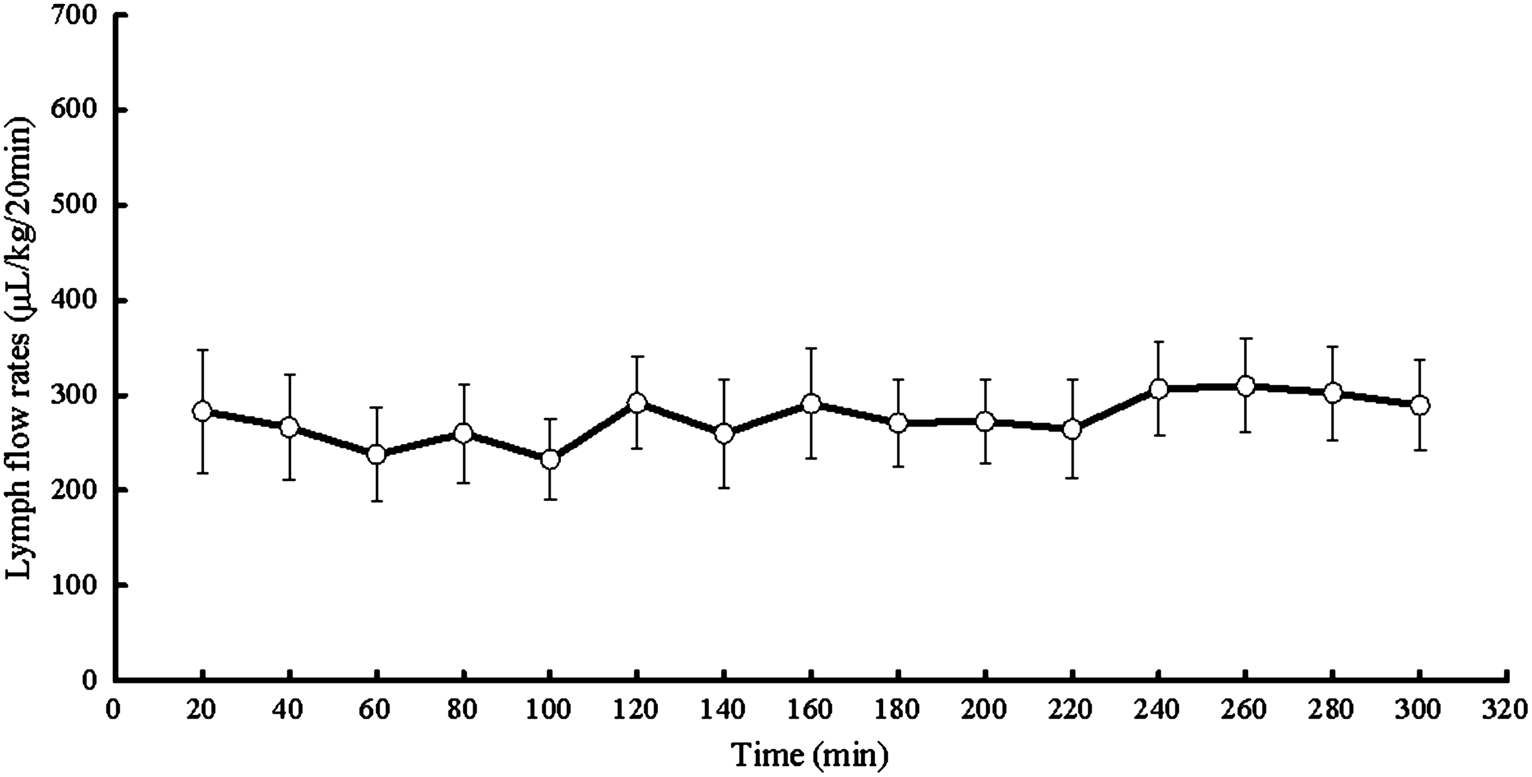
Time course of mesenteric lymph flow rate changes in normal animals. Mesenteric lymph showed a steady flow rate over 5 h. Values represent the means±SEM of 7 experiments.
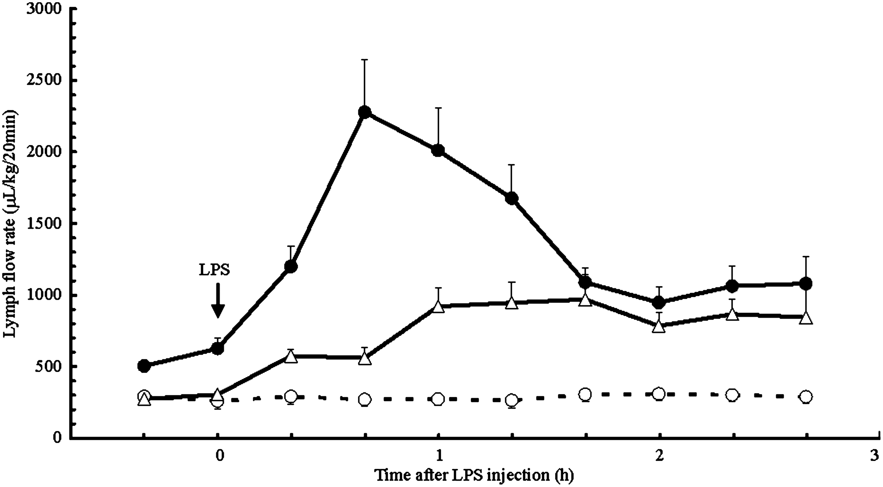
Effects of LPS on mesenteric lymph flow rates in guinea pigs. Guinea pigs were injected with 1 mg/kg LPS (open triangles), 10 mg/kg LPS (closed circles), or vehicle (open circles) as indicated by the arrow. LPS administration significantly and immediately increased the lymph flow rates. Mean values±SEM are shown (n=5–7 per group).
Evans blue leakage to lymph
Following i.v. injection of Evans blue, the dye gradually migrated into the lymph in the control animals (Fig. 4). In contrast, the Evans blue migration began immediately after injection of LPS in the LPS-treatment groups. Injection of LPS increased the lymph content of Evans blue dose-dependently and significantly. The Evans blue content 20 min after injection of 1 mg/kg LPS was 1.9±0.7 μg/20 min, and it was 6.3±1.3 μg/20 min 20 min after injection of 10 mg/kg LPS (both P<0.01 vs. vehicle). The lymph content of Evans blue increased maximally by 4.8-fold over the vehicle group 80 min after the 1 mg/kg LPS injection (9.8±2.8 μg/20 min) and by 11.3-fold over the vehicle group 60 min after the 10 mg/kg LPS injection (19.4±4.4 μg/20 min). Significant differences were found between the vehicle-treated and the 1 and 10 mg/kg LPS-treated groups (P=0.0002 and P<0.0001, respectively; repeated-measures ANOVA followed by Bonferroni post hoc test). The positive correlation between the lymph flow rate and Evans blue dye leakage is depicted in Figure 5 (r=0.947, P<0.01).
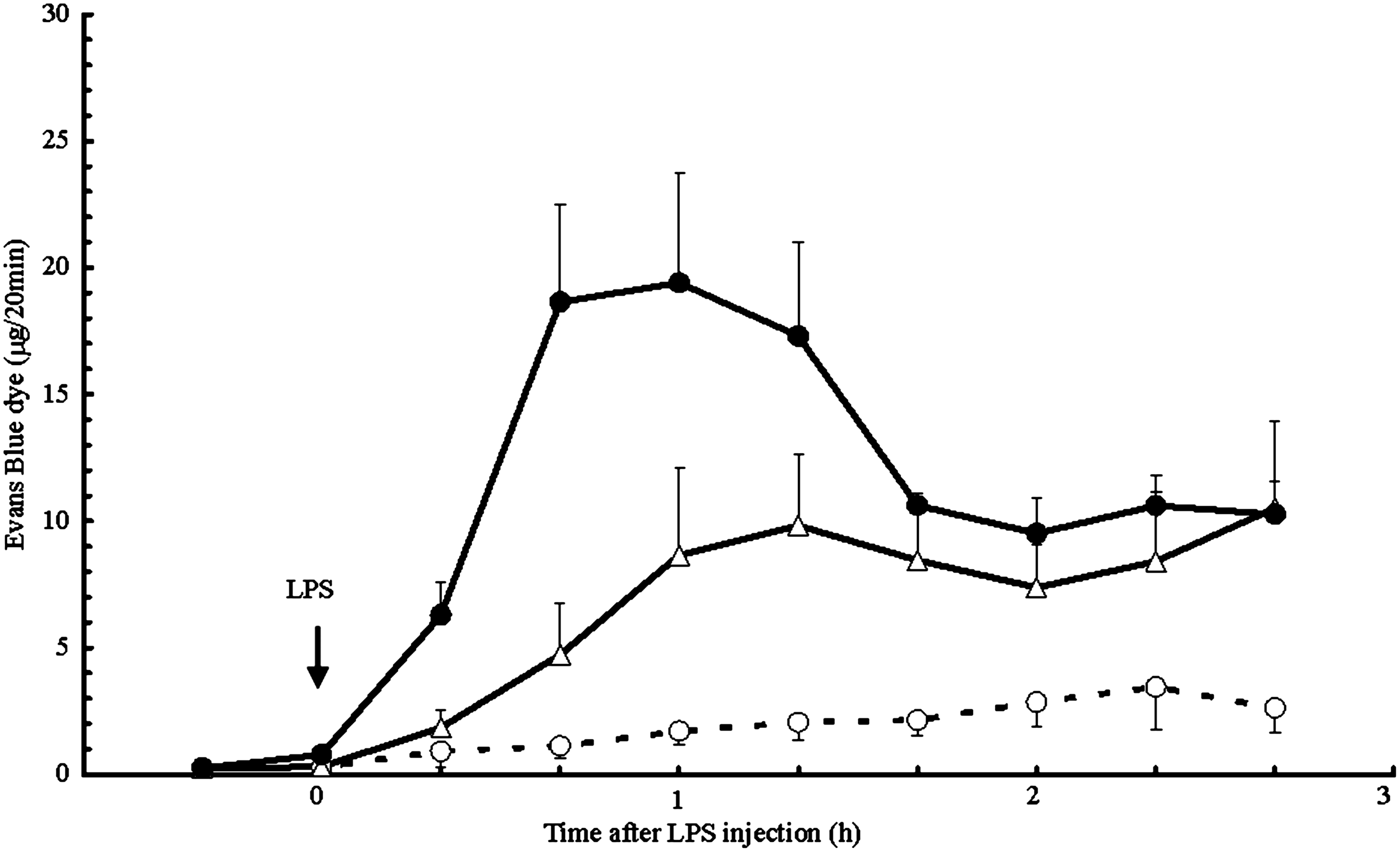
Effects of LPS on Evans blue dye leakage in guinea pigs. Guinea pigs were injected with 1 mg/kg of LPS (open triangles), 10 mg/kg of LPS (closed circles), or vehicle (open circles) as indicated by the arrow. LPS administration significantly and immediately increased Evans blue dye leakage. Mean values±SEM are shown (n=5–7 per group).
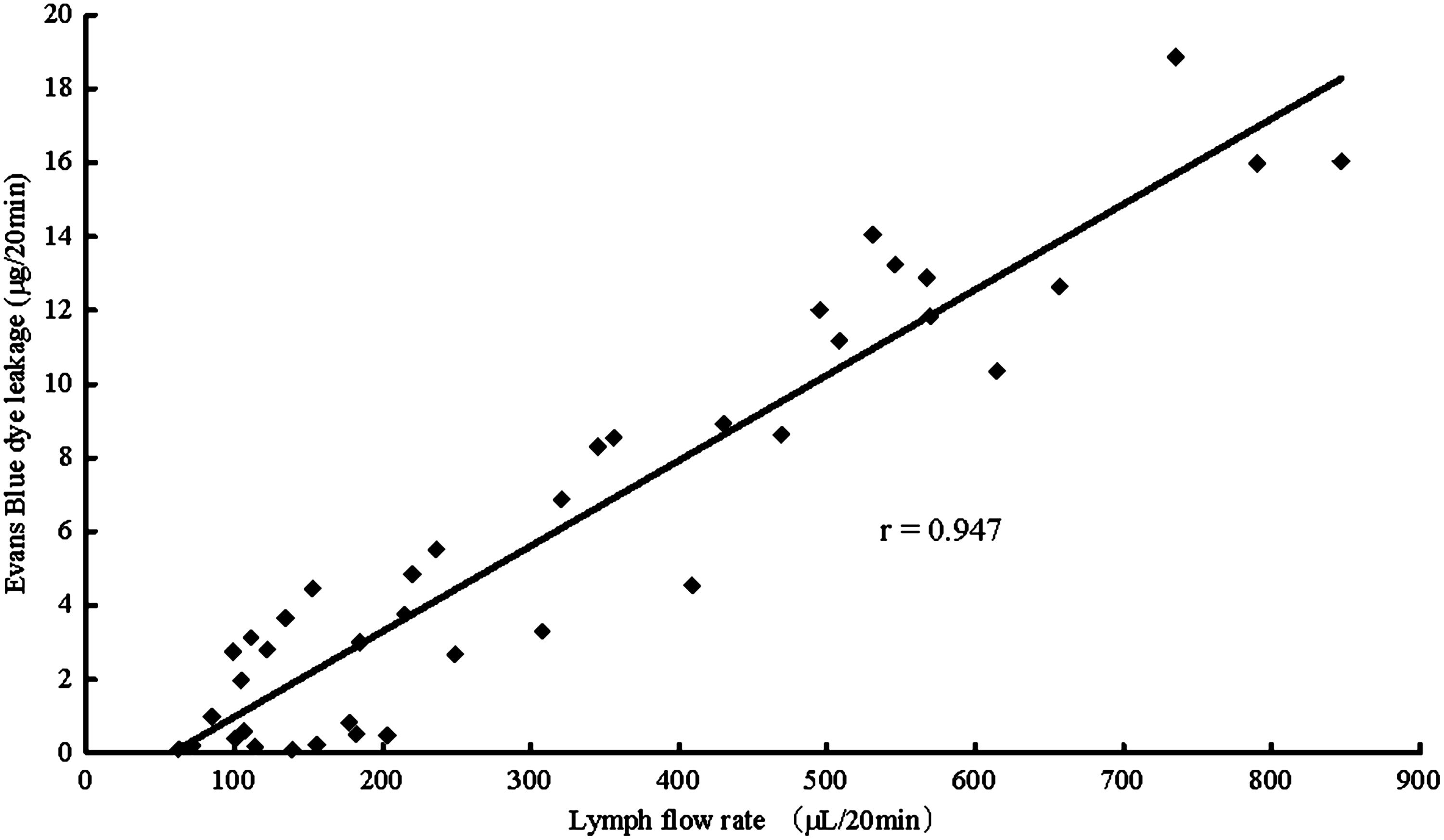
Correlation of lymph flow rates and Evans blue dye leakage. A positive correlation was observed between lymph flow rates and Evans blue dye leakage (r=0.947, P<0.01).
Effects of LPS on arterial pressure
In the vehicle group, the mean arterial pressure prior to the saline administration was 63.0±0.7 mmHg, and 20 min after saline administration it was 63.2±1.2 mmHg. Thereafter, the mean arterial pressure maintained a constant level until the termination of the experiment (Fig. 6). In the 1 mg/kg LPS group, the arterial pressure decreased shortly after LPS administration, dropping to 42.1±2.6 mmHg by 100 min. In the 10 mg/kg LPS group, the arterial pressure decreased more rapidly after LPS administration, dropping to 44.4±2.7 mmHg by 80 min. There was a significant difference (P<0.05) in the arterial pressure between the 1 and the 10 mg/kg LPS group at the 20 min point after LPS administration. When compared with the vehicle-treated group, there were significant differences in the arterial pressure between the vehicle-treated and the 1 and 10 mg/kg LPS-treated groups (both P<0.0001; repeated-measures ANOVA followed by Bonferroni post hoc test).
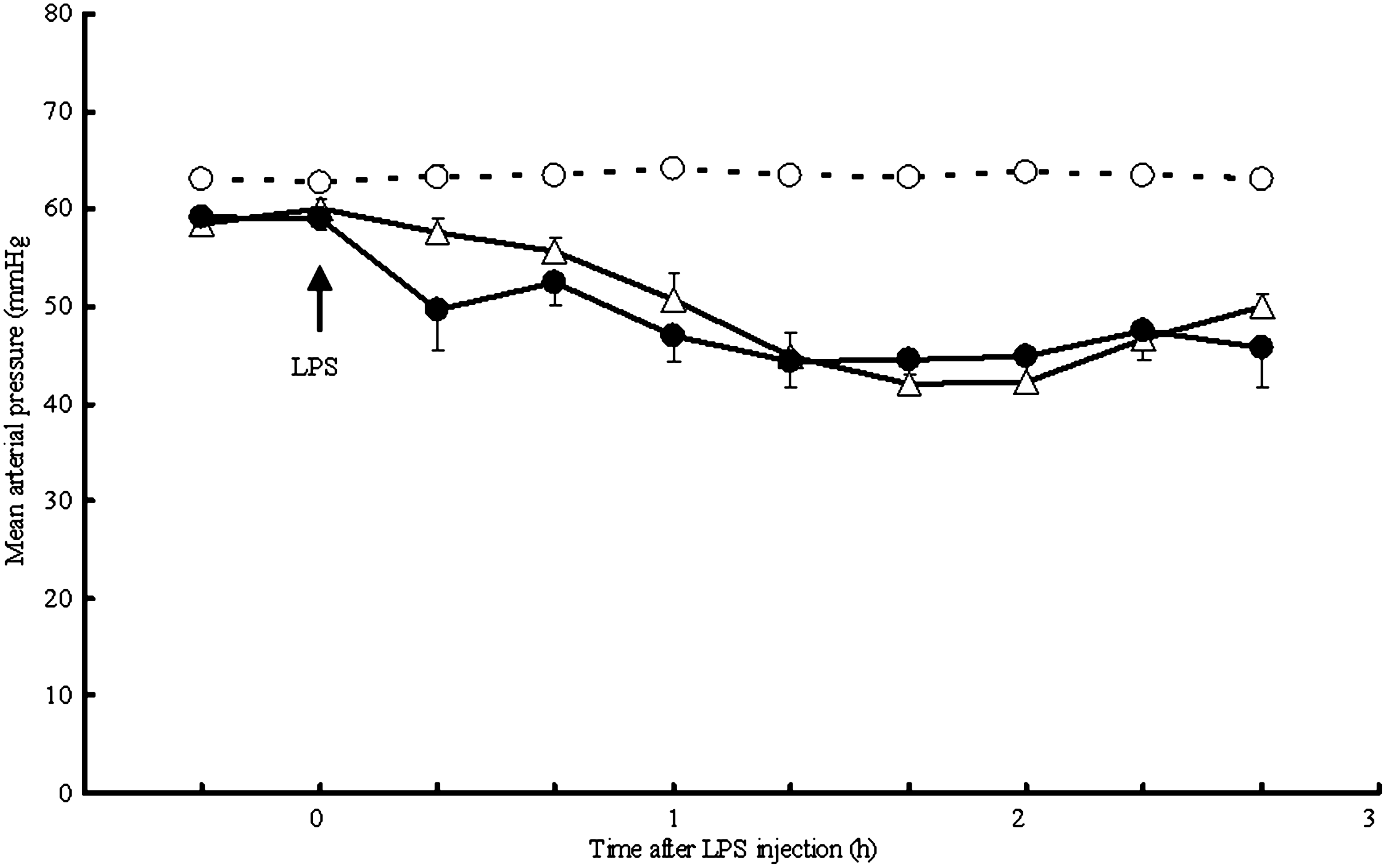
Effects of LPS on arterial pressure in guinea pigs. Guinea pigs were injected with 1 mg/kg of LPS (open triangles), 10 mg/kg of LPS (closed circles), or vehicle (open circles) as indicated by the arrow. LPS administration significantly and transiently decreased arterial pressure. Mean values±SEM are shown (n=4–6 per group).
Discussion
In our study, we demonstrate that mesenteric lymph flow increases immediately after intravenous LPS injection in guinea pigs. Additionally, the albumin content in lymph significantly increased in proportion to the increased lymph volume. These results suggest that LPS leads to the activation of vascular endothelial cells immediately after its administration, which is followed by increased permeability, resulting in serum albumin leakage into the interstitium. Large molecules, such as albumin, were easily drained into the lymphatic system via open junctions in the lymph vessels. As a result, the osmotic pressure increased and the interstitial fluid entered the lymphatic capillaries, which increased the lymph flow rates. We observed that LPS injection decreased the blood pressure. It was unclear whether the decline in blood pressure influenced the increase in the mesenteric lymph flow.
A number of factors may contribute to the increased lymph flow. In terms of endogenous factors, the activation of lymphatic pumping that is regulated by rhythmical contraction of the muscle present in the wall of the collecting lymphatics promotes lymph flow. 15 In terms of exogenous factors, the movement of intestinal muscles, pulsating movement of mesenteric vasculatures, decrease of intrathoracic pressure by inspiratory movements, and feeding promote lymph flow. However, these factors would have small effects in the present study, because the muscle tonus and blood pressure were very low during anesthesia. Furthermore, the direct action of LPS on lymphatic vessels should be considered, when LPS is intravenously administered to guinea pigs. It is reported that LPS diffuses to the lymph and directly acts on the lymphatic vessels. 16 However, even if LPS directly increases pumping of lymphatic vessels, the increase of lymph flow would never last long, because interstitial fluid drained out soon. In the present study, the increase of lymph flow lasted over 2 hours (Fig. 3). Accordingly, it appears that the major factor contributing to the increased lymph flow is albumin leakage into the lymphatic system, which is induced by endothelial hyperpermeability. Our results clearly demonstrate that lymph flow is strongly correlated with the absolute amount of albumin, and not with the concentration of albumin. This result indicates that the mesenteric lymph system is influenced by albumin quantity, not by albumin concentration.
It remains unclear whether LPS-induced vascular hyperpermeability is caused by the direct stimulation of endothelial cells or by secondary reactions via mediators from the other immune cells. As already stated, LPS causes the production and release of inflammatory cytokines. Besides, LPS provokes the release of vasoactive amines, such as histamine and serotonin stored in granular vesicles. Therefore, these mediators might potentially act on the vascular endothelial cells and cause vascular hyperpermeability. However, provided that lymph flow increased 20 minutes after LPS injection and lasted over 2 hours (Fig. 4), vascular hyperpermeability was caused by the lasting contraction of endothelial cells rather than by cytokines and vasoactive amines, because it takes over a couple of hours for cytokines to be produced, and because the biogenic amines are very short-lived. 17 Thus, it seems reasonable to suppose that LPS directly acts vascular endothelial cells and induces vascular hyperpermeability.
We had shown that the administration of 10 mg/kg LPS to guinea pigs provoked severe endotoxemia. 18 LPS is a very important causative agent of endotoxemia and sepsis. Despite recent advances in intensive care treatment, sepsis continues to be associated with a high mortality rate. Although anti-tumor necrosis factor-α, and interleukin-1 receptor blockers have been used to treat sepsis, the results of randomized controlled trials on these treatments have been unsatisfactory.19,20 Treating sepsis requires a more etiologic therapy than a symptomatic one. Since the present study showed that the lymph system is highly involved in the primary response following LPS administration, the therapeutic strategies which ameliorate the vascular leaky barrier could be used to treat inflammatory diseases and sepsis. In conclusion, the present study indicates that LPS injected intravenously to living organisms leads to endothelial hyperpermeability followed by leakage of plasma albumin, which is drained into the mesenteric lymph, resulting in increased lymph flow.
Footnotes
Author Disclosure Statement
The authors have no financial conflicts of interest.