




Abstract
Abstract
Purpose:
To make a lymphatic specimen of the dorsum of the hand for educational and clinical purposes.
Methods:
A total of four hands from two unembalmed human cadavers were used. Under a surgical microscope, 6% hydrogen peroxide was employed to detect the lymphatic vessels commencing from fingers. A 30-gauge needle was inserted into vessels and injected with a barium sulphate mixture. Each specimen was radiographed, photographed, and dissected to demonstrate the lymphatic vessels on the dorsum of the hand. After being embalmed in 4% formalin for 3 months, specimen was then sealed in a plexiglass container for display.
Results:
An average of sixteen lymph collecting vessels were found in the subcutaneous tissue of the dorsum of the hand. The diameter of the vessels varied from 0.2 mm to 0.6 mm. Vessels were the continuation of lymph vessels arising from fingers. They travelled meanderingly in the subcutaneous tissue and traversed over or under the veins when they met.
Conclusion:
Actual and accurate lymphatic distributions of the dorsum of the hand have been described and displayed. The information upgrades our anatomical knowledge and the results will be of benefit for the lymphatic education and clinical application.
Introduction
S
In the last decade, a new technique with radio-opaque materials as the injection medium has been used for the lymphatic studies in human cadavers.14–24 Results including new discoveries might help to clarify some unexpected findings in the clinic. Studies accurately revealed the lymphatic pathways by the radiographs and photographs but the displayable lymphatic specimen has not been made for medical educational use.
In this study, a new method has been described to make a lymphatic specimen of the dorsum of the hand. It will fill the gap in the field of the lymphatic system in our anatomic museum for educational and clinical purposes.
Materials and Methods
The study was performed with appropriate institutional ethics approval. Four hands were transected at the level measuring two inches above wrist joins from two unembalmed human cadavers, one male and one female, aged 68 and 72.
Lymphatic mapping commenced on the bilateral side of each finger. 0.5 mL of 6% hydrogen peroxide (Zhonglian Chemical Co., Ltd, Suzhou, China) was injected into the dermis and subcutaneous tissue. The epidermis and dermis were incised carefully and gently under a surgical microscope (Leica M651, Leica Microsystems Ltd, Heerbrugg, Switerland). The distended lymphatic vessel was identified in the subcutaneous tissue of the finger. A 30-gauge needle (Zhejiang KDL Medical Equipment Group Ltd., Wenzhou, China) was inserted into the vessel, which was then injected with a radio-opaque barium sulphate mixture (barium sulphate 15 g; Shanghai Silian Industry Co. Ltd., China; milk powder 5 g; Heinz Ltd., Qingdao, China; concentrated poster color: dark green 3 g; Liaoyuan Arts and Stationery Ltd., Hunang, China; water 20 mL) (Fig. 1). Lymphatic vessels were thus traced, photographed, and radiographed (Fuji FCR IP Cassette, and Fuji Computed Radiography Processor: Fuji Film Corporation, Tokyo, Japan) to demonstrate lymphatic distribution on the dorsum of the hand. Subsequently the radial and ulnar arteries in the transection of the specimen were found and cannulated. Each blood vessel was perfused by 10 mL of 4% formalin. The specimen was then soaked in 4% formalin solution for 3 months, and was sealed in a plexiglass container with the solution for display afterwards.

Initial injecting site in the lateral side of the left middle finger.
The final results were transferred to computer (Dell Vostro 200: Dell Computer Inc. Chinese Division, P.R. China) for image analysis using the DICOM viewer (OsiriX Imaging Software, osirux@orisix-viewer.com) and Adobe Photoshop (Adobe Photoshop CS5, Adobe Systems Software Co., Ltd, Beijing, China).
Results
An average of sixteen lymph collecting vessels (range 12 to 18) were found in the subcutaneous tissue of the dorsum of the hand (Figs. 2 and 3). The mean vessel diameter was 0.3 mm (range 0.2 to 0.6 mm). The neighboring vessels arising from fingers converged in the web spaces of the hand, except for the vessels on the medial and lateral borders of the thumb and little finger. They then branched, diverged, converged, and anastomosed with or crossed over neighboring vessels, forming a large lymphatic network. They travelled meanderingly in the subcutaneous tissue and traversed over or under the veins when they met.

A lymphatic specimen of the hand. A color version of this figure is available in the online article at www.liebertpub.com/lrb.
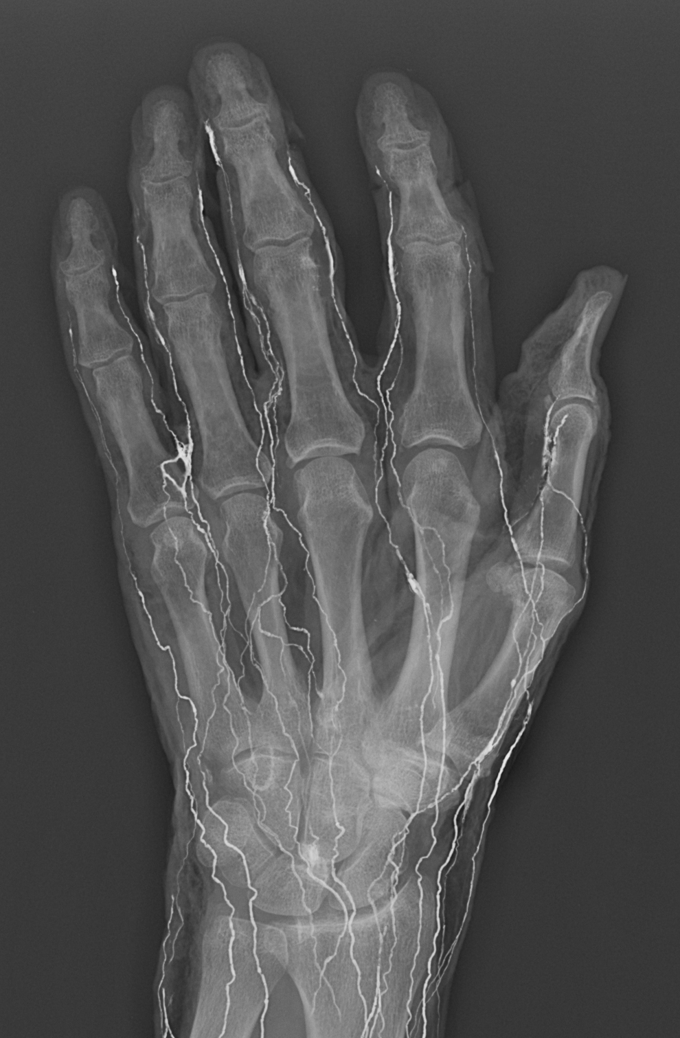
A radiograph showing the superficial lymphatic distribution of the dorsum of the left hand.
The colour of the mixture filled in the lymphatic vessels did not fade after the specimen was soaked in 4% formalin solution more than 6 months (Fig. 2).
Discussion
Since Aselli identified the lymphatic vessels in a postprandial canine in 1622, 1 the lymphatic system has been studied by using many different methods for scientific and clinical needs.2–13 In the last decade, a new technique with radio-opaque materials as the injection medium has been developed for accurately revealing the lymphatic pathways in human cadavers,14–24 and results could be recorded by radiographs and photographs. But the displayable lymphatic specimen has still not been made for further study and educational purpose.
In this study, a new method to make a lymphatic specimen of the dorsum of the hand has been described. During the experiment, lead oxide mixture was tried as an injectant. But the color of the injectant faded after the specimen was soaked in 4% formalin solution for a month. Oil paint color was difficult to mix in with the barium sulphate mixture. Only poster color paint mixed well in the barium sulphate mixture, and color does not fade for a longer time after soaking in 4% formalin solution (the specimen in Fig. 2 had been sealed in a plexiglass box for more than 3 months). After embalmment by 4% formalin, the specimen could still be radiographed and the result was satisfactory (Fig. 3). Displayed with specimens of the other systems of the human body, the lymphatic specimen could fill in the gap of the lymphatic system in our anatomical museum.
The results from this study have presented the number, size, and distribution of lymphatic vessels, on the dorsum of the hand using radiography and photography. The detailed relationship between the lymphatics and venous has been shown. It could be an alternative site for the lymphatico-venous anastomoses to treat the secondary lymphedema in the upper limb.25,26
Furthermore, the method may be applied to different regions of the human cadaver for displaying the lymphatic system, including deep lymphatics. In fact, pilot studies of deep lymphatics of the palm and forearm have been recently performed by the authors. Deep lymphatic vessels have been found and partially injected. It is believed that it is possible to reveal the deep lymphatics in limbs using the technique.
Summary
Actual and accurate lymphatic distribution of the dorsum of the hand have been described and displayed. The information upgrades our anatomical knowledge and the results will be of benefit for the lymphatic education and clinical application.
Footnotes
Author Disclosure Statement
No competing financial interests exist.