
Abstract
Abstract
Background:
Intra-articular wear particulate migration from the knee joint has been studied in various animal models as well as postmortem in patients who received total knee joint replacement. However, there still exists a need for a simple, yet analogous animal model for tracking the migration of wear debris from the knee joint, especially through the draining lymph nodes.
Methods and Results:
To fill this need, a proof-of-concept porcine model was developed for particle migration from the knee joint into the surrounding lymphatic system. Vitreous carbon particles were deposited both intra-articularly and extracapsularly in a bilateral manner to the hind limbs in pigs (n = 6). The regional/draining lymph nodes were qualitatively assessed weekly by a veterinarian by manual palpation to detect any enlargement or change in consistency when compared to the initial assessment before the surgical procedure. At 6 weeks, the draining lymph nodes were harvested and processed for histology. Microscopic evaluation showed carbon particle migration from the knee into 100% of the iliac lymph nodes, 50% of the inguinal lymph nodes, and 0% of the popliteal lymph nodes.
Discussion:
Overall, this study established a needed animal model for evaluating carbon particle migration to the draining lymph nodes from the knee joint.
Introduction
Intra-articular wear particulate migration from orthopedic implants in the knee joint has been studied in a variety of animal models such as sheep,1,2 rabbits, 3 guinea pigs,4,5 and mice. 6 In addition, wear particle migration has also been studied postmortem in patients who received total knee joint replacement. 7 Each of these models possesses benefits (e.g., low cost) and detriments (e.g., anatomic differences of bone structure and lymphatic system compared to humans, difficulty with manufacturing appropriately sized implants, and the retrospective nature of a postmortem analysis), and so there still exists a need for a simple, but analogous animal model for tracking migration of wear debris from the knee joint and surrounding tissues, especially through the draining lymph nodes. In reviewing the relevant comparative anatomy and in vivo surgical model literature, the possibility that common domestic pigs (Sus scrofa domesticus) may provide a more clinically relevant model for particle migration became apparent. Specifically, the porcine knee is more similarly sized to the human knee compared with smaller animals and other large animals, except for bovine knees,8,9 has similar lymphatic structures and drainage characteristics,10–12 and has been validated as a model for the evaluation of arthroscopy skills. 13
A rigorous discussion of the various transportation pathways that particles may take as they migrate from inside or near to the joint space into and then throughout the lymphatic system is beyond the scope of this article. Sieving by the synovial extracellular matrix is probably only effective for clearing the smallest particles (<0.05 μm),14,15 whereas cellular transport by macrophages is the most likely candidate for larger wear particles as those particles found in draining lymph nodes are often associated with multinucleated giant cells.1,3
The primary goal of this work was the creation of a proof-of-concept porcine model of particle migration from the knee into the draining lymph nodes. The envisioned application of this animal model was for evaluating particles emanating from orthopedic implants.
Materials and Methods
Six male adolescent Yorkshire pigs (Sus scrofa domesticus) weighing 40–50 kg were used for the study, which was conducted under an approved Institutional Animal Care and Use Committee (IACUC) protocol at an independent testing laboratory (CBSET, Inc., Bedford, MA). This study was performed in compliance with Food and Drug Administration Good Laboratory Practice (GLP) regulations. The test protocol was approved by the appropriate governing committee, while quality assurance monitors conducted in-process inspections at critical phases of the study, including, but not limited to, material implantation and material recovery.
All animals were quarantined and underwent standard veterinary examinations to confirm their health status before study assignment. Once deemed healthy, they were released for the study. None was rejected and both knees of each animal were used for implantation. Equal doses of vitreous carbon particles were placed in both the joint and the extracapsular space following joint capsule closure.
The vitreous carbon particles were prepared by mixing 0.06 g of Goodfellow Vitreous Carbon (VC006010/6; sized 0.4 to 12 μm) powder, 0.45 g of Goodfellow Vitreous Carbon (VC006015/2; sized 20 to 50 μm) powder, 0.81 g of Goodfellow Vitreous Carbon (VC006021/1; sized less than 300 μm) powder, and ∼10 mL of sterile saline. This range of particle sizes was chosen to match the particle size profile for a prototype implant that was not evaluated in this study (unpublished data), although this range also roughly coincides with the previously published sizes of wear particles found in the lymph nodes (<0.1 to 50 μm), found in vivo following the death of patients with prior hip and knee implants. 7 The saline/carbon particle mixture was sterilized and shaken vigorously before injection. Particles sized below 100 μm have been shown to migrate more readily into the lymphatic system than particles sized above this threshold16–18 and indeed, the human lymphatic system has network vessels that range in diameter between 100 and 300 μm 19 Carbon debris has been shown to produce synovitis and nonspecific foreign body reactions, but these are not of sufficient severity or duration to alter the mechanical properties of knee cartilage. 20
Surgical procedure
The animals were fasted before the implantation surgery, Telazol® (Zoetis Services LLC, Parsippany, NJ; 4 mg/kg, intramuscular [IM]) administered as a pre-anesthetic, and isoflurane anesthesia (delivered in 100% oxygen) administered to effect through mask/nosecone until the animals were in a plane of anesthesia that facilitated endotracheal intubation. Once sufficiently anesthetized, the animals were intubated and maintained on continuous inhalant 2.5% isoflurane anesthesia delivered to effect in 100% oxygen for the remainder of the procedure. A peripheral intravenous (IV) catheter was placed for administration of supportive IV fluids. Accepted veterinary care standards were followed. The incision sites were aseptically prepared and draped for the procedure. A parapatellar incision was made and carried down to the capsule. A moderate (∼2 cm) capsulotomy was performed and the patella retracted. Using a sterile syringe with no needle, half of the saline/particle mixture (∼5 mL) was injected into the joint space and a water-tight capsular closure performed with appropriate sutures. Then, the remaining particles were injected onto the capsule (i.e., superficially) and the skin closed over this site. In this way, approximately half the particles were placed inside the joint and half placed in the soft tissues surrounding the joint. Wear debris from orthopedic implants could theoretically originate from either location. The same steps were then repeated on the contralateral side. The postoperative care for all animals was the same. Buprenorphine (0.01–0.03 mg/kg, IM) was administered at least once every 12 hours to provide analgesic coverage through at least the first 72 postoperative hours and then as needed based on clinical assessment.
Six weeks after the implantation procedures, the animals were administered Telazol and anesthetized by isoflurane inhalant before being euthanized by an overdose of potassium chloride solution (IV) in accordance with accepted American Veterinary Medical Association (AVMA) guidelines.
Immediately after euthanasia, the knees were harvested. The prior surgical incision on both knees with the underlying tissue was excised en bloc and examined for visible extracapsular debris (particulates). This examination included the skin, subcutaneous tissue, and the joint capsule. Then, the patellar tendon was detached from the tibia and reflected proximally to fully expose the joint. The joint was examined for visible debris and any indication of an inflammatory reaction. Articular and meniscal cartilage damage, if present, were noted. Any inflammatory synovitis, deposits in the fat pad, or other intra-articular abnormalities was noted. These specimens and the joint contents were documented photographically and harvested for histology. In addition, right and left surface inguinal, popliteal, and iliac lymph nodes were harvested and visually examined.
Histology
The harvested lymph nodes were fixed in 10% neutral buffered formalin before being bisected, the cut faces placed side by side, and embedded in paraffin. In this manner, each slide incorporated two sections of tissue. Slices were taken approximately 0.6 mm apart in the block and the resulting sections were stained with hematoxylin-eosin (H&E). The incision site tissue sections and any other soft tissues of the knee with macroscopic changes were similarly processed and assessed.
Light microscopy was used for characterization of the host response and evaluation of the lymph tissue for foreign material using standard illumination and polarized light, the presence of inflammation and inflammatory cell subtypes, any disruption of tissue architecture, and signs of tissue injury (such as cell injury and death). The veterinary pathologist (T.R.M.) was blinded to the treatment group at the time of evaluation. The following scoring matrix was used: 0 is no observable change; 1 is a minimal or nearly imperceptible feature or change in the tissue; 2 is a mild to moderate or easily identifiable or notable feature or change in the tissue; and 3 is a marked or severe change (prominent to overwhelming feature or change in the tissue).
The study endpoints included the presence of the following: visible carbon debris during necropsy, cellular evidence of inflammation in the lymph nodes, and carbon particles in the lymph nodes. Descriptive statistics were performed on the inflammation scores.
Results
All animals survived to the 6-week time point, while gaining weight throughout the study with no changes in ambulation. While there were some observations of transient inguinal lymph node enlargement, all lymph nodes were deemed within normal limits before necropsy. Regions of black discoloration (such as would be expected in the presence of carbon particles) in the subcutaneous layer and/or joint capsule were macroscopically observed during tissue harvest (Fig. 1A–D) for 4/6 animals, but articular and meniscal cartilage damage were not observed.
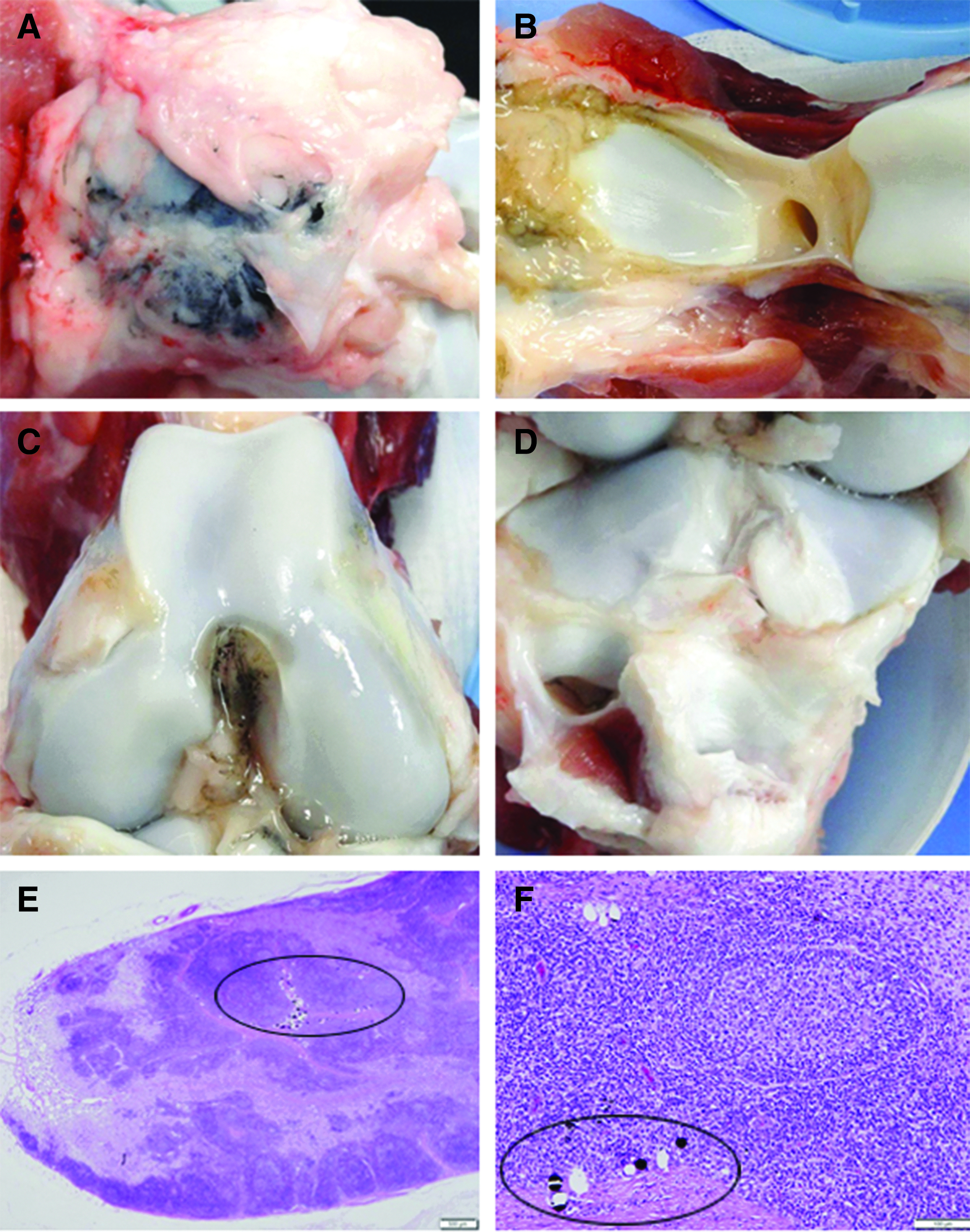
Representative macroscopic images of right knee joint superficial to joint capsule
Histologic assessment found that carbon particles migrated to 100% of the iliac lymph nodes (Fig. 1E, F) and 50% of the inguinal lymph nodes, but none (0%) of the popliteal lymph nodes, while inflammation and inflammatory cell types were found more often in the iliac nodes compared to the inguinal and popliteal nodes (Table 1). In a region of interest, the size range of carbon particles was between 2.4 and 49.2 μm (Fig. 1F). In looking at the individual iliac lymph nodes on these six animals, the presence of carbon particles was strongly associated with the presence of inflammation (12/12), histiocytes (12/12), multinucleated giant cells (12/12), but neutrophils (2/12) and eosinophils (2/12) were found bilaterally in a single animal (#47042). Interestingly, the iliac lymph nodes for this same animal exhibited mild/moderate inflammation in contradistinction to the remaining five animals that demonstrated only minimal inflammation. A similarly strong association between the presence of carbon particles and the presence of inflammation, eosinophils, and multinucleated giant cells was found for the inguinal lymph nodes. Eosinophils were found bilaterally in two animals, one where carbon particles were found and one where they were not found (animal #47042). Finally, none of the popliteal lymph nodes showed any presence of inflammation or inflammatory cell types. In no case did histology provide any evidence of cell injury or death.
Mean ± Standard Deviation, Median, and Incidence of Inflammation, Inflammatory Cell Types, and Carbon Particles
Endpoint scores: 0, no observable change; 1, minimal—a nearly imperceptible feature/change in the tissue; 2, mild/moderate—an easily identifiable and/or notable feature/change in the tissue; 3, marked/severe—prominent to overwhelming feature/change in the tissue.
Discussion
The most important finding of this GLP study is the validation of a porcine model described here, which allows for the assessment of particle migration to the draining lymph nodes from the intra-articular and extracapsular spaces of the knee joint. Carbon particles migrated from the knee into the iliac lymph nodes 100% of the time and the inguinal lymph nodes in 50%, but did not migrate to the popliteal nodes (0%). Further characterization of the model (such as a rigorous measurement of the particle size(s) that successfully migrate) is warranted, although this porcine model even as currently detailed can be useful for evaluating migration of orthopedic device-generated particles in the knee.
Margevicius et al. 1 evaluated ligament replacement devices and the resulting foreign particle migration of ultra-high molecular weight polyethylene (UHMWPE), polylactic acid, carbon, polytetrafluoroethylene, aramid, polyester, and polyethylene (PE) in an ovine knee model. Carbon particles were found in 43% of the iliac lymph nodes (57% in the ipsilateral node and 29% in the contralateral node). This value is lower than the percent migration found in this study, but the effect of slow (yet constant) particle generation versus a single bolus of particles on migration kinetics is unknown.
In a study by Meachim and Brooke, 4 no migration of bone cement particles was found. Ipsilateral popliteal and inguinal lymph nodes were free of all foreign particles at time points between 3 months and 1 year following injections into the knee joints of guinea pigs. Interestingly, no particles were found in the swine popliteal lymph nodes in this study. It is not known why these lymph nodes, which are in close proximity to the knee joint, do not appear to collect foreign particles introduced to the knee; however, it is possible that the particles migrate completely through these lymph nodes in less than 6 weeks. Lalor et al. 3 found “some PE particles” in the popliteal lymph nodes of rabbits at 2 and 4 weeks following intra-articular injection of PE particles. However, Ito and Suami 21 found that the region of drainage into the popliteal lymph nodes was predominantly on the dorsal/posterior aspect of the hind limb in a mapping study performed on an in vivo porcine model. In this case, particles placed within the knee capsule and more superficially (such as in this study) might not migrate at all to the popliteal lymph nodes in swine.
Urban et al. 7 studied the para-aortic lymph nodes of cadavers with prior knee replacement surgery and found either PE or metallic particles in 67% of patients. This is lower than the incidence reported in this study for iliac lymph nodes and higher than the incidence reported for inguinal and popliteal lymph nodes. One reason for the discrepancy might be the distance that these various lymph nodes are from the knee joint, although Pan et al. 22 showed that there are two divergent lymphatic drainage systems (medial and lateral groups) in human lower limbs.
There are several limitations to this study. While the similarities between the porcine and human lymphatic systems have been previously documented,10,11 there are also important differences, such as reduced number and size of lymphatic vessels as well as some difference in lymphatic drainage pathways in swine.21,23 The results from this animal model may not be directly comparable to the clinical experience. Also, only one time point was evaluated for this study. Based on an unpublished pilot study, this appeared to be an appropriate length of time to bring the animals to term, but it is possible that some carbon particles had already cleared the lymph nodes that were examined, while other particles might take longer to reach the lymph nodes. We acknowledge that the kinetic process for transporting particles to the lymph nodes might differ from that for transporting the particles out of the lymph nodes. Taking only a single “snapshot” in time of the lymph node likely does not tell the whole picture and future work should target not only additional time points but also an understanding of the particle transport mechanisms into and out of the lymph nodes. As expected, multinucleated giant cells were closely associated with the carbon particles found in the lymph nodes for this model. Finally, while the benefit of using carbon spheres as particles has the advantage that the particles are readily identifiable both macroscopically and microscopically, other particles that are more relevant to orthopedic devices (e.g., titanium, stainless steel, UHMWPE, and silicone) might migrate at different rates based on their size, shape, and composition. There is some evidence that wear particle migration to the lymph nodes is more closely tied to the chemical makeup of the particles rather than their size. 24
Six weeks after intra-articular and pericapsular implantation, carbon particles migrated from the porcine knee into the iliac lymph nodes 100% of the time and the inguinal lymph nodes in 50%, but were not found in the popliteal nodes (0%). This study validates a porcine model for the evaluation of carbon particle migration from the knee into the draining lymph nodes.
Footnotes
Acknowledgment
This study was funded by DePuy Synthes Mitek Sports Medicine (Johnson & Johnson).
Author Disclosure Statement
D.B.S. and T.R.M. are both employees of Johnson & Johnson; F.A.B. has received research or institutional support previously from Johnson & Johnson; And B.G.Z. and R.M. are both employees of the CRO where this study was performed. This study does not describe a product developed or marketed by Johnson & Johnson.