
Abstract
Background:
Lymphedema (LE) is a chronic progressive protein-rich edema of the soft tissues. Measurement of extracellular fluid of the affected limbs is widely used in detecting LE; however, quantification of the skin alterations and early tissue changes in LE lacks approaches.
Methods and Results:
Ninety-one patients with LE were assessed. Measurement of transepidermal water loss (TEWL), skin stiffness (SF), and percentage water content (PWC) was assessed on five predetermined skin sites. The value of TEWL, SF, and PWC increased significantly in lymphedematous skin compare with controls, indicating damaged function and texture of the affected skin. Both PWC ratio and SF ratio strongly correlated with LE stage. High correlations were found among instruments assessing tissue fluid.
Conclusions:
Assessment of the skin parameters has contributed new information about the functional and structural alterations in chronic lymphedematous skin. Quantification of skin properties changes could be a valuable supplement to diagnosis and evaluation of chronic LE.
Introduction
Lymphedema (LE) is a chronic progressive protein-rich edema of soft tissues resulting from abnormal functioning of the lymphatic circulation. 1 Based on the causes, LE can be classified into primary LE, which is usually considered as developmental anomaly of the lymphatic system, and secondary LE, which includes causes such as radical cancer surgery, radiotherapy, trauma, and filariasis. Fluid accumulation, inflammatory cell infiltration, and increased fibroblast activity begin at the very early stage of LE. With progression, thickening, and fibrosis of skin, adipose tissue deposition is generally involved in the superficial tissue.2–4 The hardening of the skin and subcutis further exacerbates impaired lymph transport.
LE has significant impact on quality of life since it often involves extremities and leads to limb heaviness, tension, hyperpigmentation, altered sensation, and high risks of infection, skin disorders, and functional limitations. 5 Therefore, accurate assessment of skin conditions, including early tissue changes, is crucial for detecting latent or incipient LE and the effective management of LE. Early and accurate detection may help to reduce the risk of developing clinically manifest LE. In those cases of long standing LE, detailed assessment of lymphedematous skin and its response to treatment may help in monitoring the development of the disease and selecting the optimal treatment regimen.
Bioimpedance (BI) analysis is able to measure small changes in the extracellular fluid (ECF) composition of a limb and has currently been widely used for detecting LE.6,7 However, detailed alteration of the function and texture of the skin and subcutis has been overlooked. Currently, noninvasive and quantitative assessment of skin properties involves measurement of skin stiffness (SF), tissue dielectric constant (TDC), and transepidermal water loss (TEWL). SF is an indicator of mechanical properties of the skin. It is usually measured through sensing the force (Newton, N) against the indurated tissue and can be applied to evaluate the degree of fibrosis of the progressive LE. 8 Besides fibrosis, tissue water changes accompany LE. Since TDC value (percentage water content [PWC]) of human skin measured at a frequency of 300 MHz is directly proportional to skin water content, local tissue water is quantifiable using the TDC measurements. Previous study has shown that measurements of TDC are useful for documenting LE and the early detection of incipient LE.9,10 TEWL [measures as g/(m2·h)], another noninvasive approach, is a physiological characteristic reflecting epidermal permeability barrier function. 11 Measurement of TEWL has been used in a variety of clinically related applications, including wound healing, sympathetic skin response, and evaluation of scar maturation.12,13 Increased TEWL value is considered as a characteristic feature of disturbed skin barrier. 14 Although chronic lymph stasis is known to disrupt skin function, alter skin innate immunity, and prone to infections and immune-mediated skin disorders,15,16 alteration of TEWL has only been studied in podoconiosis, 17 the TEWL change in secondary LE of extremities has not been investigated. In addition, there have been only limited studies comparing the mentioned skin parameters in assessing LE. Thus one goal of this study was to evaluate skin barrier function changes in patients with secondary LE. A second goal was to investigate the correlation among TDC, SF, TEWL, and BI values in assessment of chronic lymphedematous skin, and finally to determine the feasibility of these methods in evaluating skin conditions in patients with secondary LE.
Materials and Methods
Patients
Ninety women patients (mean age of 55 years, range: 39–64 years) with unilateral chronic LE of extremities were included in the study from September 2016 to March 2018. All patients were diagnosed with secondary LE, 34 with upper limb LE and 56 with lower extremity LE. All had undergone radical surgery and completed radiotherapy and/or chemotherapy for breast, endometrial, ovarian, or cervical cancer. The mean duration of the disease was 4 years (range: 0.5–19 years). According to the International Society of Lymphology staging system, 18 the number of patients diagnosed with stages I, II, III, and IV LE in the limbs was 3, 46, 36, and 5, respectively. The patients' clinical characteristics are summarized in Table 1. All subjects provided written informed consent. This clinical trial was approved by the Ethics Committee of the Shanghai Ninth People's Hospital.
Patient Characteristics (n = 90)
Values are presented as mean (range) or number (%).
LE, lymphedema.
Bioelectrical impedance analysis of ECF
The ECF in the limbs was measured by using an eight-polar bioelectrical impedance analysis instrument (Inbody 3.0; Biospace, Seoul, Korea), which has been reported to provide accurate estimates of extracellular water.19–21 Measurements were performed in accordance with the manufacturer's instructions. Measurement data were recorded in kilogram (kg). Difference between affected and contralateral limb was calculated. The excess ECF ratio was defined as (ECF of lymphedematous limb − ECF of the control limb)/ECF of the control limb × 100%.
SF measurement
SF was measured as induration value in the affected and contralateral limbs by using the SkinFibroMeter (Delfin Technologies) (Fig. 1A), which is expressed in Newtons (N). Five successive valid measurements need to be made to calculate the mean induration value for a single measurement site. A total of five sites were measured as indicated in Figure 1B: at the dorsal midline across the first web space of the hand, as well as the anterior and posterior midline across the midupper arm and forearm level of the upper limb; at 5 cm above the second plantar toe joint, as well as the lateral midline across the midthigh and calf level of the lower limb. The mean value of these five readings was calculated to indicate the overall SF for each limb. The excess SF ratio was defined as (mean induration value of lymphedematous limb − mean induration value of the control limb)/mean induration value of the control limb × 100%.

Instrument used for skin examinations in this study
TDC measurement
TDC was measured by using LymphScanner (Delfin Technologies) (Fig. 1A). The device contains a force-controlled probe, through which PWC can be assessed by touching the measurement site of the skin. Five sites of each limb were measured as already described. Three repeated measurements were made to calculate an average PWC value for a single measurement site. The mean PWC value of these five sites was calculated for each limb. The excess PWC ratio was defined as (mean PWC value of lymphedematous limb − mean PWC value of the control limb)/mean PWC value of the control limb × 100%.
TEWL measurement
The VapoMeter (Delfin Technologies) (Fig. 1A) is used for the evaluation of skin barrier function by measuring TEWL and evaporation rate, expressed in grams per square meter per hour [g/(m2·h)]. Patients were asked to sit still for at least 15 minutes before measurement in the examination room with a room temperature maintained at 25°C and humidity of ∼45%. Five sites of each limb were measured as already described, with three repeated measurements made to calculate an average value for each site. The mean TEWL value of these five sites was calculated for each limb. The excess TEWL ratio was defined as (mean TEWL value of lymphedematous limb°− mean TEWL value of the control limb)/mean TEWL value of the control limb × 100%.
Statistical analysis
Statistical analysis of the data was performed using paired sample t-test and one-way analysis of variance followed by Bonferroni's post hoc tests. The Spearman correlation coefficient was used to express correlations between LE stage and measured parameters. Pearson correlation coefficient was used to analyze correlations between two skin parameters. p Values were obtained at the significance level of 0.05. Statistical analysis was done using SPSS17.0 software (IBM, Armonk, NY).
Results
The value of ECF, SF, PWC, and TEWL in lymphedematous and control limb
Mean value of ECF, SF, PWC, and TEWL in the lymphedematous and control limb was calculated. All of these values were significantly higher in lymphedematous than in control limb, as shown in Figure 2. The values of each parameter separated by upper and lower extremities are listed in Table 2. These results indicating increased fluid accumulation, skin fibrosis, and damaged skin barrier function under lymphedematous condition.

Lymphedematous limbs had higher value of ECF
The Value of Extracellular Fluid, Skin Stiffness, Percentage Water Content, and Transepidermal Water Loss in Lymphedema and Control Limbs
p < 0.01; bp < 0.05.
ECF, extracellular fluid; PWC, percentage water content; SF, skin stiffness; TEWL, transepidermal water loss.
Correlations between the excess ECF, SF, PWC, and TEWL ratio with the severity of LE
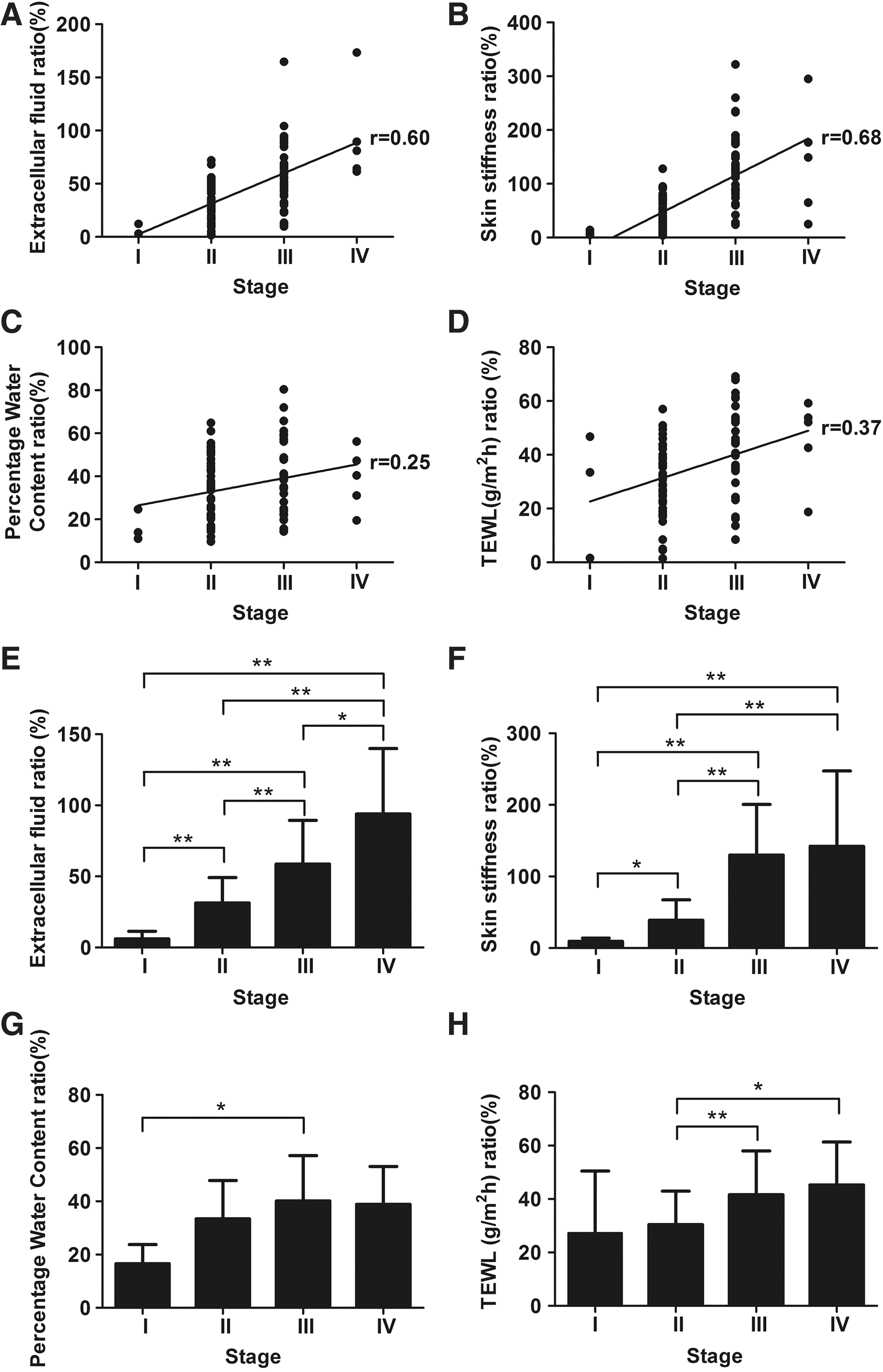
Correlations between the stage of LE and the excess ratio of ECF, SF, PWC, and TEWL were analyzed. As shown in Figure 3A–D, the excess ratio of ECF, SF, PWC, and TEWL correlates positively with the stage of LE. Strong correlations were found between ECF ratio and LE stage (r = 0.60), as well as between SF ratio and LE stage (r = 0.63). The correlation analysis performed with PWC ratio and TEWL ratio showed weak correlations with LE stage (r = 0.26 and r = 0.37, respectively). Comparisons of the mean ECF, SF, PWC, and TEWL ratio between different stages of LE showed that the ratio of these parameters tends to increase from stage I to stage III, with the excess ratio of ECF and SF showing significantly higher value in stage III LE than in either stage II or stage I LE (Fig. 3E–H).
Correlations between the ratio of excess ECF
Correlations between the excess ratio of SF, PWC, and TEWL with ECF in the affected limb
Correlations between the excess ratio of SF, PWC, and TEWL with ECF were analyzed. SF and PWC ratio of lymphedematous limbs were significantly positively correlated with ECF ratio (r = 0.61, r = 0.62) (Fig. 4A, B). The strength of these correlations is strong. Correlation analyses between the TEWL ratio and ECF ratio show relatively weak correlation (r = 0.42) (Fig. 4C).

Correlations between the excess ratio of skin stiffness
Correlations among the measured skin parameters
Pearson's correlation analysis between the measured skin parameters of SF, PWC, and TEWL ratios was performed. The correlation coefficient between measurement of SF and PWC, SF and TEWL, as well as PWC and TEWL was 0.435, 0.289, and 0.530, respectively. Despite the significantly positive correlations among these parameters, the strength of the correlations is weak.
Discussion
In chronic LE, sustained lymph stagnation engenders a series pathological changes of the soft tissue, among which, skin and subcutis are the most widely affected. 22 However, current knowledge of the skin alterations in LE based mainly on biopsies and histological evaluations of the lymphedematous tissues and detailed functions and texture changes of the affected skin has been overlooked in clinical examination of LE. In this, by using more sensitive and convenient diagnostic equipment, we performed a comprehensive examination of skin property changes in chronic lymphedematous limb and evaluated the diagnostic value of these skin parameters in assessing secondary LE.
As BI analysis is commonly used by clinicians and considered as an ideal approach for detection of LE, we used BI analysis as a reference method to evaluate the severity of LE. Both the value of ECF measured by BI analysis and the value of PWC measured by LymphScanner reflect the extent and amount of fluid accumulation. Although these two values are significantly increased in lymphedematous limb, strong correlations were found between LE stage with ECF ratio but not PWC ratio, whereas compared with ECF ratio, PWC ratio showed a higher value in stage I (16.57%) and stage II (33.40%) LE, but the value increased less obviously in late-stage LE. This could be explained by the progressive accumulation of the fluid with increased severity of LE. In early stage of edema, the accumulation of lymphatic fluid begins at the skin dermis, which could be accurately detected by LymphScanner with a sensitive probe of a measurement depth up to 2.5 mm. With progression, fluid accumulation extends from the dermis to deep fascia, leading to greatly increased thickness of the skin and subcutis, which might be beyond the maximum measurement depth of the probe and thus unable to reflect the severity of late-stage LE. Compared with LymphScanner that measures PWC of a certain layer of the tissue, BI analysis measures the ECF change in the whole limb, thus could be more suitable in reflecting the severity and progression of the disease.
Although BI analysis is often suggested as a diagnostic tool to measure fluid volume in LE, a recent study revealed that diagnosing early stage LE through BI analysis leads to an excessively high rate of false-negative results, indicating the insufficient sensitivity of BI analysis. 7 Besides, since BI analysis measures ECF of the whole limb, localized changes may not be detected. In contrast, Lymphscanner shows high sensitivity for detection of early stage LE and measurement of water content of superficial LE. In addition, by rapid skin scanning, localized swelling or the extent of LE can be easily assessed.
Chronic inflammation and progressive fibrosis are characteristic pathological features of LE. SF value is accurate and reliable for the evaluation of skin fibrosis. Measurement and analysis of SF showed increased SF value with increasing severity of LE. Consistent with our previous study, 8 high correlations were found between excess SF ratio with both LE stage and ECF ratio, indicating decreased tissue compliance and increased skin fibrosis and hardening with disease progression. These results, in turn, reflect that tissue fibrosis progresses throughout the development of the disease and plays a critical role in the pathophysiology of the disease.
On clinical examination, patients with chronic LE often have dry hyperkeratotic skin, especially in advanced stage LE. 23 TEWL, an indicator of skin barrier function, was measured with VapoMeter. TEWL value increased significantly in lymphedematous skin, which indicates impaired skin barrier system due to long-term lymph stasis. The compromised skin barrier function may result in increased risks of skin irritation, sensitization, or infection in the affected limb. The TEWL ratio tends to increase with disease severity, although the correlation between TEWL and stage of LE or the ECF value was weak. This might be due to the high interindividual variability of TEWL parameter. Indeed, it has been reported that a variety of variables originating from individuals would influence TEWL parameters, such as age, gender, sweating, and skin surface temperature. 24 Interestingly, we also observed that TEWL was much lower in extremely hyperkeratotic skin sites of stage IV LE than in normal skin (data not shown), which probably indicates significantly severe skin thickness or damages. Impaired skin barrier function explains the dry itchy skin symptoms in LE as well as reflects the susceptibility to inflammation and other skin disorders. Thus skincare regimens should be given to LE patients for skin protection such as maintenance of skin moisture and bandaging with high air permeability material.
We observed that there are high inter-individual differences of the absolute value of the measured parameters among both upper and lower control limbs (data not shown). We, therefore, employ the “excess ratio” of each parameter for analysis to reflect the skin property change in lymphedematous limb relative to the contralateral control limb. Although patients with unilateral LE were included to evaluate the “excess ratio” of the measured parameters, we cannot exclude latent conditions in the case of lower extremity LE. Skin property alterations in latent or preclinical lymphedema need further investigation.
This study evaluated comprehensively the impaired skin properties in patients with chronic LE, including increased skin water content, SF, and reduced skin barrier function. These physical parameters of skin changes further our understanding of the pathophysiological alterations behind LE. Quantification of skin changes in chronic LE provides diagnostic valuable information of the skin condition in the affected limb. Further studies on the assessment of skin properties in LE after complex decongestive therapy or lymphatic surgeries should be considered.
Footnotes
Author Disclosure Statement
No competing financial interests exist.
Funding Information
This study was supported by the National Natural Science Foundation of China (Grant No. 81272146).