
Abstract
Renal lymphatic abnormalities are rare, and the understanding of pathophysiology involving renal lymphatics is limited. Symptoms can include hypertension, hematuria, proteinuria, chyluria, and abdominal and lumbar pain. Imaging techniques specific to the renal lymphatics have not been clarified. We review the intrahospital imaging evaluation/workup and clinical course of a 6-year-old male who presented to our institution with a large perirenal cyst. His presentation presented a diagnostic and management challenge. The cyst was determined to be lymphatic in origin and required multiple interventional radiology and surgical procedures for management.
Introduction
Pathophysiology of the kidney generally involves the arterial system, collecting system, or the renal parenchyma. Abnormalities of the renal lymphatic system are infrequently reported and even less frequently investigated. Successful in vivo imaging of human renal lymphatics has never been reported.
The vascular system of the kidneys is the same as elsewhere within the body. The renal lymphatics are found in conjunction with vascular structures, as well as within the cortex and medulla. 1 A previous report has described the lymphatic distribution in the human kidney due to distension by gastric carcinoma. 2
Renal lymphangiectasia (or renal lymphangioma) is a rare disorder, where there is dilation of perirenal, peripelvic, and intrarenal lymphatics. It might have a familial component, or can be acquired. It can be found in children or adults. 3 Imaging of the renal lymphatics is difficult, and most analyses of renal lymphatic issues occur after tissue removal, given the identification of a sensitive immunostain.
Podoplanin (D2-40) has become an established immunostain to identify lymphatic cells and is strongly expressed in the cytoplasm of lymphatic endothelial cells. 4 First identified in rats, Podoplanin is a 43-kDa membrane glycoprotein of podocytes. 5
Following approval by our institutional ethics review board, we reviewed and now present the case of a 6-year old without significant medical history who presented with right-sided flank pain and intermittent emesis. We demonstrated that he has normal central lymphatic magnetic resonance imaging (MRI) and we performed direct MRI renal lymphangiography to visualize his lymphatic channels preoperatively. Dilated lymphatics were confirmed pathologically with D2-40 staining after nephrectomy.
Case Report
A 6-year-old boy with no medical history presented with a large abdominal mass complaining of abdominal pain with intermittent nonbilious emesis and lumbar back pain for 3 weeks before presentation. He denied any fever, chills, or weight loss. There was no history of hematuria, frequency, or dysuria. An ultrasound was performed that demonstrated a large simple-appearing intra-abdominal cyst. On physical examination, he was found to have abdominal distension, diffuse tenderness, but no peritonitis. Laboratories were unremarkable.
An abdominal and pelvic computed tomography (CT) scan was obtained and he was diagnosed with a large right upper quadrant retroperitoneal cyst adjacent to a stretched out but otherwise radiographically normal-appearing right kidney (Fig. 1). There was significant mass effect on the surrounding organs. The cyst measured 16.9 × 14.6 × 11.8 cm and the fluid measured 4 Hounsfield units consistent with simple fluid. There was no contrast accumulation that would have suggested this collection to be a urinoma from a urinary leak. A definitive source/origin of the cyst was not ascertained by the CT scan. A few days later, a percutaneous drain was placed in interventional radiology (IR) to relieve the patient's back pain and allow for diagnostic sampling of the fluid (Fig. 2). This catheter drained 3L of simple-appearing fluid to start with and then plateaued at 1–1.5 L of fluid output daily.

Sagittal abdominal computed tomography scan of flank. Deformed right kidney and perinephric cyst.

Fluoroscopic image. Large cyst cavity, filling in entirety.
Laboratory analysis from the initial fluid drained revealed normal triglycerides (<10), with lymphocytes rising from 37% initial laboratory evaluation to 86% on repeat analyses. Fluid creatinine was normal and multiple comparisons of urine creatinine with serum creatinine were identical. He did not respond to two rounds of sclerotherapy with doxycycline (∼13 mg/mL Doxy, performed 2 days apart). With each sclerotherapy cycle, it was notable that he had severe postprocedural pain that required pain management consultation and heavy narcotics to resolve. He was then taken to the operating room for cystectomy, with plans to leave the kidney intact.
At surgery, a very large cyst was emanating from the renal capsule, also tightly adherent to the undersurface of the liver, diaphragm, psoas muscle, and lateral abdominal wall (Fig. 3A, B). It contained a large volume of liquid, which visibly was actively draining from the cyst walls into the cyst. After complete resection of the cyst, the renal surface was noted to be “leaking/oozing” and was thoroughly coagulated with argon beam. The adrenal gland was also noted to be attached to the cyst wall and could not be separated, and therefore, an adrenalectomy was performed. There was no active fluid leaking identified after coagulation. A surgical Jackson-Pratt drain was placed and the incision closed. Three days postoperation, drainage persisted from the surgical drain, plateauing at ∼1L/day. Postoperatively, his hospital course was further complicated by progressive weight loss over the course of his admission that now required total parenteral nutrition for nutritional support.

At this point, with a diagnosis of lymphatic fluid confirmed, the multidisciplinary Vascular Anomalies Care Team debated further treatments. Possible options discussed at that time included the following:
Renal biopsy in attempt to identify abnormal dilation of renal lymphatic channels. Magnetic resonance (MR) lymphangiogram in attempt to visualize an abnormal central to renal-directed lymphatic channel, which might be amenable to direct access and embolization. Rapamycin (sirolimus) to slow the lymphatic flow from the kidney into the retroperitoneal space. Repeat sclerotherapy.
Thereafter, sirolimus was initiated at dose 0.8 mg/m2/dose q12 hours with a goal level 8–12 ng/mL.
MR lymphangiography (MRL) was performed (bilateral inguinal lymph node injection), which demonstrated a normal central lymphatic system with no visualized renal-directed branches. Our MRL technique is previously described and remains similar to those prior reports. 6 For this imaging examination, a Siemens Vida, 3T, was utilized.
We used 25-gauge hypodermic needles to access groin lymph nodes. A test injection of saline or dilute contrast/gadolinium was performed under sonographic evaluation to confirm appropriate placement and nodal tolerance. The patient was moved into the MR scanner, and imaging sequences were obtained. During the appropriate sequences, diluted gadolinium (Magnevist®; Bayer HealthCare Pharmaceuticals, Wayne, NJ) was slowly hand-injected into the accessed nodes by a practitioner with the patient inside the magnet room. Imaging sequences used were as follows: Coronal Thrive fat sat mask, Coronal Thrive fat sat (after gadolinium injection), axial STIR, and axial echo planar imaging.
After normal MRL (Fig. 4), the patient returned to the inpatient unit. Drain output 1.5 weeks after initiation of sirolimus remained greater than 1 L/day. Of note, at all points of care, the child also had pain when the percutaneous drainage catheter was intermittently clamped.
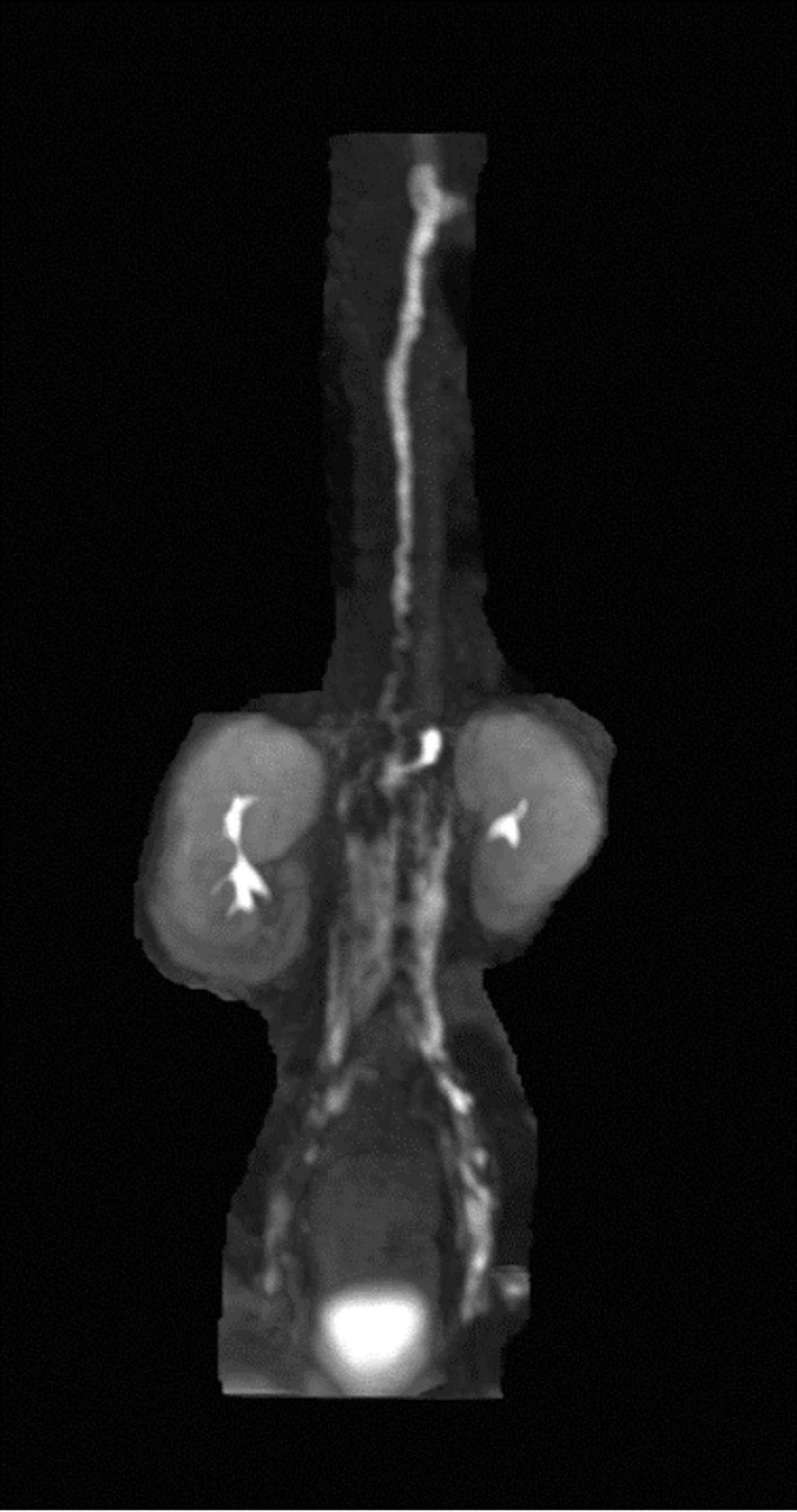
Reconstructed MRL. Normal central lymphatic channels/thoracic duct. MRL, magnetic resonance lymphangiography.
The working diagnosis at this point was that the renal lymphatic drainage was disconnected (presumptively congenital) from the retroperitoneal/central lymphatic channels and that the child would require nephrectomy for definitive therapy. However, after multiple discussions between the care-giving teams and the patient's family, the family felt that if there was potential that medical or percutaneous therapies might work to preserve the kidney, they would be amenable to exploring those options.
We then discussed direct renal injection in attempt to opacify the renal lymphatic system and see if we could visualize intrarenal lymphatics and their path of outflow. In a similar manner to the initial MRL, the patient was returned to the MR suite. Anesthesia was initiated and he was placed in a prone position. After sterile prep and drape, under direct sonographic visualization, two 22-gauge Chiba needles were advanced. Initially, two needles were advanced to the right middle and lower pole corticomedullary junctions (Fig. 5A–E).

The patient was then wheeled into MRI in the prone position with the Chiba needles in position. While obtaining dynamic sequences, contrast was injected (diluted 1 mL Gd:4 mL normal saline for a final concentration of 20% Gd). Contrast was noted to immediately opacify the perirenal space. Due to imaging artifact from the Chiba needles, it was not clear if the needles remained in position. Therefore, the patient was removed from the MRI scanner. Ultrasound demonstrated that the needles had partially withdrawn. Again under sonographic visualization, the needles were repositioned using a transgressive approach (lateral to medial intraparenchymal). This allowed the Chiba needles longer purchase within the renal parenchyma to help avoid dislodgement. As previously, the patient was returned to the MRI scanner and contrast was injected while real-time images were obtained. During this cycle, it was apparent that there was immediate outflow of contrast from the parenchyma to the perinephric space. Gadolinium was noted to opacify the adjacent percutaneous drainage catheter, which was not opacified on the preinjection imaging. Ultimately, we were unable to identify any outflow of gadolinium from the renal parenchyma to the central lymphatic channels. In addition, we did not see direct injection of contrast into the arterial or venous structures of the kidney, although we did see filling of the central venous system and bilateral renal collecting systems. Due to the presumed disconnection of the renal from retroperitoneal/central lymphatics, we surmised that there was likely little that could be done to salvage the kidney.
Subsequent to the MRI examination, the sirolimus was discontinued and the patient underwent nephrectomy nearly 4 weeks after his initial operation. The subcostal incision was re-entered, and massive amounts of fluid were evacuated. The kidney was re-exposed. Active and rapid oozing of cystic fluid was seen coming from the anterior surface of the kidney (Fig. 6). A complete right nephrectomy was performed. Minimal leakage was noted from the perinephric space. Fibrin glue was placed in the renal bed.
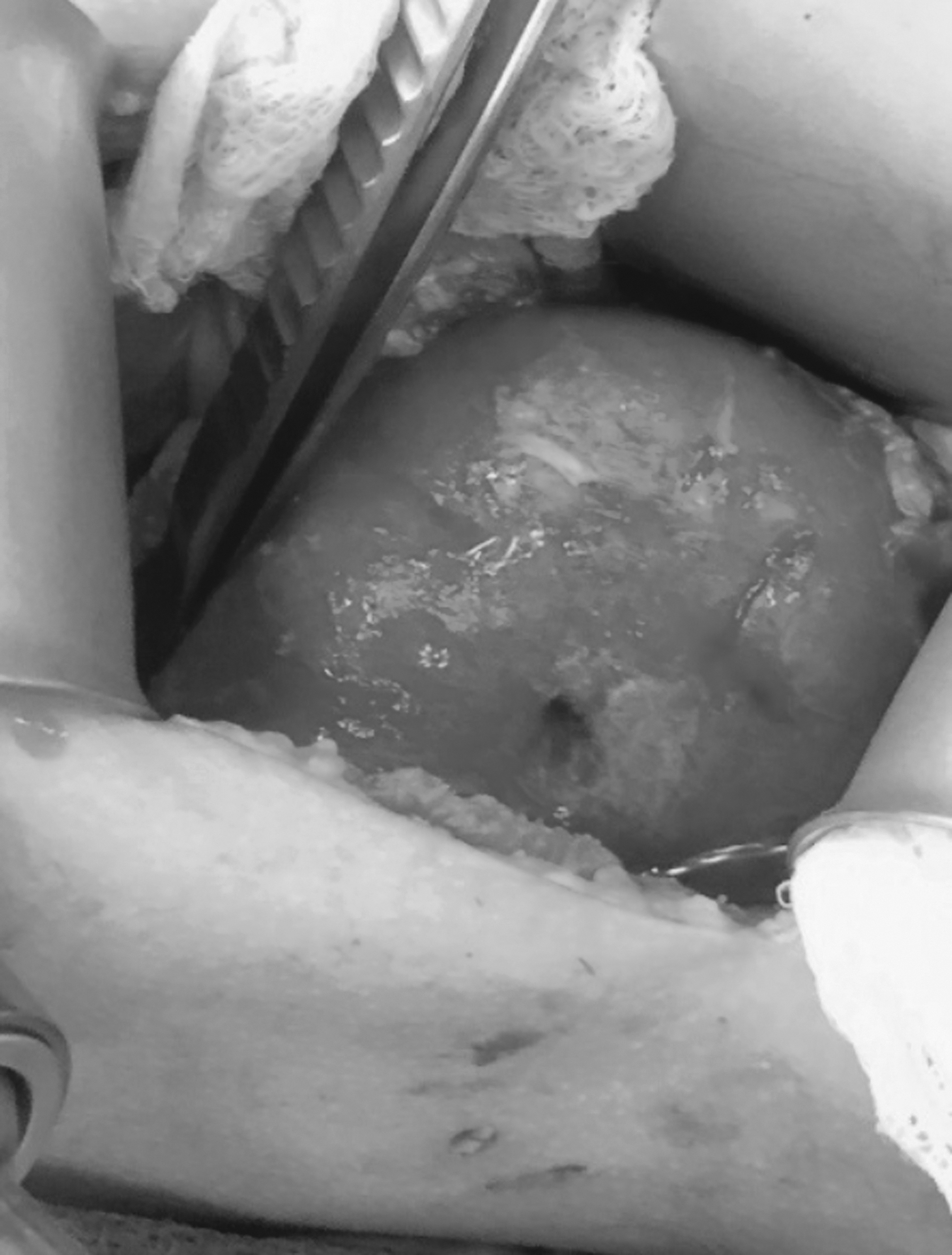
Intraoperative nephrectomy picture.
Histopathologic analysis of the right kidney showed subcapsular granulation tissue and fibrinous material from prior surgical intervention. Dilated, but normal number of, lymphatic vessels were present within the hilar and interlobular connective tissue as highlighted on immunostains for CD31 (a pan-endothelial cell marker) and D2-40 (a lymphatic marker) (Fig. 7).

Dilated lymphatic vessels in the kidney.
Postoperatively, the child did well with no evidence of recurrence or reaccumulation of fluid in his abdomen. He was discharged on postnephrectomy day 5. Currently, more than 1 year after the unilateral nephrectomy, the child recovered completely and there has been complete resolution of lymphatic fluid accumulation.
Discussion
Renal lymphangiectasia is a rare benign lymphatic malformation of the renal system, which may involve the renal parenchyma, capsule, and perinephric tissue.7,8 The exact etiology of this disease process is unknown. Typically, renal lymphatics drain into the retroperitoneal lymphatics. An obstruction or failure to drain into the larger lymphatic system can cause dilation of lymphatic ducts leading to perinephric or nephric unilocular or multilocular collections.
The clinical presentations are variable with many being asymptomatic. Patients have presented with abdominal pain, fever, ascites, hematuria, hypertension, and ipsilateral flank pain.9,10 There are also rare pediatric cases reporting chronic renal insufficiency and bilateral nephromegaly in the neonate.11,12
Based on the clinical, radiographic, and pathologic findings, we deduce that our patient's right renal lymphatics were disconnected from the retroperitoneal and central conducting lymphatic system. As previously mentioned, this was likely congenital given his age and lack of other discernable insults.
Initially, an IR-guided drain was placed to decompress the cyst and allow for sclerotherapy, which was not successful for this patient. However, throughout the hospital course, the child could not tolerate capping the drain, which suggested the cyst's fluid collection reaccumulated at a high rate. Therefore, we proceeded with exploratory laparotomy with the goal of preserving his kidney. With the lymphatic cyst drained and then resected, the pressure against the kidney was removed and allowed free flow of renal lymphatic fluid into the retroperitoneum. Interrogation of the central lymphatics was performed to exclude a large lymphatic branch going from the central system to the retroperitoneum. No such anatomy was identified.
The use of direct gadolinium injection into the renal parenchyma was intended to identify potential renal lymphatic outflow, but we did not directly visualize intrarenal lymphatics. We hypothesize that the immediate outflow of contrast into extrarenal space followed the in vivo course of lymphatic flow, and added secondary support for our hypothesis. There are no reported protocols by which to image the intraparenchymal lymphatics. Our personal communication identified one laboratory in which MR visualization of intrarenal lymphatics was successful in a pig (S. Fishman, MD, Boston Children's Hospital, Personal Communication).
Our technique to image the intrarenal lymphatics may provide future value where there is a clinical question related to the integrity of renal lymphatic outflow. A second operation was performed as a planned nephrectomy. Intraoperatively, there were oozing and pooling of clear lymph on the renal parenchyma at a fairly rapid rate when the entire kidney was exposed. The patient's symptoms resolved after nephrectomy.
The proposed treatment for renal lymphangiectasia is drain placement for decompression followed by sclerotherapy. Rapamycin (sirolimus) might also have a role in management. Cystectomy and nephrectomy are surgical options when medical or less invasive procedures have failed to control lymphatic drainage.
Footnotes
Author Disclosure Statement
None of the authors of this work has any conflicts of interest or financial relationships to disclose.
Funding Information
This research did not receive any specific grant from funding agencies in the public, commercial, or not-for-profit sectors.