
Abstract
Currently, there are many methods to evaluate the effectiveness of manual lymph drainage in the treatment of lymphedema, that is, limb volume measurement, bio-electrical impedance measurement, computer tomography, and ultrasound imaging. However, it is difficult for these methods to accurately address the lymph flow generated by manual lymph drainage. Therefore, we aimed at developing a concise and accurate method to measure the lymph flow through the thoracic duct in human subjects, which is applicable for evaluating the effectiveness of manual lymph drainage. In the present mini-review, we demonstrate the developed method in detail and its scientific evidence for the effectiveness obtained with animal and human clinical experiments. In rat in vivo experiments, intragastric administration of distilled water significantly increased mesenteric flow, which was transported via the cisterna chyli and then the thoracic duct. The manual massage on the cisterna chyli in the anesthetized rabbit significantly accelerated the lymph flow through the thoracic duct, resulting in marked hemodilution. Abdominal respiration in the supine position in human subjects produced similar hemodilution, with a marked decrease in the concentration of vasopressin in the blood. On this basis, we developed a new method to accurately measure the lymph flow through the thoracic duct by using changes in the concentration of vasopressin in the blood. In addition, with changes in urine osmolarity depending on the concentration of vasopressin in the blood, we developed a more concise and noninvasive method for evaluating the lymph flow through the thoracic duct in human subjects. These methods may be applicable for evaluating the effectiveness for the manual lymph drainage in the patients with lymphedema.
Physiological and Immunological Significance of Manual Lymph Drainage
Regarding the lymphatic functions, the transport and drainage of hydrophilic substances, including plasma protein through the lymphatic system, play pivotal roles in maintaining the homeostasis of the internal environment between the cells in tissues in collaboration with the exchange of substances through blood capillaries and venules.1–11 On maintaining immunological defense responses in the internal environment, the lymphatic system contributes to transporting the immune-related cells and cytokines and supporting the lymph node-related immunological responses.12–16
In addition, lymph drainage is very important for maintaining physiological defense mechanisms for the promotion of health in human subjects.11–16 The smaller-sized lymph vessels and lymph nodes play a crucial role in increasing the concentration of albumin in the lymph during the lymph traveling through the lymphatic system.17–23 The excretion of nonselective lymphocytes from lymph nodes is positively correlated with the concentration of albumin in the lymph passing through the afferent lymph vessels of lymph nodes. 23 With the findings, manual lymph drainage in human subjects may accelerate innate immunity via an activation of the lymph transport.
Considering the current studies in lymphatic physiology,1–11 innate immunology,12–15 and oncology,24–27 we have proposed a new lymphology that combined with the inspired knowledge of the earlier mentioned academic disciplines in terms of biological defense mechanisms. 11
On the other hand, the initial clinical signs of diseases, such as inflammation, tumors, and circulatory disorders, including infarction and thrombosis, appear as functional and morphological abnormalities of the internal environment in tissues. These abnormalities manifest themselves as defense reactions by immune responses and irregular actions of the transport and drainage of lymph such as lymph edema.11,16
Clinical Application of Manual Lymph Drainage in the Treatment of Lymphedema
The manual lymph drainage is currently used for treatment and clinical management of the patients with lymphedema.28–30 Lymphedema is a chronic condition involving localized fluid retention and tissue swelling caused by the dysfunction of the lymphatic system. The physiotherapy with the manual lymph drainage using elastic vantage becomes important to treat and control the lymphedema of upper and lower extremities in patients.31,32 For evaluating the effectiveness of manual lymph drainage, it is very important to accurately measure the lymph flow from the extremities with lymph edema. Volume measurements of the extremities are commonly used to address lymph flow from the extremities with lymphedema. In addition, the increasing use of microsurgical techniques, such as lymphaticovenular anastomosis 33 and vascularized lymph node transfer, 34 in the treatment of lymphedema has highlighted the importance of accurate limb volume measurements. Without an accurate assessment of the limb volume, it is difficult to assess the impact of these microsurgical treatments. However, the difficulties in accurately measuring the limb volumes are suggested by many techniques available: limb volume measurement,28,29 near-infrared fluorescence imaging, 35 computer tomography, 37 ultrasound imaging, 36 bio-electrical impedance measurement, 38 and various methods of water displacement in the edematous tissues. 39 Overall, no concise and accurate clinical methods for evaluating the efficacy of surgical and manual lymph drainage treatments for lymphedema in patients are currently available. We, therefore, investigated animal and clinical human experiments to develop novel methods for assessing the lymph flow through the thoracic duct in human subjects.40–43 which are applicable for evaluating the effectiveness of surgical and manual lymph drainage. In this review, we demonstrated the findings of animal and human experiments to address the lymph flow through the thoracic duct, which contribute toward developing the new methods and showed the potential application of the methods for evaluating the skill of therapists in the treatment of lymphedema.
Water Intake Increases Mesenteric Lymph Flow and the Flux of Albumin and Interleukin-22 in the Lymph
To clarify the physiological properties of the jejunal-originated lymph vessels and cisterna chyli, we first aimed at investigating the effects of water intake on the mesenteric lymph flow and the composition of lymph in anesthetized rats and rabbits. 40 The jejunal microcirculation, compared with that in other organs, has specific properties such as the movement of large amounts of albumin from the venular walls to tissues with its related higher tissue osmotic pressure on the venular side. 40 It is also known that the jejunal-originated mesenteric lymph flow is greater than those in other organs.10,11 Consistent with these properties, the mesenteric collecting lymph vessels show heart-like spontaneous contractions,10,11 resulting in the large amounts of lymph transport into cisterna chyli. However, it is still unsolved as to how these physiological properties of the jejunal microcirculation contribute to the absorption and transport of the consumed water and water-soluble substances. In the background, we investigated the effects of water intake on the jejunal-originated lymph flow, and the total flux of albumin and long-chain fatty acids in the lymph. In addition, we evaluated the distribution and activity of innate lymphoid cells (ILC)-3 in the gut walls, and water intake-mediated changes in the total flux of ILC-3-secreted interleukin (IL)-22 in the lymph. 40
Thus, to collect the lymph from the jejunal-originated mesenteric lymph vessel, a small polyethylene catheter was inserted centrifugally into an efferent lymph vessel. The greatest amount of the lymph was collected in the first 15 minutes after intragastric administration of 3 mL distilled water. The total flux of albumin transported through the lymph vessel was significantly increased after 1 hour after the administration of distilled water. Similar to the finding of albumin, the total flux of long-chain fatty acids included with ω3, ω6, and ω9 in the lymph was increased significantly at 1 hour after the intragastric administration of distilled water. In contrast, the intragastric administration of distilled water resulted in a significant increase in both the cell density of lymphocytes and the total flux of cells through the lymph vessel. No significant time-dependent changes in the ratio between T and B lymphocytes were observed in the lymph after the intragastric administration of distilled water. 40
To confirm the crucial role of plasma albumin movement in the water intake-mediated increase in the mesenteric lymph flow, we investigated the time-dependent changes in the lymph color in the lymph vessels and cistern chyli in rabbits that were pretreated with intravenous administration of Evans blue dye. Fig. 140 (the permission from American Physiological Society with proprietary rights) shows representative photomicrographs of the time-dependent changes in blue color of lymph within mesenteric lymph vessels and cisterna chyli of rabbits in the control physiological condition (without water intake). Two minutes after intravenous administration of Evans blue dye, the color of lymph in the mesenteric lymph vessels became slightly blue. Approximately 15 minutes after Evans blue dye administration, the cisterna chyli was stained clearly blue. Intragastric administration of distilled water significantly shortened the time of color changes in the lymph. 40
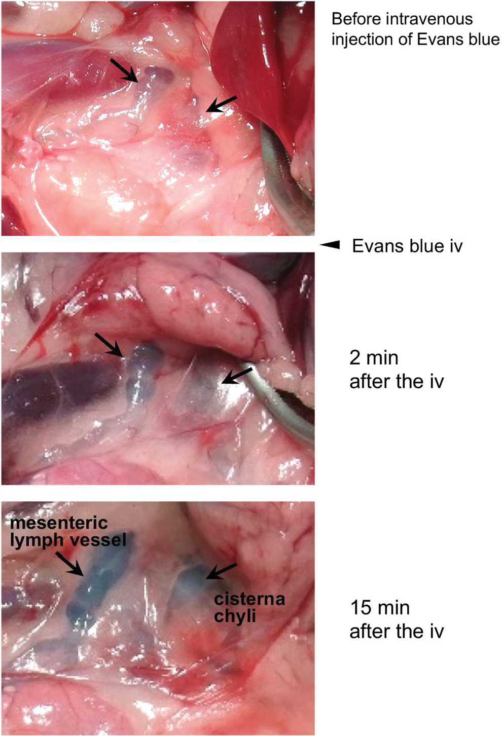
Representative photomicrographs of color changes in rabbit mesenteric lymph vessels and cisterna chyli on intravenous administration (iv) of Evans blue dye under control condition without water intake (the permission from American Physiological Society with proprietary rights).
To assess whether water intake could play crucial roles in gut-associated innate immunity, we next evaluated the distribution and dynamics of ILC-3 in the lamina propria of rat jejunal and ileal walls. There was marked heterogeneity in the distribution of ILC-3 between the jejunum and ileum. Thus, ILC-3 was localized mainly in the lamina propria of the upper part of rat jejunum. Similar to the flow cytometry results, the expression of IL-22 mRNA was significantly higher in the upper part of jejunum. However, no significant effect was observed on the IL-22 mRNA expression in the lamina propria of jejunum by 2 hours of water intake. In contrast, the total flux of IL-22 through the lymph vessel significantly increased at 1 hour after the administration. 40
Manual Massage on Cisterna Chyli in Rabbit Accelerates the Lymph Flow Through Thoracic Duct
To investigate whether the increase of lymph into cisterna chyli accelerates the lymph flow through the thoracic duct, we aimed at addressing the lymph flow through the thoracic duct in anesthetized rabbits by using a Sonazoid-based contrast-enhanced ultrasound (CEUS)-guided method. 41 On confirmation of the lymph flow with the CEUS-guided method, we also examined the effects of manual massage on the cisterna chyli on the concentrations of total protein (TP), red blood cells (RBCs) and hemoglobin (Hb), and hematocrit (Ht) in rabbit blood. To address the CEUS-guided images of the lymph flow through the thoracic duct in the left venous angle, 0.3 mL Sonazoid was injected into the cisterna chyli and then the cisterna chyli was gently massaged for 30 seconds. Fig. 2A 41 (the permission form Clarivate with proprietary rights) shows representative CEUS-guided images of the rabbit thoracic duct in the region near the left venous angle, and the internal jugular and subclavian veins. In all cases (n = 15), clear images of the thoracic ducts were obtained within ∼10 seconds of the injection of Sonazoid into the cisterna chyli. To confirm the lymph flow thoracic duct, the flash replenishment with a higher mechanical index (>1.0) was done to rupture the Sonazoid microbubbles. Fig. 2B 41 shows a representative flash replenishment image.

Representative microphotographs of CEUS-guided contrast-enhanced images of rabbit left venous angle, including the thoracic duct, left internal jugular vein, and left subclavian vein. The panel of
To examine the effects of manual massage pressed on the cisterna chyli on changes in the concentrations of TP, Alb, RBC, and Ht in the blood, we first addressed whether the manual massage increases the lymph flow through the thoracic duct by using the Sonazoid-based CEUS-guided images. The contrast-enhanced images of the thoracic duct became clearer at several seconds after the massage. Next, to evaluate the effects of the manual massage on the concentrations of TP, Alb, RBC, and Ht, we collected blood samples from the internal jugular vein before and 30 minutes after the manual massage on the cisterna chyli in rabbits. Fig. 3A 41 (the permission form Clarivate with proprietary rights) shows representative recordings of the effects of manual massage on changes in TP, Alb, RBC, and Ht in the rabbit blood. The manual massage on the cisterna chyli produced significant reductions of TB, Alb, RBC, and Ht, resulting in marked hemodilution (Fig. 3B 41 ).

Abdominal Respiration Accelerates the Hemodilution in Human Subjects
On the findings that the manual massage on rabbit cisterna chyli produced significant decreases in TB, Alb, RBC, and Ht in the blood, which were suggested a marked hemodilution. 42 We aimed at evaluating whether abdominal respiration, which may press the cisterna chyli, accelerates the lymph flow through the thoracic duct and then induces marked hemodilution in human subjects, similar to hemodilution in rabbits. A total of 48 healthy volunteers (45.2 ± 7.2 years old, 26 male and 22 female) participated in the clinical observation study. 42 This study was a single-blinded, randomized, and controlled human trial. The ethical committee for human studies at the School of Medicine Shinshu University approved the study. All subjects gave their written and oral informed consent. All study data and procedures were managed in accordance with the concepts outlined in the Declaration of Helsinki.
To evaluate the effects of manual lymph drainage on the blood concentration of TP, Alb, RBC, white blood cells (WBCs), platelet (PLT), and Ht in human subjects, they had manual lymph drainage with their facial, upper, or lower extremities for 5 minutes in the supine position, followed with 30 minutes rest. The parameters of blood samples and the circular measurements of right ankle and knee skin surface were investigated before and after the manual lymph drainage. In the control study (without manual lymph drainage), the 30 minutes rest only in the supine position produced significant reductions of TP and Alb only in all subjects. In contrast, the TP, Alb, RBC, and Ht were significantly decreased by the 30 minutes rest in the supine position with the 5 minutes massage of upper or lower extremity. However, the clinical studies did not produce a significant change in the concentration of vasopressin in the blood in all participants.
Therefore, to increase the lymph flow through the thoracic duct, we next designed additional experiments to evaluate the effects of abdominal respiration at 30 minutes rest in the supine position on the concentration of vasopressin in the blood and micturition desire. Fig. 4A 42 (the permission from Clarivate with proprietary rights) shows representative recordings of TP, Alb, RBC, Ht, WBC, PLT, concentration of antidiuretic hormone (ADH), and circular skin measurements of right ankle (R ankle) and right knee (R knee) in all participants. Fig. 4B 42 shows the summarized data before and after the abdominal respiration. Fig. 4C 42 shows the summarized data before and after the 30 minutes rest in the supine position only without abdominal respiration. The 30 minutes rest in the supine position without abdominal respiration produced hemodilution with decreases in TP, Alb, and Ht. Abdominal respiration in 30 minutes rest at the supine position induced more marked hemodilution with larger decreases in TP, Alb, RBC, Ht, and R knee. In addition, the abdominal respiration produced a significant decrease in the concentration of antidiuretic hormone (ADH, Fig. 4D), which may be related to the abdominal respiration-mediated marked hemodilution. Fig. 4E 42 demonstrates the relationship between the level of micturition desire (ordinate) and abdominal respiration-mediated reduction in the blood concentration of vasopressin (abscissa). In summary, changes in the concentration of vasopressin compared with before and after the abdominal respiration are suitable parameters for evaluating the lymph flow through the thoracic duct in human subjects. Thus, the lowering of vasopressin concentration in the blood and the increasing miction desire may become accurate tools for evaluating the efficacy of manual lymph drainage in human subjects. The study 42 that fluid shifts in lymphedema patients with manual drainage were reflected in the increased plasma volume may be compatible with our conclusion.
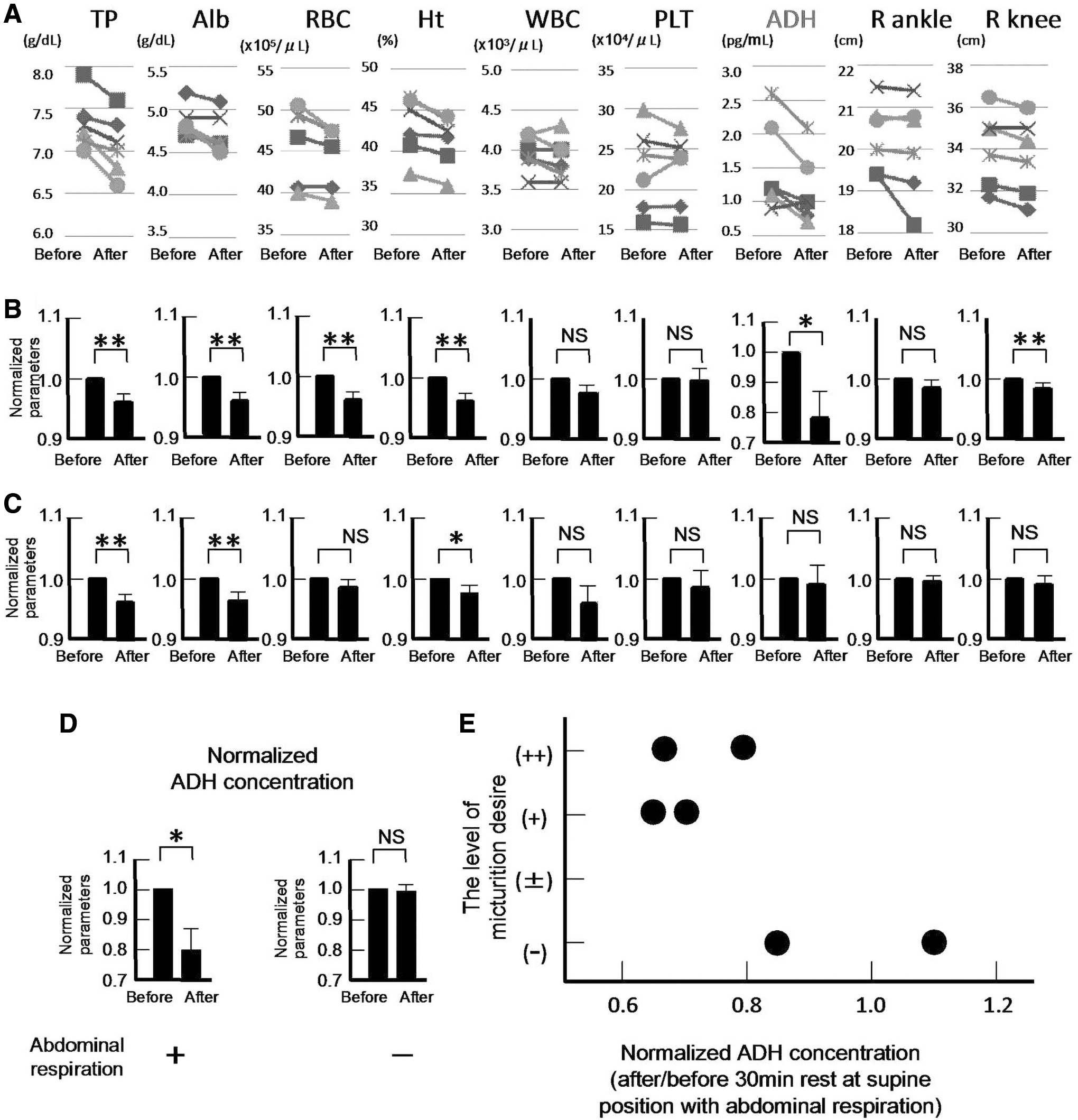
Abdominal respiration produced marked hemodilution and reduction for the concentration of antidiuretic hormone in human subjects.
Noninvasive and Concise Method for Evaluating the Lymph Flow Through Thoracic Duct Using Urine Osmolarity
Based on our previous studies,40–42 we conducted human clinical observational experiments to establish a harmless and concise method using the urine osmolarity for measuring the lymph flow through the thoracic duct. When completed, the method using urine osmolarity may provide a more concise and useful method for evaluating the effectiveness of manual lymph drainage by lymphedema physicotherapists. This study was a single-blinded, randomized, and controlled human experiment. The volunteers were given 250 mL of distilled water and had rest for 10 minutes in a supine position. Blood samples were taken pre- and post-rest to measure changes in the concentration of TP, Alb, RBC, Hb, and ADH in the blood. Urine samples were also collected to measure the [Na+], [Cl−], and osmolarity in the same experimental protocol. Water intake of 250 mL with 10 minutes rest in the supine position without abdominal respiration produced a significant reduction of vasopressin concentration in the blood with marked hemodilution. The urine [Na+], [Cl−], and osmolarity were simultaneously decreased in all participants. The approximate linear relationship between the normalized concentration of vasopressin obtained with after and before 10 minutes rest with water intake, and the normalized urine osmolarity with after and before the procedure (Fig. 543, the permission from Clarivate with proprietary rights) was confirmed. In summary, the urine osmolarity obtained with 250 mL water intake and 10 minutes rest in the supine position becomes a concise and useful parameter for monitoring changes in the concentration of vasopressin in the blood, which may be used to evaluate the lymph flow through the thoracic duct in human subjects. In fact, a linear relationship between vasopressin concentration in the blood ranging from 0.5 to 3.0 pg/mL and urine osmolarity ranging from 0 to 450 mOsm/L is well known in human subjects. 43 In this experiment, the concentrations of vasopressin in the blood for the four participants were 2.2, 2.5, 3.0, and 2.0 pg/mL, respectively, before starting the experiment. The corresponding values of vasopressin on each participant obtained after 10 minutes rest in a supine position decreased to 1.1, 1.8, 2.1, and 1.3 pg/mL, respectively. The urine osmolarities of these participants were also reduced from 520 to 201, 354 to 194, 370 to180, and 601 to 240 mOsm/L, respectively.
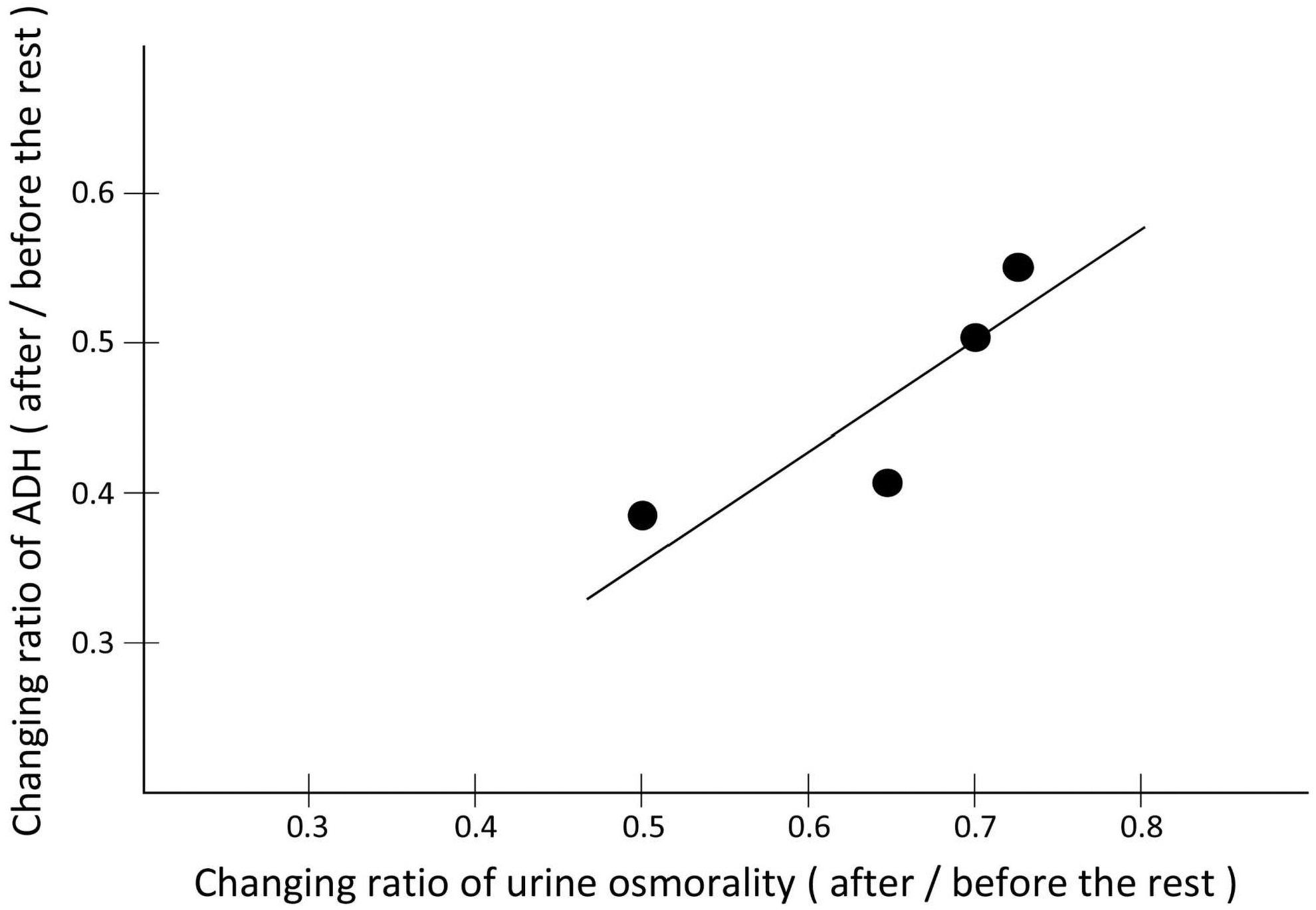
The relationship between the normalized ratio of ADH concentration in the blood obtained at after or before 10 minutes rest in the supine position with 250 mL water intake, and the normalized ratio of urine osmolarity obtained at after or before the same procedure (the permission from Clarivate with proprietary rights).
Conclusions
Regarding innate immunity, water intake and abdominal respiration at rest in the supine position in human subjects increased the lymph flow through the thoracic duct and ILC-3 released IL-22 in the mesenteric lymph. These findings suggest that the water intake and abdominal respiration may contribute toward maintaining an advanced state of innate immunity in physiological conditions. On the assessment of the treatment with lymphedema, the changes in urine osmolarity obtained before and after manual lymph drainage in lymphedema patients with water intake may become a concise and noninvasive parameter for evaluating the effectiveness of manual drainage.
Ethics
The ethical committee for human studies in the School of Medicine at Shinshu University approved the study (document no. 805, 2015; no. 4348, 2019), and all patients gave written and oral informed consent. All study data and procedures were managed in accordance with the concept outlined in the Declaration of Helsinki. Animal experiments in the present study were performed with the approval of the Shinshu University Animal Care and Use Committee.
Data Availability Statement
The data that support the findings of this study are available from the corresponding author on reasonable request.
Footnotes
Authors' Contributions
T.O. and Y.K. wrote the article. T.O., Y.K., M.H., and T.W.-A. revised the article. All authors approved to submit the article.
Author Disclosure Statement
No competing financial interests exist.
Funding Information
The Department of Innovation of Medical and Health Sciences Research at Shinshu University School of Medicine was established and is supported financially by donations from BOURBON, Co., Ltd (Kashiwazaki, Niigata, Japan) and Aizawa Hospital (Matsumoto, Nagano, Japan). The authors declare that this study received funding from BOURBON Co. Ltd. The funder was not involved in the study design, collection, analysis, interpretation of data, the writing of this article, or the decision to submit it for publication.