
Abstract

Keynote Articles
Itoh, F. and T. Watabe (2022). “TGF-beta signaling in lymphatic vascular vessel formation and maintenance.” Front Physiol 13: 1081376. E-Pub12/15/2022
Transforming growth factor (TGF)-beta and its family members, including bone morphogenetic proteins (BMPs), nodal proteins, and activins, are implicated in the development and maintenance of various organs. Here, we review its role in the lymphatic vascular system (the secondary vascular system in vertebrates), which plays a crucial role in various physiological and pathological processes, participating in the maintenance of the normal tissue fluid balance, immune cell trafficking, and fatty acid absorption in the gut. The lymphatic system is associated with pathogenesis in multiple diseases, including lymphedema, inflammatory diseases, and tumor metastasis. Lymphatic vessels are composed of lymphatic endothelial cells, which differentiate from blood vascular endothelial cells (BECs). Although TGF-beta family signaling is essential for maintaining blood vessel function, little is known about the role of TGF-beta in lymphatic homeostasis. Recently, we reported that endothelial-specific depletion of TGF-beta signaling affects lymphatic function. These reports suggest that TGF-beta signaling in lymphatic endothelial cells maintains the structure of lymphatic vessels and lymphatic homeostasis, and promotes tumor lymphatic metastasis. Suppression of TGF-beta signaling in lymphatic endothelial cells may therefore be effective in inhibiting cancer metastasis. We highlight recent advances in understanding the roles of TGF-beta signaling in the formation and maintenance of the lymphatic system.
This manuscript provides a review elucidating the role of TGF-beta in various aspects of lymphatic vessel differentiation, development and maintenance, as noted in published Figure 2 displayed here. This may be relevant in tumor lymphangiogenesis and metastases, as well as other lymphatic diseases. Further research will ideally lead to targeted therapeutic agents.
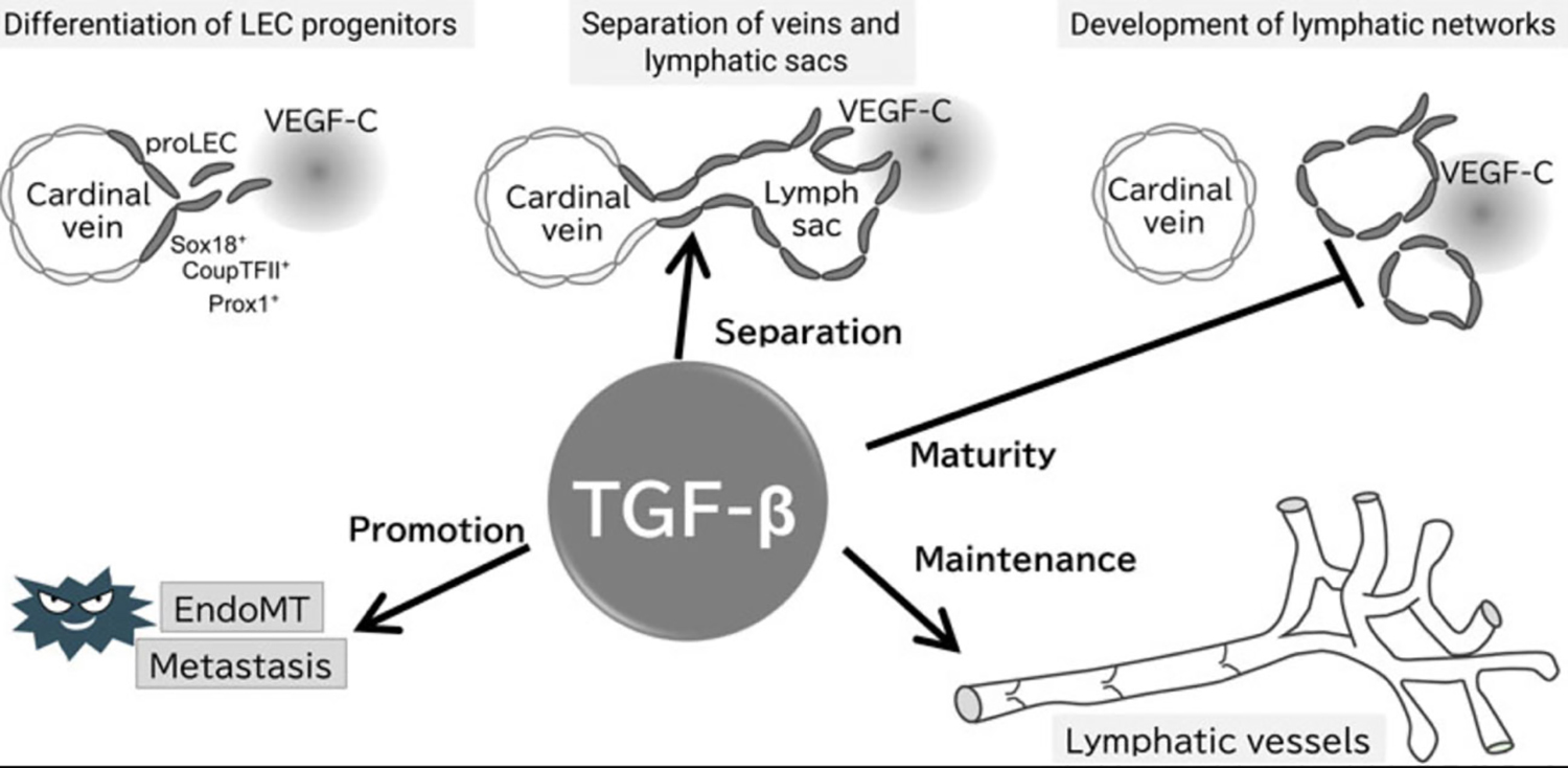
Proposed working model of TGF-β signaling in the formation and maintenance of the lymphatic system. Evidence for the presence of each step of the proposed model is provided in the text; TGF-β does not affect the early development of lymphatic vessels, whereas is important for their separation from veins, maturation, and maintenance of lymphatic function. TGF-β also promotes tumor lymphatic metastasis via EndoMT of LECs.
Of note, this journal has a series of manuscripts categorized under the rubric “New Lymphology combined with angiology, oncology, innate immunology, and basic medical sciences.” https://www.frontiersin.org/research-topics/37065/new-lymphology-combined-with-angiology-oncology-innate immunology-and-basic-medical-sciences#articles
Yu, W. X., et al. (2022). “Kinesin-5 Eg5 is essential for spindle assembly, chromosome stability and organogenesis in development.” Cell Death Discov 8(1): 490. E-Pub 12/13/2022
Chromosome stability relies on bipolar spindle assembly and faithful chromosome segregation during cell division. Kinesin-5 Eg5 is a plus-end-directed kinesin motor protein, which is essential for spindle pole separation and chromosome alignment in mitosis. Heterozygous Eg5 mutations cause autosomal-dominant microcephaly, primary lymphedema, and chorioretinal dysplasia syndrome in humans. However, the developmental roles and cellular mechanisms of Eg5 in organogenesis remain largely unknown. In this study, we have shown that Eg5 inhibition leads to the formation of the monopolar spindle, chromosome misalignment, polyploidy, and subsequent apoptosis. Strikingly, long-term inhibition of Eg5 stimulates the immune responses and the accumulation of lymphocytes in the mouse spleen through the innate and specific immunity pathways. Eg5 inhibition results in metaphase arrest and cell growth inhibition, and suppresses the formation of somite and retinal development in zebrafish embryos. Our data have revealed the essential roles of kinesin-5 Eg5 involved in cell proliferation, chromosome stability, and organogenesis during development. Our findings shed a light on the cellular basis and pathogenesis in microcephaly, primary lymphedema, and chorioretinal dysplasia syndrome of Eg5-mutation-positive patients.
I found this manuscript interesting, as it to describes, via studying Eg5, which when mutated, causes MLCRD (microcephaly, primary lymphedema, and chorioretinal dysplasia syndrome (MLCRD; OMIM 152950). Eg5, a Kinesin-like protein [KIF11] on chromosome 10q23.33 is important in mitosis, crosslinking miotic spindles to preserve polarity. While this has been known to affect neuronal function, the researchers in this article assess the role of Eg5 in organ development overall. Specific to the lymphatic system, their findings demonstrated EG5 inhibition led to activation of an immune response with lymphocytosis and an increased number of splenic lymphocytes. While this group was able to demonstrate interruption of retinal, neural and renal development in Egf inhibited zebrafish models, its role in the development of low limb lymphedema in humans remains elusive.
Basic Science and Reviews
Geng, X., et al. (2022). “Lack of embryonic homozygous or adult heterozygous lymphatic phenotypes for a Sos1 mutation and lack of lymphatic embryonic phenotypes for a homozygous Cx47 mutation in mice.” Lymphology 55(3): 129–134.
We have studied the lymphatic phenotypes of 2 mutations, known to cause abnormalities of lymphatics in humans, in mice. The Cx47 R260C mutation (variably penetrant in humans heterozygous for it and causing limb lymphedema) had an adult mouse phenotype of hyperplasia and increased lymph nodes only in homozygous condition but we did not find any anatomical phenotype in day 16.5 homozygous embryos. Mice harboring the Sos1 mutation E846K (causing Noonan's in man which occasionally shows lymphatic dysplasia) had no adult heterozygous phenotype in lymphatic vessel appearance and drainage (homozygotes are early embryonic lethals) while day 16.5 heterozygous embryos also had no detectable anatomical phenotype.
Han, D., et al. (2023). “Lymphatic uptake of biotherapeutics through a 3D hybrid discrete-continuum vessel network in the skin tissue.” J Control Release: 11942. E-Pub 01/09/2023
Subcutaneous administration is a common approach for the delivery of biotherapeutics, which is achieved mainly through the absorption across lymphatic vessels. In this paper, the drug transport and lymphatic uptake through a three-dimensional hybrid discrete-continuum vessel network in the skin tissue are investigated through high-fidelity numerical simulations. We find that the local lymphatic uptake through the explicit vessels significantly affects macroscopic drug absorption. The diffusion of drug solute through the explicit vessel network affects the lymphatic uptake after the injection. This effect, however, cannot be captured using previously developed continuum models. The lymphatic uptake is dominated by the convection due to lymphatic drainage driven by the pressure difference, which is rarely studied in experiments and simulations. Furthermore, the effects of injection volume and depth on the lymphatic uptake are investigated in a multi-layered domain. We find that the injection volume significantly affects the rate of lymphatic uptake through the heterogeneous vessel network, while the injection depth has little influence, which is consistent with the experimental results. At last, the binding and metabolism of drug molecules are studied to bridge the simulation to the experimentally measured drug clearance. We provide a new approach to study the diffusion and convection of drug molecules into the lymphatic system through the hybrid vessel network.
Hettrick, H. and F. Aviles (2023). “Microgravity and Lymphatics: Why Space Programs Need Lymphedema Physiology Specialists.” Lymphat Res Biol. E-Pub 01/09/2023
Background: The resurgence of space travel in the recent years, both through formally trained astronauts on the International Space Station and the civilian space race to send astrocivilians to Low Earth Orbit and beyond, beckons the need to understand the role of the lymphatic system and role of endothelial glycocalyx when subjected to gravitational alterations. Methods and Results: A comprehensive narrative review of the literature explores a call to action for research and countermeasure development to support the health and well-being of humans subjected to space flight, with particular attention to the role of the lymphatic system and endothelial glycocalyx. Emerging evidence suggests a link between the dysfunction experienced with various physiological processes in microgravity, highlighting the need for more research exploring the role of the lymphatic system in the extremes of gravity and countermeasure development to reduce dysregulation. Conclusion: The synergistic and interdependent relationship of these structures are fundamental to health in space and on Earth.
Hsiao, H. Y., et al. (2022). “The Impacts of Lymph on the Adipogenesis of Adipose-Derived Stem Cells.” Plast Reconstr Surg. E-Pub 12/20/2022
BACKGROUND: The pathophysiology of adipose proliferation or differentiation in extremity lymphedema has not been thoroughly studied. This study investigated the impacts of the lymph harvested from lymphedematous limbs on the adipogenesis of adipose-derived stem cells (ASCs). METHODS: ASCs were isolated from the adipose tissue of normal extremities and cultured with lymph collected from Cheng's Lymphedema Grade III-IV patients or adipogenic differentiation medium (ADM) and further subjected to differentiation and proliferation assay. The expression of adipogenesis genes was examined by RT-PCR to investigate the effect of lymph on ASCs. The level of adipogenic cytokines in the lymph was also evaluated. RESULTS: The size of the adipocytes were significantly larger in lymphedema fat tissue compared to that in normal fat tissues (p < 0.00). The adipogenesis of ASCs cultured in lymph was significantly enhanced than in ADM (p = 0.008) on Day 10, suggesting the adipogenesis of ASCs was promoted under the lymph-cultured environment. The expression of adipogenesis genes, PPARgamma (p = 0.02), C/EBPalpha (p = 0.008); FABP4: (p = 0.004), LPL (p = 0.003), were statistically elevated when the ASCs were cultured with lymph. The insulin content in lymph was statistically higher in the lymph (p < 0.001) than that in plasma. CONCLUSIONS: The adipogenesis of ASCs was promoted under the lymph-cultured environment with statistically increased adipogenesis genes of PPARgamma, C/EBPalpha, FABP4, and LPL. The excess lymph accumulated in the lymphedematous extremity contained a greater insulin/IGF-2. These adipogenic factors promoted the expression of early adipogenesis genes and led ASCs to undergo adipogenesis and differentiated into adipocytes. CLINICAL RELEVANCE STATEMENT: The accumulation of adipose tissue in lymphedema region was contributed from the content of excess lymph.
Huang, S., et al. (2023). “Three-dimensional mapping of hepatic lymphatic vessels and transcriptome profiling of lymphatic endothelial cells in healthy and diseased livers.” Theranostics 13(2): 639–658.
Rationale: Hepatic lymphatics are essential for liver homeostasis and immune function. However, the 3D structure and spatial distribution of hepatic lymphatic vessels (LVs) need to be confirmed. Moreover, the molecular information of hepatic lymphatic endothelial cells (LyECs) needs to be further studied. The bottleneck is the lack of specific markers or labeling methods for hepatic lymphatic endothelial cells (LyECs) Methods: Here, we proposed a method for the spatiotemporal sequential injection of antibodies (STSI-Ab) to selectively label hepatic LyECs in vivo. In addition, we also developed an efficient hepatic LyEC sorting method and performed deep transcriptome sequencing on hepatic LyECs. Results: The STSI-Ab method achieved selective labeling of the mouse hepatic lymphatic network. Three-dimensional fluorescence imaging results of the STSI-Ab mouse liver lobe clearly showed that hepatic LVs entangled with the portal vein but were not present in the central vein. The imaging data inspired a novel hepatic lobule structure model with an added set of LVs in the portal area. Furthermore, deep transcriptome sequencing of isolated hepatic LyECs and Masson's trichrome staining results suggested that hepatic LyECs might be an important source of collagen fibers deposited in the portal area during the process of liver fibrosis and bile duct ligation (BDL). Conclusions: We proposed an STSI-Ab method for selectively labeling hepatic LVs, distinguishing the hepatic LVs from other vessels, and mapping their 3D structure. This study opens an avenue for understanding hepatic lymphatic structure and it will be very beneficial to the study of hepatic LyEC functions.
Kong, A. M., et al. (2022). “Engineering transplantable human lymphatic and blood capillary networks in a porous scaffold.” J Tissue Eng 13: 20417314221140979. E-Pub 12/26/2022
Due to a relative paucity of studies on human lymphatic assembly in vitro and subsequent in vivo transplantation, capillary formation and survival of primary human lymphatic (hLEC) and blood endothelial cells (hBEC) +/- primary human vascular smooth muscle cells (hvSMC) were evaluated and compared in vitro and in vivo. hLEC +/- hvSMC or hBEC +/- hvSMC were seeded in a 3D porous scaffold in vitro, and capillary percent vascular volume (PVV) and vascular density (VD)/mm(2) assessed. Scaffolds were also transplanted into a sub-cutaneous rat wound with morphology/morphometry assessment. Initially hBEC formed a larger vessel network in vitro than hLEC, with interconnected capillaries evident at 2 days. Interconnected lymphatic capillaries were slower (3 days) to assemble. hLEC capillaries demonstrated a significant overall increase in PVV (p = 0.0083) and VD (p = 0.0039) in vitro when co-cultured with hvSMC. A similar increase did not occur for hBEC + hvSMC in vitro, but hBEC + hvSMC in vivo significantly increased PVV (p = 0.0035) and VD (p = 0.0087). Morphology/morphometry established that hLEC vessels maintained distinct cell markers, and demonstrated significantly increased individual vessel and network size, and longer survival than hBEC capillaries in vivo, and established inosculation with rat lymphatics, with evidence of lymphatic function. The porous polyurethane scaffold provided advantages to capillary network formation due to its large (300–600 mum diameter) interconnected pores, and sufficient stability to ensure successful surgical transplantation in vivo. Given their successful survival and function in vivo within the porous scaffold, in vitro assembled hLEC networks using this method are potentially applicable to clinical scenarios requiring replacement of dysfunctional or absent lymphatic networks.
Lampejo, A. O., et al. (2023). “Lymphatic/blood vessel plasticity: motivation for a future research area based on present and past observations.” Am J Physiol Heart Circ Physiol 324(1): H109–H121. E-Pub 12/02/2022
The lymphatic system plays a significant role in homeostasis and drainage of excess fluid back into venous circulation. Lymphatics are also associated with a number of diseases including lymphedema, tumor metastasis, and various lymphatic malformations. Emerging evidence suggests that lymphatics might have a bigger connection to the blood vascular system than originally presumed. As these two systems are often studied in isolation, several knowledge gaps exist surrounding what constitutes lymphatic vascular plasticity, under what conditions it arises, and where structures characteristic of plasticity can form. The objective of this review is to overview current structural, cell lineage-based, and cell identity-based evidence for lymphatic plasticity. These examples of plasticity will then be considered in the context of potential clinical and surgical implications of this evolving research area. This review details our current understanding of lymphatic plasticity, highlights key unanswered questions in the field, and motivates future research aimed at clarifying the role and therapeutic potential of lymphatic plasticity in disease.
Liu, X., et al. (2023). “Promoting Lymphangiogenesis and Lymphatic Growth and Remodeling to Treat Cardiovascular and Metabolic Diseases.” Arterioscler Thromb Vasc Biol 43(1): e1–e10. E-Pub 12/01/2022.
Lymphatic vessels are low-pressure, blind-ended tubular structures that play a crucial role in the maintenance of tissue fluid homeostasis, immune cell trafficking, and dietary lipid uptake and transport. Emerging research has indicated that the promotion of lymphatic vascular growth, remodeling, and function can reduce inflammation and diminish disease severity in several pathophysiologic conditions. In particular, recent groundbreaking studies have shown that lymphangiogenesis, which describes the formation of new lymphatic vessels from the existing lymphatic vasculature, can be beneficial for the alleviation and resolution of metabolic and cardiovascular diseases. Therefore, promoting lymphangiogenesis represents a promising therapeutic approach. This brief review summarizes the most recent findings related to the modulation of lymphatic function to treat metabolic and cardiovascular diseases such as obesity, myocardial infarction, atherosclerosis, and hypertension. We also discuss experimental and therapeutic approaches to enforce lymphatic growth and remodeling as well as efforts to define the molecular and cellular mechanisms underlying these processes.
Lyu, Q. and K. Ley (2022). “How Lymphatic Endothelial Cells Destabilize Regulatory T Cells.” Arterioscler Thromb Vasc Biol. E-Pub 12/29/2022.
Miyazaki, T., et al. (2022). “Hypercholesterolemic Dysregulation of Calpain in Lymphatic Endothelial Cells Interferes With Regulatory T-Cell Stability and Trafficking.” Arterioscler Thromb Vasc Biol. E-Pub 12/15/2022.
BACKGROUND: Although hypercholesterolemia reportedly counteracts lymphocyte trafficking across lymphatic vessels, the roles of lymphatic endothelial cells (LECs) in the lymphocyte regulations remain unclear. Previous studies showed that calpain-an intracellular modulatory protease-interferes with leukocyte dynamics in the blood microcirculation and is associated with hypercholesterolemic dysfunction in vascular endothelial cells. METHODS: This study investigated whether the calpain systems in LECs associate with the LEC-lymphocyte interaction under hypercholesterolemia using gene-targeted mice. RESULTS: Lipidomic analysis in hypercholesterolemic mice showed that several lysophospholipids, including lysophosphatidic acid, accumulated in the lymphatic environment. Lysophosphatidic acid enables the potentiation of calpain systems in cultured LECs, which limits their ability to stabilize regulatory T cells (Treg) without altering Th1/Th2 subsets. This occurs via the proteolytic degradation of MEKK1 and the subsequent inhibition of TGF (transforming growth factor)-beta1 production in LECs. Targeting calpain systems in LECs expanded Tregs in the blood circulation and reduced aortic atherosclerosis in hypercholesterolemic mice, concomitant with the reduction of proinflammatory macrophages in the lesions. Treg expansion in the blood circulation and atheroprotection in calpain-targeted mice was prevented by the administration of TGF-beta type-I receptor inhibitor. Moreover, lysophosphatidic acid-induced calpain overactivation potentiated the IL (interleukin)-18/NF-kappaB (nuclear factor kappaB)/VCAM1 (vascular cell adhesion molecule 1) axis in LECs, thereby inhibiting lymphocyte mobility on the cells. Indeed, VCAM1 in LECs was upregulated in hypercholesterolemic mice and human cases of coronary artery disease. Neutralization of VCAM1 or targeting LEC calpain systems recovered afferent Treg transportation via lymphatic vessels in mice. CONCLUSIONS: Calpain systems in LECs have a key role in controlling Treg stability and trafficking under hypercholesterolemia.
Moisio, O., et al. (2022). “Preclinical Evaluation of a Humanized Antibody Against Common Lymphatic Endothelial and Vascular Endothelial Receptor-1, (89)Zr-Desferrioxamine-Bexmarilimab, in a Rabbit Model of Renal Fibrosis.” J Nucl Med. E-Pub 12/27/2022
Bexmarilimab is a new humanized monoclonal antibody against common lymphatic endothelial and vascular endothelial receptor-1 (CLEVER-1), and is in clinical trials for macrophage-guided cancer immunotherapy. In addition to cancer, CLEVER-1 is also associated with fibrosis. To facilitate prospective human PET studies, we preclinically evaluated (89)Zr-labeled bexmarilimab in rabbits. Methods: Bexmarilimab was conjugated with desferrioxamine (DFO) and radiolabeled with (89)Zr. Retained immunoreactivity was confirmed by flow cytometry. Distribution kinetics of intravenously administered (89)Zr-DFO-bexmarilimab (0.1 mg/kg) for up to 7 days in a rabbit model of renal fibrosis mediated by unilateral ureteric obstruction (UUO). The in-vivo stability of (89)Zr-DFO-bexmarilimab was evaluated by sodium dodecyl sulfate-polyacrylamide gel electrophoresis in combination with autoradiography. Additionally, we estimated the human radiation dose from data obtained in healthy rabbits. Results: (89)Zr-DFO-bexmarilimab cleared rapidly from the blood circulation and distributed to the liver and spleen. At 24 hours post-injection, PET/CT, ex-vivo gamma counting and autoradiography demonstrated that there was significantly higher (89)Zr-DFO-bexmarilimab uptake in UUO-operated fibrotic renal cortex, characterized by abundant CLEVER-1-positive cells, than in contralateral or healthy kidneys. The estimated effective dose for a 70-kg human was 0.70 mSv/MBq. Conclusion: The characteristics of (89)Zr-DFO-bexmarilimab support future human PET studies to, for example, stratify patients for bexmarilimab treatment, evaluate the efficacy of treatment, or monitor disease progression.
Nagahara, A. I., et al. (2022). “Networked lymphatic endothelial cells in a transplanted cell sheet contribute to form functional lymphatic vessels.” Sci Rep 12(1): 21698. E-Pub 12/15/2022.
This study evaluated whether cell sheets containing a network of lymphatic endothelial cells (LECs) promoted lymphangiogenesis after transplantation in vivo. Cell sheets with a LEC network were constructed by co-culturing LECs and adipose-derived stem cells (ASCs) on temperature-responsive culture dishes. A cell ratio of 3:2 (vs. 1:4) generated networks with more branches and longer branch lengths. LEC-derived lymphatic vessels were observed 2 weeks after transplantation of a three-layered cell sheet construct onto rat gluteal muscle. Lymphatic vessel number, diameter and depth were greatest for a construct comprising two ASC sheets stacked on a LEC/ASC (3:2 ratio) sheet. Transplantation of this construct in a rat model of femoral lymphangiectomy led to the formation of functional lymphatic vessels containing both transplanted and host LECs. Further development of this technique may lead to a new method of promoting lymphangiogenesis.
Negrini, D. (2022). “Morphological, Mechanical and Hydrodynamic Aspects of Diaphragmatic Lymphatics.” Biology (Basel) 11(12). E-Pub 12/12/2022
The diaphragmatic lymphatic vascular network has unique anatomical characteristics. Studying the morphology and distribution of the lymphatic network in the mouse diaphragm by fluorescence-immunohistochemistry using LYVE-1 (a lymphatic endothelial marker) revealed LYVE1(+) structures on both sides of the diaphragm-both in its the muscular and tendinous portion, but with different vessel density and configurations. On the pleural side, most LYVE1(+) configurations are vessel-like with scanty stomata, while the peritoneal side is characterized by abundant LYVE1(+) flattened lacy-ladder shaped structures with several stomata-like pores, particularly in the muscular portion. Such a complex, three-dimensional organization is enriched, at the peripheral rim of the muscular diaphragm, with spontaneously contracting lymphatic vessel segments able to prompt contractile waves to adjacent collecting lymphatics. This review aims at describing how the external tissue forces developing in the diaphragm, along with cyclic cardiogenic and respiratory swings, interplay with the spontaneous contraction of lymphatic vessel segments at the peripheral diaphragmatic rim to simultaneously set and modulate lymph flow from the pleural and peritoneal cavities. These details may provide useful in understanding the role of diaphragmatic lymphatics not only in physiological but, more so, in pathophysiological circumstances such as in dialysis, metastasis or infection.
Nurlaila, I., et al. (2022). “Acquired lymphedema: Molecular contributors and future directions for developing intervention strategies.” Front Pharmacol 13: 873650. E-Pub 10/25/2022.
Lymphedema is a debilitating chronic disease that mostly develops as an adverse reaction to cancer treatment modalities such as chemotherapy, surgery, and radiotherapy. Lymphedema also appears to be a deteriorating consequence of roundworm infections, as best represented by filariasis. According to its origin, lymphedema is classified as primary lymphedema and acquired lymphedema. The latter is an acquired condition that, hitherto, received a considerably low attention owing to the less number of fatal cases been reported. Notably, despite the low mortality rate in lymphedema, it has been widely reported to reduce the disease-free survival and thus the quality of life of affected patients. Hence, in this review, we focused on acquired lymphedema and orchestration of molecular interplays associated with either stimulation or inhibition of lymphedema development that were, in vast majority, clearly depicted in animal models with their specific and distinct technical approaches. We also discussed some recent progress made in phytochemical-based anti-lymphedema intervention strategies and the specific mechanisms underlying their anti-lymphedema properties. This review is crucial to understand not only the comprehensive aspects of the disease but also the future directions of the intervention strategies that can address the quality of life of affected patients rather than alleviating apparent symptoms only.
Ohhashi, T., et al. (2022). “Physiological Roles of Lymph Flow-Mediated Nitric Oxide in Lymphatic System.” Lymphat Res Biol. E-Pub 12/26/2022
It is known that nitric oxide (NO) is a gas and synthesized from l-arginine by the NO synthase (NOS) in vascular endothelial cells. The diffused NO activates the guanosine monophosphate, which initiates a series of intracellular events, leading to physiological response such as vasodilation. There are three different types of NOS, namely endothelial constitutive NOS (ecNOS), neuronal NOS (nNOS), and cytokine-inducible NOS (iNOS). The ecNOS and nNOS are expressed constitutively at low levels and can be activated rapidly by an increase in cytoplasmic calcium ions. In contrast, the iNOS is induced when macrophages are activated by cytokine, resulting in the induction of pathophysiological effects. Lymph flow is known to stimulate the release of NO from lymphatic endothelial cells (LEC) and then produce the relaxation of lymphatic smooth muscle cells. The NO also plays a key role in the control of lymphatic pump activity in vivo. Many studies have shown the NO-mediated findings in various kinds of lymph vessels. However, there is no or little study to demonstrate the effects of lymph flow on the molecular expression of ecNOS mRNA and the protein. In addition, little study is available for clarifying the relationship between NO and sympathetic nerve fibers in the regulation of lymph transport and production. Therefore, in this review, the experimental findings of lymph flow-mediated increases in the ecNOS mRNA and the protein in LEC are demonstrated in detail. In addition, the roles of NO and aminergic nerve fibers in the physiological control system of lymph transport and production are discussed.
Selahi, A. and A. Jain (2022). “Engineered models of the lymphatic vascular system: Past, present, and future.” Microcirculation: e12793. E-Pub 22/22/2022
The lymphatic vascular system is crucial for optimizing body fluid level, regulating immune function, and transporting lipid. Relative to the experimental models to investigate blood vasculature, there are significantly fewer tools to explore lymphatics. Although in vivo studies have contributed to major discoveries in the field, finding and characterizing lymphatic specific markers has opened the door to isolating lymphatic vessels and cells for building ex vivo and in vitro platforms. These preparations have enabled the study and analysis of lymphatic vasculature in various physiological and pathophysiological conditions leading to a better understanding of cellular expressions and signaling. In this review, a broad range of ex vivo and in vitro engineered models are highlighted and categorized based on the major lymphatic function they model including contractile function, inflammation, drainage and immune regulation, lymphangiogenesis, and tumor-lymphatic interactions. Then, the novel 3D engineered tissues are introduced consisting of acellularized scaffolds and hydrogels to form vessels and cellular structures close to in vivo morphology. This paper also compares traditional in vitro methods with recent technologies and elaborates on the inherent advantages and limitations of each preparation by critically discussing simplest to most complex tissue-cellular structures. It concludes with an outlook of the lymphatic vasculature models and the possible future direction of contemporary tools, such as organ-on-chips.
Singh, R., et al. (2023). “Dichotomous effects of in vivo and in vitro ionizing radiation exposure on lymphatic function.” Am J Physiol Heart Circ Physiol 324(1): H155–H171. E-Pub 12/02/2022
On the one hand, lymphatic dysfunction induces interstitial edema and inflammation. On the other hand, the formation of edema and inflammation induce lymphatic dysfunction. However, informed by the earlier reports of undetected apoptosis of irradiated lymphatic endothelial cells (LECs) in vivo, lymphatic vessels are commonly considered inconsequential to ionizing radiation (IR)-induced inflammatory injury to normal tissues. Primarily because of the lack of understanding of the acute effects of IR exposure on lymphatic function, acute edema and inflammation, common sequelae of IR exposure, have been ascribed solely to blood vessel damage. Therefore, in the present study, the lymphatic acute responses to IR exposure were quantified to evaluate the hypothesis that IR exposure impairs lymphatic pumping. Rat mesenteric lymphatic vessels were irradiated in vivo or in vitro, and changes in pumping were quantified in isolated vessels in vitro. Compared with sham-treated vessels, pumping was lowered in lymphatic vessels irradiated in vivo but increased in vessels irradiated in vitro. Furthermore, unlike in blood vessels, the acute effects of IR exposure in lymphatic vessels were not mediated by nitric oxide-dependent pathways in either in vivo or in vitro irradiated vessels. After cyclooxygenase blockade, pumping was partially restored in lymphatic vessels irradiated in vitro but not in vessels irradiated in vivo. Taken together, these findings demonstrated that lymphatic vessels are radiosensitive and LEC apoptosis alone may not account for all the effects of IR exposure on the lymphatic system.NEW & NOTEWORTHY Earlier studies leading to the common belief that lymphatic vessels are radioresistant either did not characterize lymphatic pumping, deemed necessary for the resolution of edema and inflammation, or did it in vivo. By characterizing pumping in vitro, the present study, for the first time, demonstrated that lymphatic pumping was impaired in vessels irradiated in vivo and enhanced in vessels irradiated in vitro. Furthermore, the pathways implicated in ionizing radiation-induced blood vessel damage did not mediate lymphatic responses.
Sung, C., et al. (2023). “Lymphatic endothelial cell RXRalpha is critical for 9-cis-retinoic acid-mediated lymphangiogenesis and prevention of secondary lymphedema.” FASEB J 37(1): e22674.
Secondary lymphedema is a debilitating disease characterized by abnormal soft tissue swelling and caused by lymphatic system dysfunction. Despite a high prevalence of secondary lymphedema after cancer treatments, current management is supportive and there are no approved therapeutic agents that can thwart disease progression. We have previously demonstrated that 9-cis-retinoic acid (9-cisRA) has the potential to be repurposed for lymphedema as it mitigates disease by promoting lymphangiogenesis at the site of lymphatic injury. Although the efficacy of 9-cisRA has been demonstrated in previous studies, the mechanism of action is not completely understood. In this study, we demonstrate that when RXRalpha is specifically deleted in lymphatic endothelial cells, 9-cisRA fails to induce lymphangiogenesis in vitro and prevent pathologic progression of postsurgical lymphedema in vivo. These findings demonstrate that downstream nuclear receptor RXRalpha plays a critical role in the therapeutic efficacy of 9-cisRA in postsurgical lymphedema.
Suzuki, J., et al. (2022). “Hydrogen Sulfide Attenuates Lymphedema Via the Induction of Lymphangiogenesis Through a PI3K/Akt-Dependent Mechanism.” J Am Heart Assoc 11(21): e026889. E-Pub 10/26/2022
Background Accumulating evidence suggests that hydrogen sulfide ( H(2)S ), an endogenously produced gaseous molecule, plays a critical role in the regulation of cardiovascular homeostasis. However, little is known about its role in lymphangiogenesis. Thus, the current study aimed to investigate the involvement of H(2)S in lymphatic vessel growth and lymphedema resolution using a murine model and assess the underlying mechanisms. Methods and Results A murine model of tail lymphedema was created both in wild-type mice and cystathionine gamma-lyase-knockout mice, to evaluate lymphedema up to 28 days after lymphatic ablation. Cystathionine gamma-lyase-knockout mice had greater tail diameters than wild-type mice, and this phenomenon was associated with the inhibition of reparative lymphangiogenesis at the site of lymphatic ablation. In contrast, the administration of an H(2)S donor, diallyl trisulfide, ameliorated lymphedema by inducing the formation of a considerable number of lymphatic vessels at the injured sites in the tails. In vitro experiments using human lymphatic endothelial cells revealed that diallyl trisulfide promoted their proliferation and differentiation into tube-like structures by enhancing Akt (protein kinase B) phosphorylation in a concentration-dependent manner. The blockade of Akt activation negated the diallyl trisulfide-induced prolymphangiogenic responses in lymphatic endothelial cells. Furthermore, the effects of diallyl trisulfide treatment on lymphangiogenesis in the tail lymphedema model were also negated by the inhibition of phosphoinositide 3'-kinase (P13K)/Akt signaling. Conclusions H(2)S promotes reparative lymphatic vessel growth and ameliorates secondary lymphedema, at least in part, through the activation of the Akt pathway in lymphatic endothelial cells. As such, H(2)S donors could be used as therapeutics against refractory secondary lymphedema.
Wu, M., et al. (2023). “Modulation of Lymphangiogenesis in Incisional Murine Diabetic Wound Healing Using Negative Pressure Wound Therapy.” Adv Wound Care (New Rochelle). E-Pub 01/13/2023
Objective: Despite the significant function of lymphatics in wound healing, and frequent clinical use of Negative Pressure Wound Therapy (NPWT), the effect of mechanical force application on lymphangiogenesis remains to be elucidated. We utilize a murine incisional wound healing model to assess the mechanisms of lymphangiogenesis following NPWT. Approach: Dorsal incisional skin wounds were created on diabetic mice (genetically obese leptin receptor-deficient mice [db/db]; n = 30) and covered with an occlusive dressing (Control, n = 15) or NPWT (-125 mmHg, continuous, 24 h for 7 days; NPWT, n = 15). The wounds were macroscopically assessed for 28 days. Tissue was harvested on day 10 for analysis. Qualitative functional analysis of lymphatic drainage was performed on day 28 using Evans Blue staining (n = 2). Results: NPWT increased lymphatic vessel density (40 +/- 20 vs. 12 +/- 6 podoplanin [PDPN](+) and 25 +/- 9 vs. 14 +/- 8 lymphatic vessel endothelial receptor 1 [LYVE-1](+)) and vessel diameter (28 +/- 9 vs. 12 +/- 2 mum). Western blotting verified the upregulation of LYVE-1 with NPWT. Leukocyte presence was higher with NPWT (22% +/- 3.7% vs. 9.1% +/- 4.1% lymphocyte common antigen [CD45](+)) and the leukocytes were predominately B cells clustered within vessels (8.8% +/- 2.5% vs. 18% +/- 3.6% B-lymphocyte antigen CD20 [CD20](+)). Macrophage presence was lower in the NPWT group. Lymphatic drainage was increased in the NPWT group, which exhibited greater Evans Blue positivity. Innovation: The lymphangiogenic effects take place independent of macrophage infiltration, appearing to correlate with B cell presence. Conclusion: NPWT promotes lymphangiogenesis in incisional wounds, significantly increasing the lymph vessel density and diameter. This study highlights the potential of NPWT to stimulate lymphatic drainage and wound healing of surgical incisions.
Wu, Y., et al. (2023). “Borneol-driven meningeal lymphatic drainage clears amyloid-beta peptide to attenuate Alzheimer-like phenotype in mice.” Theranostics 13(1): 106–124.
Rationale: The accumulation and clearance of amyloid-beta (Abeta) peptides play a crucial role in the pathogenesis of Alzheimer's disease (AD). The (re)discovery of meningeal lymphatic vessels in recent years has focused attention on the lymphatic clearance of Abeta and has become a promising therapeutic target for such diseases. However, there is a lack of small molecular compounds that could clearly regulate meningeal lymphatic drainage to remove Abeta from the brain. Methods: We investigated the effect of borneol on meningeal lymphatic clearance of macromolecules with different molecular weights (including Abeta) in the brain. To further investigate the mechanism of borneol regulating meningeal lymphatic drainage, immunofluorescence staining, western blotting, ELISA, RT-qPCR, and Nitric Oxide assay kits were used. The cognitive function of AD mice after borneol treatment was evaluated using two behavioral tests: open field (OF) and Morris water maze (MWM). Results: This study discovered that borneol could accelerate the lymphatic clearance of Abeta from the brain by enhancing meningeal lymphatic drainage. Preliminary mechanism analysis revealed that borneol could improve the permeability and inner diameter of lymphatic vessels, allowing macromolecules to drain into the cervical lymph nodes (CLNS) and then be transported to the lymphatic circulation. To speed up the clearance of macromolecules, borneol also stimulated lymphatic constriction by lowering the level of nitric oxide in the meninges. In addition, borneol stimulated lymphangiogenesis by increasing the levels of FOXC2, VEGFC, and LYVE-1 in the meninges, which promoted the clearance rates of macromolecules. Borneol improved meningeal lymphatic clearance not only for Abeta but also for other macromolecular polymers (molecular weight in the range of 2 KD - 45 KD. Borneol ameliorated cognitive deficits and alleviated brain Abeta burden in Abeta-injected mice. Conclusions: Our findings not only provide a strategy to regulate lymphatic clearance pathways of macromolecules in the brain, but also new targets and ideas for treating neurodegenerative diseases like AD. Furthermore, our findings indicate that borneol is a promising therapeutic drug for AD.
Clinical
Bektas, M., et al. (2022). “A case of systemic lupus erythematosus presenting with intestinal lymphangiectasia-associated protein-losing enteropathy accompanying hyperinflammation.” Int J Rheum Dis. E-Pub 12/23/2022
Systemic lupus erythematosus (SLE) has the potential to affect virtually every organ; however, gastrointestinal system manifestations are relatively rare compared to other autoimmune diseases such as systemic sclerosis and inflammatory bowel disease. A 29-year-old female patient attended to the emergency room with abdominal distention, acute onset abdominal pain and constipation. She had watery chronic diarrhea (4–5 times/d) and weight loss (6 kg, 12%) for 4 months. While there was increased intestinal wall thickness, air-liquid levels were shown on abdomen computed tomography scan. The patient underwent abdominal surgery due to diagnosis of ileus. Ileocecal resection was performed and pathologic evaluation revealed intestinal lymphangiectasia. Autoimmune serology was performed with the following resulats: anti-nuclear antibody 1/3200 with homogenous pattern, anti-DNA antibody and anti-Sm/ribonucleoprotein antibodies were positive in addition to low complement levels (C3: 0.28 [0.9–1.8 g/L], C4: 0.06 [0.1–0.4 g/L]) indicating diagnosis of SLE. Development of intestinal involvement in SLE (lupus enteritis) is mainly grouped into 3 headings such as mesenteric vasculitis, pseudo-obstruction, and protein-losing enteropathy. Although the pathogenesis of intestinal lymphangiectasia remains unknown, it has been reported that immune complex-mediated visceral vasculitis may result in bowel wall and mucosal edema. To our knowledge this is the first case report accompanying hyperinflammatory response in addition to intestinal lymphangiectasia in SLE. On the other hand, clinicians should be alert for other reasons for hyperinflammatory syndromes rather than COVID-19, even during the pandemic.
Bloom, J. A., et al. (2022). “Power-assisted Liposuction for Lymphedema: A Cost-utility Analysis.” Plast Reconstr Surg Glob Open 10(11): e4671. E-Pub 11/18/2022
Lymphedema is a chronic, debilitating disease that has been described as the largest breast cancer survivorship burden. Debulking surgery has been shown to improve extremity volume, improve patient quality of life, and decrease the incidence of cellulitis in the literature. This procedure is routinely covered in numerous other developed countries, yet it is still inconsistently covered in the United States. METHODS: Extremity volumes from all patients who underwent debulking surgery of the upper extremity at two institutions between December 2017 and January 2020 with at least 12 months follow-up were included. Procedural costs were calculated using Medicare reimbursement data. Average utility scores were obtained for each health state using a visual analog scale, then converted to quality-adjusted life years. A decision tree was generated, and incremental cost-utility ratios were calculated. Sensitivity analyses were performed to evaluate our findings. RESULTS: Debulking surgery is associated with a higher clinical effectiveness (quality-adjusted life year) of 27.05 compared to conservative management (23.34), with a relative cost reduction of $74,487. Rollback analysis favored debulking surgery as the cost-effective option compared to conservative management. The resulting negative incremental cost-utility ratio of -20,115.07 favored debulking surgery and indicated a dominant strategy. CONCLUSION: Our study supports the use of debulking surgery for the treatment of chronic lymphedema of the upper extremity.
Chen, K., et al. (2022). “The Effect of Lymphangiogenesis in Transplant Arteriosclerosis.” Circulation. E-Pub 12/14/2022
BACKGROUND: Transplant arteriosclerosis is a major complication in long-term survivors of heart transplantation. Increased lymph flow from donor heart to host lymph nodes has been reported to play a role in transplant arteriosclerosis, but how lymphangiogenesis affects this process is unknown. METHODS: Vascular allografts were transplanted among various combinations of mice, including wild-type, Lyve1-CreER(T2); R26-tdTomato, CAG-Cre-tdTomato, severe combined immune deficiency, Ccr2(KO), Foxn1(KO), and lghm/lghd(KO) mice. Whole-mount staining and 3-dimensional reconstruction identified lymphatic vessels within the grafted arteries. Lineage tracing strategies delineated the cellular origin of lymphatic endothelial cells. Adeno-associated viral vectors and a selective inhibitor were used to regulate lymphangiogenesis. RESULTS: Lymphangiogenesis within allograft vessels began at the anastomotic sites and extended from preexisting lymphatic vessels in the host. Tertiary lymphatic organs were identified in transplanted arteries at the anastomotic site and lymphatic vessels expressing CCL21 (chemokine [C-C motif] ligand 21) were associated with these immune structures. Fibroblasts in the vascular allografts released VEGF-C (vascular endothelial growth factor C), which stimulated lymphangiogenesis into the grafts. Inhibition of VEGF-C signaling inhibited lymphangiogenesis, neointima formation, and adventitial fibrosis of vascular allografts. These studies identified VEGF-C released from fibroblasts as a signal stimulating lymphangiogenesis extending from the host into the vascular allografts. CONCLUSIONS: Formation of lymphatic vessels plays a key role in the immune response to vascular transplantation. The inhibition of lymphangiogenesis may be a novel approach to prevent transplant arteriosclerosis.
Co, M., et al. (2023). “Axillary Reverse Mapping in the Prevention of Lymphoedema: A Systematic Review and Pooled Analysis.” Clin Breast Cancer 23(1): e14–e19.
BACKGROUND: This is a systematic review of randomized controlled trials (RCT) comparing the use of axillary reverse mapping (ARM) with conventional technique for axillary dissection (AD) in breast cancer surgery. METHODS: This review was written in line with the PRISMA protocol. Articles were retrieved from PubMed, EMBASE, CINAHL and Cochrane databases, using keywords ..uaxillary reverse mapping..N and “axillary lymph node dissection”. Non-RCT were excluded. Abstracts were screened independently by 2 reviewers. Data from eligible studies were retrieved for qualitative synthesis and pooled analysis. 73 publications were identified for initial screening. RESULTS: 68 articles were excluded from analysis according to the pre-defined systematic review protocol. 5 RCTS with 1696 subjects were included for analysis. 802 patients received ARM, 894 patients received AD. Pooled ARM node detection rate was 84.9% (Range 79.2 - 94.9%). There was a lower rate of post-operative lymphedema in ARM group patients across all 5 RCTs. The pooled lymphedema incidence in the ARM group was 4.8% (37/766) when compared to 18.8% (164/873) in the AD group (P < .0001). Axillary recurrence rate with median followof 37 months was 1.03% (8/778) in the ARM group, which was identical to 1.03% (9/870) in the AD group (P = 1). CONCLUSION: ARM resulted in decreased incidence of lymphedema. There was no significant increase in axillary recurrence at 37 months post-operation.
Cordero, J., et al. (2022). “The Top 100 Cited Articles in the Microsurgical Treatment for Lymphedema.” J Reconstr Microsurg. E-Pub 12/23/2022
BACKGROUND: Evidence-based medicine uses the current best evidence for decisions about patient care. Lymphedema is a chronic debilitating medical condition caused by a dysfunctional lymphatic system. This study analyzes the most cited articles, including the levels of evidence, for the surgical treatment of lymphedema. METHODS: The Web of Science Sci-Expanded Index was utilized to search for surgical treatment of lymphedema. Articles were examined by three independent reviewers and the top 100 articles were determined. The corresponding author, citation count, publication year, topic, study design, level of evidence, journal, country, and institution were analyzed. RESULTS: Since 1970, the top 100 articles have been cited 7,300 times. The average citation count was 68 and standard deviation was 55. The majority were case series (71), followed by retrospective cohort (8), prospective cohort (7), retrospective case-control (5), and randomized controlled trials (2). Based on the “Level of Evidence Pyramid”, 71 articles were level IV, 13 articles were level III, and 9 articles were level II. On the Grading of Recommendations Assessment, Development, and Evaluation (GRADE) Scale, there were 71 articles with “very low,” 20 articles with “low,” and 2 articles with “moderate” quality of evidence. CONCLUSION: The top 100 cited articles were mostly case series and lacked high levels of evidence. Most studies are retrospective case series with short-term outcomes. However, low level evidence for new surgical procedures is to be expected. Current trends suggest the treatment and understanding of lymphedema will continue to improve.
Cristina de Sousa Pedrosa, B., et al. (2022). “Effects of complex decongestive therapy and aquatic physiotherapy on markers of the inflammatory process in individuals with lymphedema.” Physiother Theory Pract: 1–9. E-Pub 11/17/2022
BACKGROUND: Chronic lymphedema is a progressive and inflammatory disease caused by impaired lymphatic transport. PURPOSE: This study evaluates the effects of complex decongestive therapy (CDT) and aquatic physiotherapy on markers of the inflammatory process and lower limb volumes in individuals with lymphedema. METHODS: A randomized controlled clinical trial was carried out with three groups: patients with lymphedema submitted to CDT, patients with lymphedema submitted to aquatic physiotherapy, and control group of individuals without lymphedema. The evaluation was performed through blood count, CRP measurements, C3, C4 complement, measurement of serum levels of cytokines interferon-gamma (IFN-gamma), tumor necrosis factor-alpha (TNF-alpha), interleukins 4 (IL-4), 6 (IL-6), and 10 (IL-10), and the volume of a lower limb using the volume formula of a truncated cone. The study was registered with the Brazilian Clinical Trials Registry (RBR-4tpkszn). RESULTS: Our work showed a reduction in the TNF-alpha levels of patients in the CDT group (p = .028). Significant differences were found between the control group and the CDT group for IL-10 (p = .049) and Monocytes (p = .039). No significant reduction in limb volume was found. CONCLUSION: Our results show that CDT was able to significantly reduce the inflammatory marker TNF-alpha in patients with lymphedema, suggesting an anti-inflammatory effect of the therapy.
Dip, F., et al. (2022). “Use of fluorescence imaging during lymphatic surgery: A Delphi survey of experts worldwide.” Surgery 172(6S): S14–S20.
BACKGROUND: Fluorescence imaging with indocyanine green is increasingly used during lymphedema patient management. However, to date, no guidelines exist on when it should and should not be used or how it should be performed. Our objective was to have an international panel of experts identify areas of consensus and nonconsensus in current attitudes and practices in fluorescence imaging with indocyanine green use during lymphedema surgery patient management. METHODS: A 2-round Delphi study was conducted involving 18 experts in the use of fluorescence imaging during lymphatic surgery, all asked to vote on 49 statements on patient preparation and contraindications (n = 7 statements), indocyanine green dosing and administration (n = 10), fluorescence imaging uses and potential advantages (n = 16), and potential disadvantages and training needs (n = 16). RESULTS: Consensus ultimately was reached on 40/49 statements, including consistent consensus regarding the value of fluorescence imaging with indocyanine green in almost all facets of lymphedema patient management, including early detection, assessing disease extent, preoperative work-up, surgical planning, intraoperative guidance, monitoring short- and longer-term outcomes, quality control, and resident training. All experts felt it was very safe, while 94% felt it should be part of routine care and that indocyanine green was superior to colored dyes and ultrasound. Nonetheless, there also was consensus that limited high-quality evidence remains a barrier to its widespread use and that patients should still be provided with specific information and asked to sign specific consent for both fluorescence imaging and indocyanine green. CONCLUSION: Fluorescence imaging with or without indocyanine green appears to have several roles in lymphedema prevention, diagnosis, assessment, and treatment.
Ghesh, L., et al. (2022). “Perinatal presentations of non-immune hydrops fetalis due to recessive PIEZO1 disease: A challenging fetal diagnosis.” Clin Genet. E-Pub 12/01/2022
Hydrops fetalis is a rare disorder associated with significant perinatal complications and a high perinatal mortality of at least 50%. Nonimmune hydrops fetalis (NIHF) is more frequent and results from a wide variety of etiologies. One cause of NIHF is lymphatic malformation 6 (LMPHM6) due to biallelic loss-of-function (LoF) variants in PIEZO1. Most individuals are diagnosed postnatally and only few clinical data are available on fetal presentations. We report six novel biallelic predicted LoF variants in PIEZO1 identified by exome sequencing in six fetuses and one deceased neonate from four unrelated families affected with LMPHM6. During the pregnancy, most cases are revealed by isolated NIHF at second trimester of gestation. At post-mortem examination ascites, pleural effusions and telengectasies can guide the etiological diagnosis. We aim to further describe the perinatal presentation of this condition which could be underdiagnosed.
Glassman, G. E., et al. (2022). “Measuring Biomechanical Properties Using a Noninvasive Myoton Device for Lymphedema Detection and Tracking: A Pilot Study.” Eplasty 22: e54. E-Pub 11/10/2022
BACKGROUND: Improved techniques for lymphedema detection and monitoring of disease progression are needed. This study aims to use the noninvasive MyotonPRO Device to detect differences in biomechanical skin characteristics in patients with breast cancer-related lymphedema (BCRL). METHODS: The handheld Myoton device was used to measure skin parameters including dynamic skin stiffness, oscillation frequency (tone), mechanical stress relaxation time, and creep in 11 women diagnosed with BCRL. Seven anatomical sites were measured bilaterally for each participant. The average values in the affected arms were compared with those in the contralateral unaffected arms. RESULTS: Among the 11 female participants with unilateral BCRL Stages 0 to II, the combined averages for dynamic skin stiffness and frequency measurements were decreased in the affected arms when compared with those for the contralateral control arms (ratio <1). The median ratio of stiffness (affected to unaffected control arm) was 0.91 (interquartile range [IQR] 0.78–1.03) while frequency was 0.94 (IQR 0.89–1.0). Skin relaxation time and creep averages were increased in the affected arms. The relaxation time median ratio (affected to unaffected control arm) was 1.07 (IQR 1.02–1.14) and the median ratio of creep was 1.06 (IQR 1.03–1.16). CONCLUSIONS: This study suggests the Myoton can detect differences in skin biomechanical parameters of the affected and unaffected arms in patients with BCRL. Larger studies are needed to draw strong conclusions.
Hara, H. and M. Mihara (2022). “Lymphatic refill in ultrasound and lymphatic washout after lymphaticovenous anastomosis.” Microsurgery. E-Pub 11/23/2022
BACKGROUND: Lymphaticovenous anastomosis (LVA) drains lymph accumulated in the lymphatic vessels into the veins (lymphatic washout). A method to identify the ideal lymphatic vessels to achieve washout has not been established. This study examined the relationship between lymphatic washout, lymphatic ultrasonographic findings, and surgical outcomes. METHODS: We reviewed consecutive patients who underwent LVA for lower limb lymphedema between September 2020 and March 2021. Patients who lacked data were excluded. Preoperative ultrasonography was performed to measure the lymphatic diameter. After the probe was pressed against the skin and released, the reaction of the lymphatic vessels was classified as either refilled, crushed, undecidable, or solid. Intraoperatively, whether lymphatic washout was observed or not, was recorded and compared to preoperative findings using the chi-square test. In 54 limbs from 32 patients, the total number of LVA, number of anastomoses with washout, number of refills detected by ultrasound, and severity of lymphedema were compared with the surgical result (postoperative limb volume change) by multiple regression analysis (49 limbs whose pre-or postoperative circumference data were lacking or who underwent intensive compression therapy postoperatively were excluded). RESULTS: Sixty-five patients were reviewed. After excluding six patients with missing data, 59 patients (103 limbs) were included. The median patient age was 63 years (interquartile range, 51–76 years). We performed LVA at 217 sites (mean, 2.1 anastomoses per limb). “Refilled” lymphatics were observed at 156 sites (71.6%) and significantly thicker than those classified as “undecidable” (p = .020 in the lower leg and p < .001 in the thigh). In the thigh, “refilled” lymphatics had a higher rate of a washout than those classified as “undecidable.” In Pearson's correlation coefficient for the surgical result, as the number of washout positive LVA increased, the limb volume tended to decrease postoperatively (correlation coefficient: -0.25). However, multiple regression analysis did not identify any factors that significantly affected the surgical outcomes. CONCLUSION: “Refilled” lymphatic vessels had a higher rate of intraoperative lymphatic washout after anastomosis.
Hettrick, H., et al. (2023). “Selecting appropriate compression for lymphedema patients: American Vein and Lymphatic Society position statement.” Phlebology: 2683555221149619. E-Pub 01/06/2023
BACKGROUND: Lymphedema is a significant and disabling disorder affecting millions of people worldwide. Compression therapy is an important component of lifelong treatment but the specifics of appropriate compression garment selection and prescribing is not always well understood by practitioners and payers. METHOD: An expert panel of the American Vein and Lymphatic Society was convened to write a Position Statement with explanations and recommendations for the appropriate compression therapy to be used in the treatment of lymphedema patients. RESULT: A Position Statement was produced by the expert panel with recommendations for documentation and compression therapy treatment. Their recommendations were reviewed, edited, and approved by the Guidelines Committee of the society. CONCLUSION: This societal Position Statement provides a useful document for reference for medical care providers for the appropriate compression therapy selection and treatment of patients with lymphedema.
Hickstein, D. D. and K. R. Calvo (2022). “The Spectrum of GATA2 Deficiency Syndrome.” Blood. E-Pub 12/01/2022
Inherited or de novo germline heterozygous mutations in the gene encoding the transcription factor GATA2 lead to its deficiency; this results in a constellation of clinical features including infections with non-tuberculous mycobacterial, bacterial, fungal, and human papilloma virus infections, lymphedema, pulmonary alveolar proteinosis, and myelodysplasia. The onset, or even the presence, of disease is highly variable, even in kindreds with the identical mutation in GATA2. The clinical manifestations result from the loss of a multilineage progenitor which gives rise to B-lymphocytes, monocytes, Natural Killer (NK) cells and dendritic cells, leading to cytopenias of these lineages, and subsequent infections. The bone marrow failure is typically characterized by hypocellularity; dysplasia may be absent or subtle, but typically evolves into multilineage dysplasia with prominent dysmegakaryopoiesis, followed in some instances by progression to myeloid malignancies, specifically myelodysplastic syndrome (MDS), acute myelogenous leukemia (AML), and chronic myelomonocytic leukemia (CMML). The latter three malignancies often occur in the setting of monosomy 7, trisomy 8, and acquired mutations in ASXL1 or STAG2. Importantly, myeloid malignancy may represent the primary presentation of disease without recognition of other syndromic features. Allogeneic hematopoietic stem cell transplantation (HSCT) results in reversal of the phenotype. There remain important unanswered questions in GATA2 deficiency including: 1) why do some family members remain asymptomatic despite harboring deleterious mutations in GATA2, 2) what are the genetic changes that lead to myeloid progression, 3) what causes the apparent genetic anticipation, and 4) what is the role of preemptive HSCT.
Jonsson, C., et al. (2022). “Circumferential Measurements to Calculate Lower Limb Volume in Persons with Lymphedema: What Segment Length Is to Be Recommended?” Lymphat Res Biol. E-Pub 11/24/2022
Introduction: Circumferential measurements (CMs) every 4th cm are commonly used to assess lower limb volume (LLV), but fewer measurements would be less time-consuming. The aim of this study was therefore to establish the agreement between LLV measurements derived from CM every 4th cm (V4), 8th cm (V8), and 12th cm (V12), and to evaluate the intrarater test-retest reliability for each of the three measurement methods in persons with lower limb lymphedema (LLL). Methods and Results: Forty-two persons with unilateral or bilateral LLL were measured twice, 2 weeks apart. Volume measurements for the V4, V8, and V12 methods were derived using CM. The agreement was evaluated using intraclass correlation coefficient (ICC(3.1)) and Bland-Altman graphs including 95% limits of agreement (LOA). The reliability was evaluated using ICC(2.1) and standard error of measurement (SEM%) and smallest real difference (SRD%). The agreement was high for the V4 and V8 methods (ICC 0.999), and for the V4 and V12 methods (ICC 0.998). The graphs revealed slightly higher agreement between the V4 and V8 than between the V4 and V12 methods visualized by the 95% LOA (-117 to 62 and -236 to 132 mL, respectively). For all three measurement methods, the test-retest reliability was high (ICC 0.993–0.995) and the measurement error low (SEM%: 1.2%-1.4% and SRD%: 3.4%-3.8%). Conclusions: The higher agreement between the V4 and V8 methods than between V4 and V12, and the high test-retest reliability in LLV measurements support the V8 method to replace the V4 method in persons with LLL.
Karlsson, T., et al. (2022). “Gluteal lymphoedema associated with lower extremity lymphoedema: A preliminary study with indocyanine green lymphography and magnetic resonance imaging.” J Plast Reconstr Aesthet Surg 76: 88–93.
INTRODUCTION: Indocyanine green (ICG) lymphography studies have identified that one in three to five patients with cancer-related lower extremity lymphoedema (LEL) demonstrated dermal backflow extending to the gluteal region. This study aimed to further characterize gluteal lymphoedema using contemporaneous magnetic resonance imaging (MRI). PATIENTS AND METHODS: Twenty-eight patients with unilateral advanced LEL who underwent both ICG lymphography and MRI prior to any surgical procedure were included in this study. The patients were divided into two groups with/without gluteal lymphoedema by the presence of dermal backflow on ICG lymphography. MRI was used to evaluate tissue changes. RESULTS: Ten patients demonstrated gluteal lymphoedema on ICG lymphography and had a higher incidence of skin hypertrophy in the gluteal region. However, no difference in excess leg volume was found between the two groups. A trend of increasing gluteal subcutaneous tissue in the affected side was identified in patients with gluteal lymphoedema with a median increase of 20% compared with an 11% increase in the non-gluteal lymphoedema group. The excess gluteal subcutaneous tissue was positively correlated to ipsilateral excess leg volume. CONCLUSION: The gluteal lymphoedema group on ICG lymphography had skin thickening in the gluteal region and was likely identified in the secondary cancer-related group. Surgical and conservative management options for gluteal lymphoedema need to be considered in advanced LEL.
Lajmi, Y., et al. (2022). “Two novel variations p.(Ser1275Thr) and p.(Ser1275Arg) in FLT4 causing prenatal hereditary lymphedema type 1.” Birth Defects Res. E-Pub 12/20/2022
BACKGROUND: Hereditary lymphedema 1 is a rare congenital condition, characterized by the development of chronic swelling in body parts. It is highly variable in expression and age of onset with different presentations: from feet edema to hydrops fetalis. This affection is genetically heterogeneous with autosomal dominant inheritance and incomplete penetrance due to a mutation in the FLT4 gene in most cases. CASES: In our study, we report on two fetuses harboring congenital lymphedema with FLT4 variation and review the prenatal confirmed ones of the literatures. Our cases were selected within fetuses explored by exome sequencing in a diagnosis setting. Prenatal ultrasonography showed hydrops fetalis in one case and an increased nuchal translucency with hydrothorax in the other. Comparative genomic hybridization array on amniocentesis was normal in both cases. Exome sequencing identified a variation p.(Ser1275Thr) and p.(Ser1275Arg) in fetus 1 and fetus 2 in the FLT4 gene, respectively. A de novo mutation at the same codon was reported in prenatal literature suggesting possible genotype phenotype correlation. CONCLUSION: Cystic hygroma/hydrops fetalis are possible manifestations of several disorders. This study illustrates how the integration of exome sequencing in prenatal clinical practice can facilitate the diagnosis and genetic counseling of heterogeneous developmental affections.
Mills, M., et al. (2022). “Image registration and subtraction in dynamic magnetic resonance lymphangiography (MRL) of the legs.” BJR Case Rep 8(4): 20210237. E-Pub 04/25/2022
Dynamic contrast-enhanced magnetic resonance lymphangiography (DCE-MRL) is regularly reported as unable to depict lymphatic vessels in healthy limbs. In this study, we aim to improve lymph vessel conspicuity with appropriate registration and subtraction of a reference baseline image. Five unaffected individuals and a single unilateral primary lymphoedema patient were recruited to undergo fat suppressed 3D T (1) weighted spoiled gradient echo imaging of the lower limbs at 3.0 T. Images were quality assessed by two physicians and a medical physicist following registration via one of six registration pipelines, and subtraction of the first post-contrast dynamic image (PC1). Wilcoxon non-parametric testing was performed to compare image quality ranking vs the unregistered images and inter-rater reliability estimated using intraclass correlation coefficient. Signal enhancement curves were also computed in lymphatic vessels for two participants. Subtraction images were considered to improve lymphatic visibility, and three registration pipelines significantly (p < 0.05) outranked those without registration. Those registered to PC1 with an affine and elastic approach were rated best quality (p = 0.006). Moderate inter-rater reliability was observed (intraclass correlation coefficient = 0.71) and signal enhancement behaviour appears affected by registration when motion is evident across the DCE-MRL series. We therefore conclude that lymphatic vessel visibility in DCE-MRL images can be improved with registration and baseline subtraction.
Sedbon, T., et al. (2022). “Spontaneous Lymph Flow Restoration in Free Flaps: A Pilot Study.” J Clin Med 12(1). E-Pub 12/28/2022
BACKGROUND: Oncologic excision and trauma can be responsible for major defects and lymphedema. Free flaps are commonly used for reconstruction. We aimed to determine if lymphatic flow between flap and recipient site can be restored without lymphatic surgery. METHODS: 15 free flaps were performed in different patients in our center. Infrared-based lymphography was used to plan surgery. Indocyanine green (ICG) was injected in the flap's subdermal tissue and also at the edges of the skin defect. Circumferential lymphatic channels were marked 5 min after the ICG injection. Fluorescent images were recorded with an infrared camera system. The flap inset was obtained by putting side to side the flap markings and the recipient site markings. Infrared-based lymphography was performed on every patient one year after surgery. Spontaneous lymph flow restoration was judged positive if lymphatic connections were observed between the flap and the recipient site. RESULTS: seven free ALT and eight DIEP flaps were performed. All ALT flaps were designed following the limb axis which is the lymphatic axiality. Spontaneous lymph flow restoration was observed for the seven ALT flaps. Eight DIEP flaps were designed upside down and one was designed following the lymph axiality. Spontaneous lymph flow restoration was only observed for the one designed following the lymph axiality. CONCLUSIONS: designing reconstructive free flap regarding lymph axiality seems to improve spontaneous lymph flow restoration between flap and recipient site without any specific lymphatic surgery.
Shakir, A., et al. (2023). “Health Disparities in Patients Seeking Physiological Surgical Treatment for Lymphedema.” Plast Reconstr Surg 151(1): 217–224.
BACKGROUND: Previous studies have demonstrated racial disparities in breast cancer treatment and secondary lymphedema. However, no studies have yet examined the effects of race and socioeconomic status on physiological surgical treatment for lymphedema. The authors aimed to evaluate whether disparities exist within patients seeking physiological surgical lymphedema treatment. METHODS: A retrospective review was performed of patients presenting for physiological surgical treatment of lymphedema from 2013 to 2019. Data on demographics, medical history, socioeconomic factors, lymphedema, and treatments were collected. RESULTS: A total of 789 patients (712 women and 77 men) seeking physiological surgical treatment of lymphedema were selected. Their mean age was 54.4 +/- 13.4 years. A total of 620 patients (78.5%) self-reported as White, 120 (15.2%) as Black, 17 (2.2%) as Asian, five (0.6%) as Hispanic, and eight (2.4%) as multiracial. A total of 566 patients (71.7%) met criteria for surgical candidacy. White race was associated with increased rates of surgical candidacy compared with Black race (46.6% versus 77.2%; P < 0.0001). Compared with White patients, Black patients presented with a longer symptom duration (11.07 versus 6.99 years; P < 0.001), had a higher body mass index (mean, 34.5 versus 28.1; P < 1 x 10 -10 ), had a higher International Society of Lymphology stage ( P < 0.05), and were less likely to have maximized medical treatment for lymphedema (30.8% versus 55.4%; P < 0.01). CONCLUSIONS: This study demonstrates racial disparities in patients seeking physiological surgical treatment for lymphedema. Black patients present later with more severe disease, receive less nonsurgical treatment before consultation, and are less likely to meet criteria for physiological surgery. Improved patient and provider education on lymphedema and appropriate diagnosis and nonsurgical treatment is of primary importance to address this disparity.
Suzuki, Y., et al. (2022). “Measurement of lymphatic vessel depth using photoacoustic imaging.” Lasers Surg Med. E-Pub 12/30/2022
OBJECTIVES: Information regarding the depth of lymphatic vessel is important for lymphatic surgeons because rapid identification of functional lymphatic vessels and veins is necessary to perform good lymphaticovenular anastomosis, which is a surgical procedure for lymphedema cases. Photoacoustic lymphangiography (PAL) may be useful for such identification because it allows the assessment of the depth of lymphatic vessels. Thus, we aimed to measure the lymphatic vessel depth using images obtained by PAL. METHODS: This study included healthy individuals and patients with lymphedema. In all participants, indocyanine green dissolved in dextrose was injected subcutaneously into the first and fourth webs of the foot and the lateral malleolus, and PAL was performed on the medial side of the lower leg. The lymphatic vessel depth was measured from the ankle joint, 10 cm above the medial malleolus, and 20 cm above the medial malleolus on PAL in the cross-sectional view and was compared between the participant groups. RESULTS: The healthy group (mean age, 43.3 +/- 12.9 years) included 21 limbs of 4 male and 16 female healthy individuals (bilateral limbs of 1 patient were considered). The lymphedema group (mean age, 62.0 +/- 11.7 years) included 17 limbs of 3 male and 14 female patients with lymphedema. The average lymphatic vessel depths from the ankle joint, 10 cm above the medial malleolus, and 20 cm above the medial malleolus were 2.6, 4.7, and 5.6 mm in the healthy group and 3.6, 7.3, and 7.4 mm in the lymphedema group, respectively. Lymphatic vessels were significantly deeper in the lymphedema group than in the healthy group at all measurement locations. CONCLUSIONS: Using PAL, we determined the lymphatic vessel depth in living bodies. By searching for the lymphatic vessels based on our findings, even surgeons who are relatively inexperienced with lymphatic surgery may be able to identify functional lymphatic vessels more efficiently.
Van Heumen, S., et al. (2023). “LED-based photoacoustic imaging for preoperative visualization of lymphatic vessels in patients with secondary limb lymphedema.” Photoacoustics 29: 100446. E-Pub 12/28/2023
Lymphedema is the accumulation of protein-rich fluid in the interstitium (i.e., dermal backflow (DBF)). Preoperative imaging of the lymphatic vessels is a prerequisite for lymphovenous bypass surgical planning. We investigated the visualization of lymphatic vessels and veins using light-emitting diode (LED)-based photoacoustic imaging (PAI). Indocyanine-green mediated near-infrared fluorescence lymphography (NIRF-L) was done in fifteen patients with secondary limb lymphedema. Photoacoustic images were acquired in locations where lymphatic vessels and DBF were observed with NIRF-L. We demonstrated that LED-based PAI can visualize and differentiate lymphatic vessels and veins even in the presence of DBF. We observed lymphatic and blood vessels up to depths of 8.3 and 8.6 mm, respectively. Superficial lymphatic vessels and veins can be visualized using LED-based PAI even in the presence of DBF showing the potential for pre-operative assessment. Further development of the technique is needed to improve its usability in clinical settings.
Wolf, S., et al. (2022). “Is Lymphedema a Systemic Disease? A Paired Molecular and Histological Analysis of the Affected and Unaffected Tissue in Lymphedema Patients.” Biomolecules 12(11). E-Pub 11/11/2022
Secondary lymphedema is a chronic, debilitating disease and one of the most common side effects of oncologic surgery, substantially decreasing quality of life. Despite the progress conducted in lymphedema research, the underlying pathomechanisms remain elusive. Lymphedema is considered to be a disease affecting an isolated extremity, yet imaging studies suggest systemic changes of the lymphatic system in the affected patients. To evaluate potential systemic manifestations in lymphedema, we collected matched fat and skin tissue from the edematous and non-edematous side of the same 10 lymphedema patients as well as anatomically matched probes from control patients to evaluate whether known lymphedema manifestations are present systemically and in comparison to health controls. The lymphedematous tissue displayed various known hallmarks of lymphedema compared to the healthy controls, such as increased epidermis thickness, collagen deposition in the periadipocyte space and the distinct infiltration of CD4+ cells. Furthermore, morphological changes in the lymphatic vasculature between the affected and unaffected limb in the same lymphedema patient were visible. Surprisingly, an increased collagen deposition as well as CD4 expression were also detectable in the non-lymphedematous tissue of lymphedema patients, suggesting that lymphedema may trigger systemic changes beyond the affected extremity.
Oncology
Blum, N. and M. P. Harris (2023). “Localized heterochrony integrates overgrowth potential of oncogenic clones.” Dis Model Mech. E-Pub 01/09/2023
Somatic mutations occur frequently and can arise during embryogenesis resulting in the formation of a patchwork of mutant clones. Such mosaicism has been implicated in a broad range of developmental anomalies, however their etiology is poorly understood. Patients carrying a common somatic oncogenic mutation in either PIK3CA or AKT1, can present with disproportionally large digits or limbs. How mutant clones, carrying an oncogenic mutation that often drives unchecked proliferation can lead to controlled and coordinated overgrowth is unknown. We use zebrafish to explore the growth dynamics of oncogenic clones during development. In a subset of clones, we observe a local increase in proportion of the fin skeleton closely resembling overgrowth phenotypes in patients. We unravel the cellular and developmental mechanisms of these overgrowths and pinpoint the cell type and timing of clonal expansion. Coordinated overgrowth is associated with rapid clone expansion during early pre-chondrogenic phase of bone development inducing a heterochronic shift that drives the change in bone size. Our study details how development integrates and translates growth potential of oncogenic clones, thereby shaping the phenotypic consequences of somatic mutations.
Guo, Q., et al. (2022). “Long noncoding RNA CLAN promotes lymphangiogenesis in the colorectal carcinoma.” Virchows Arch 481(6): 847–852.
Metastasis is the main cause of colorectal cancer (CRC)-related death and lymph node plays a vital role in this process. Long noncoding RNAs (lncRNAs) are emerging as an important factor of biological progress in cancers. However, lncRNAs related to CRC metastasis was rarely reported.CLAN expression data of tumor tissues and normal tissues were obtained from GEPIA database and 23 paired tumor and normal samples of patients. CLAN expression of 85 patients was carried out with RNA extracted from FFPE samples and quantified with qRT-PCR. Patients' clinical features were collected from department of Pathology of the Affiliated Hospital of Southwest Medical University. Immunohistochemistry staining was used to detect the metastasis-related proteins.CLAN was highly expressed in tumor tissues. And the expression level was not correlated with age, gender, differentiation, and location of CRC patients. Also, CLAN expression did not correlated with budding, LVI, and TILs. However, CLAN expression was strongly associated with lymph node metastasis and higher TNM stage. CLAN changed the lymphatic vessel density by promoting lymphangiogenesis but CLAN did not affect the blood vessel density.CLAN was a unique lncRNA that promoted lymphangiogenesis to accelerate CRC metastasis. CLAN might play a unique role in tumor early dissemination through lymphatic vessel.
Hayes, S. C., et al. (2022). “Do Patient-Reported Upper-Body Symptoms Predict Breast Cancer-Related Lymphoedema: Results from a Population-Based, Longitudinal Breast Cancer Cohort Study.” Cancers (Basel) 14(23). E-Pub 12/05/2022
The objectives of this work were to (i) describe upper-body symptoms post-breast cancer; (ii) explore the relationship between symptoms and upper-body function, breast cancer-related lymphoedema (BCRL), physical activity levels, and quality of life; and (iii) determine whether the presence of upper-body symptoms predicts BCRL. Nine symptoms, upper-body function, lymphoedema, physical activity, and quality of life were assessed in women with invasive breast cancer at baseline (2- to 9-months post-diagnosis; n = 2442), and at 2- and 7-years post-diagnosis. Mann-Whitney tests, unpaired t-tests, and chi-squared analyses were used to assess cross-sectional relationships, while regression analyses were used to assess the predictive relationships between symptoms at baseline, and BCRL at 2- and 7-years post-diagnosis. Symptoms are common post-breast cancer and persist at 2- and 7-years post-diagnosis. Approximately two in three women, and one in three women, reported >2 symptoms of at least mild severity, and of at least moderate severity, respectively. The presence of symptoms is associated with poorer upper-body function, and lower physical activity levels and quality of life. One or more symptoms of at least moderate severity increases the odds of developing BCRL by 2- and 7-years post-diagnosis (p < 0.05). Consequently, improved monitoring and management of symptoms following breast cancer have the potential to improve health outcomes.
Radu, S., et al. (2022). “Iatrogenic systemic lymphedema following multiple myeloma treatment.” Lymphology 55(3): 110–116.
Lymphedema is a debilitating disease characterized by abnormal lymphatic drainage, either due to primary maldevelopment of the lymphatic system or to secondary injury. The clinical features of primary and secondary lymphedema differ, with primary lymphedema more often involving progressive bilateral lower extremity disease as compared to secondary lymphedema characteristically having more localized symptoms related to the origin of injury. This case presentation describes a patient who presented with bilateral lower extremity swelling, left greater than the right, with imaging results to support the diagnosis of lymphedema. During the time he was followed in our clinic, our team witnessed rapid progression of his lymphedema despite compliance with conservative management. We believe that the primary mechanism of systemic damage to our patient's lymphatic system is the lenalidomide and bortezomib therapy prescribed to treat multiple myeloma. This review explores the relationship between lenalidomide, bortezomib, and lymphedema in efforts of understanding this unique pathology of iatrogenic lymphedema mimicking primary nature.
Rockson, S. G. (2022). “Cancer-Related Lymphedema: Primary or Secondary?” Lymphat Res Biol 20(6): 579.
Roh, S., et al. (2022). “Bioelectrical impedance analysis in patients with breast cancer-related lymphedema before and after lymphaticovenular anastomosis.” J Vasc Surg Venous Lymphat Disord. E-Pub 10/27/2022
OBJECTIVE: Bioelectrical impedance analysis (BIA) as a measure of lymphedema has been gaining popularity because of its measurement simplicity and noninvasiveness. This study was performed to investigate the effectiveness of BIA for assessment of the outcomes of lymphaticovenular anastomosis (LVA) in patients with breast cancer-related lymphedema. METHODS: This study involved 25 patients with unilateral breast cancer-related lymphedema who underwent LVA. Segmental multifrequency BIA and conventional circumferential volume measurement were performed preoperatively and 6 months postoperatively from June 2018 to June 2021 at Hiroshima University Hospital International Center for Lymphedema. The patients' clinicopathological data, operative details, and preoperative and postoperative BIA results were investigated. RESULTS: Segmental multifrequency BIA and circumferential volume measurement were strongly correlated in the lymphedema-affected upper limb both before and after LVA. The interlimb volume, interlimb extracellular water ratio (r = 0.784; P < .001), and interlimb extracellular water/total body water ratio were positively correlated (r = 0.612; P < .01), whereas the phase angle was negatively associated (r = -0.556; P < .01). CONCLUSIONS: Segmental multifrequency BIA can be a useful tool for assessing the severity of lymphedema and monitoring the outcomes of LVA.
Selcuk Yilmaz, S. and F. F. Ayhan (2023). “The Randomized Controlled Study of Low-Level Laser Therapy, Kinesio-Taping and Manual Lymphatic Drainage in Patients With Stage II Breast Cancer-Related Lymphedema.” Eur J Breast Health 19(1): 34–44.
OBJECTIVE: To compare the effects of low-level laser therapy, kinesio-taping and manual lymphatic drainage (MLD) on the affected arm volume, quality of life, arm function, neuropathic pain and shoulder mobility in patients with stage II breast cancer-related lymphedema. MATERIALS AND METHODS: Forty-five breast cancer patients with stage II lymphedema were included. The patients were randomized to three groups and treated with MLD, kinesio-taping or low-level laser therapy. After these different therapeutic modalities, all patients received multilayer compression bandaging, lymphedema remedial exercises, skin care, and a patient education program by the same lymphedema therapist. All treatments were applied 5-days a week for three weeks. The lymphedema compression garments were prescribed to all patients and follow-up visits were planned at the end of the treatment, and at four and 12 weeks. The efficacy of the treatments was evaluated by volumetric calculations based on circumferential measurements using the formula for a truncated cone, in addition to goniometric assessments for shoulder joint ROM, and questionnaires: Quick-disability of arm, shoulder and hand for arm disability; pain-detect for neuropathic pain; and quality of life for arm lymphedema (LYMQOL-arm). RESULTS: The baseline patient and disease characteristics, and outcome measures were similar between groups. All treatment modalities were found to be effective in decreasing arm volume, and improving quality of life, upper extremity disability and neuropathic pain. The percentage of decreased arm volume or treatment success was better in kinesio-taping group than in the MLD group at the end of the treatment, and at four and 12 weeks after treatment (p = 0.009, p = 0.039, and p = 0.042, respectively). CONCLUSION: Kinesio-taping led to better results than MLD and was similarly effective compared with low-level laser in stage II breast cancer-related lymphedema at the twelfth week of follow-up. Kinesio-taping and low-level laser should be considered as alternative treatments in early-moderate stages of lymphedema. After these modalities, multi-layer compression and compression bandaging remain cornerstones of lymphedema treatment.
Suarez, L., et al. (2022). “The Lymphatic Endothelium in the Context of Radioimmuno-Oncology.” Cancers (Basel) 15(1). E-Pub 12/20/2022
The study of lymphatic tumor vasculature has been gaining interest in the context of cancer immunotherapy. These vessels constitute conduits for immune cells' transit toward the lymph nodes, and they endow tumors with routes to metastasize to the lymph nodes and, from them, toward distant sites. In addition, this vasculature participates in the modulation of the immune response directly through the interaction with tumor-infiltrating leukocytes and indirectly through the secretion of cytokines and chemokines that attract leukocytes and tumor cells. Radiotherapy constitutes the therapeutic option for more than 50% of solid tumors. Besides impacting transformed cells, RT affects stromal cells such as endothelial and immune cells. Mature lymphatic endothelial cells are resistant to RT, but we do not know to what extent RT may affect tumor-aberrant lymphatics. RT compromises lymphatic integrity and functionality, and it is a risk factor to the onset of lymphedema, a condition characterized by deficient lymphatic drainage and compromised tissue homeostasis. This review aims to provide evidence of RT's effects on tumor vessels, particularly on lymphatic endothelial cell physiology and immune properties. We will also explore the therapeutic options available so far to modulate signaling through lymphatic endothelial cell receptors and their repercussions on tumor immune cells in the context of cancer. There is a need for careful consideration of the RT dosage to come to terms with the participation of the lymphatic vasculature in anti-tumor response. Here, we provide new approaches to enhance the contribution of the lymphatic endothelium to radioimmuno-oncology.
Sun, R., et al. (2023). “ALKBH5 activates FAK signaling through m6A demethylation in ITGB1 mRNA and enhances tumor-associated lymphangiogenesis and lymph node metastasis in ovarian cancer.” Theranostics 13(2): 833–848.
Background: Lymph node (LN) metastasis is common in patients with epithelial ovarian cancer (EOC) and is associated with poor prognosis. Tumor-associated lymphangiogenesis is the first stage of LN metastasis. Research on lymphangiogenesis and lymph node metastases can help develop new anti-LN-targeted therapies. Aberrant N6-methyladenosine (m6A) modifications have been reported to be linked to LN metastasis in several cancers, however, their role in EOC lymphangiogenesis and LN metastasis remains unclear. Methods: m6A levels in EOC tissues with or without LN metastases were evaluated by dot blot analysis. Real-time polymerase chain reaction (PCR) and immunofluorescence were used to examine the expression of m6A-related enzymes. Additionally, in vitro and in vivo functional studies were performed to discover the importance of the AlkB homolog 5 (ALKBH5) gene in EOC lymphatic metastasis. To identify the downstream target genes regulated by ALKBH5, we performed RNA pulldown, RNA-binding protein immunoprecipitation-quantitative PCR, co-immunoprecipitation, m6A-modified RNA immunoprecipitation-quantitative PCR, and luciferase reporter assays. Results: m6A modification was reduced in ovarian cancers with LN metastases. ALKBH5 overexpression increased tumor-associated lymphangiogenesis and LN metastasis both in vitro and in vivo. ALKBH5 overexpression also reversed the m6A modification in ITGB1 mRNA and suppressed the YTHDF2 protein-mediated m6A-dependent ITGB1 mRNA degradation, which resulted in increased expression of ITGB1 and phosphorylation of the focal adhesion kinase (FAK) and Src proto-oncogene proteins, thereby increasing LN metastasis. Furthermore, hypoxia induced the expression of hypoxia inducible factor 1 subunit alpha, which increased ALKBH5 expression and enhanced LN metastasis in EOC. Conclusions: The ALKBH5/m6A-ITGB1/FAK signalling axis is important in ovarian cancer lymphangiogenesis and LN metastasis. Antibodies that block ITGB1 and FAK kinase-inhibitors are promising anti-metastatic agents.
Ueda, K., et al. (2023). “Lymphatic flow mapping during colon cancer surgery using indocyanine green fluorescence imaging.” Minim Invasive Ther Allied Technol: 1–7.
With the development of surgical technology, indocyanine green (ICG) fluorescence navigation systems may be useful in various areas of colorectal surgery, including tumor location confirmation, bowel perfusion, ureter identification, and lymph node mapping. This review provides an overview of the current status of ICG-based navigation surgery in colorectal surgery, emphasizing its role in lymphatic flow mapping. This state-of-the-art approach will allow for appropriate oncological surgeries in the field of colorectal cancer and improve the patient's prognosis.
Vascular Anomalies
Caton, M. T., et al. (2022). “Percutaneous sclerotherapy for head and neck lymphatic malformations in neonates and infants </ = 12 months of age.” J Neurointerv Surg. E-Pub 11/22/2022
BACKGROUND: Percutaneous sclerotherapy is an effective treatment for lymphatic malformations (LM) of the head and neck in adults. The purpose of this study was to examine the indications and efficacy of sclerotherapy for head/neck LM in the neonate and infant population. METHODS: We retrospectively reviewed patients treated with percutaneous sclerotherapy for LM of the head/neck at age </ = 12 months at a single vascular anomalies clinic. The clinical, anatomic, and technical aspects of each treatment, complications, and post-treatment clinical and imaging outcomes were analyzed. RESULTS: 22 patients underwent 36 treatments during the first year of life. Median age at first treatment was 6.2 months (range 2–320 days). Severe airway compromise was the most frequent indication for treatment (31.8%). Sclerosants included doxycycline (80.5%), sodium tetradecyl sulfate (55.5%), bleomycin (11.1%) and ethanol (2.8%). There were no immediate procedure-related complications; sclerosant-related laboratory complications included transient metabolic acidosis (8.3%) and hemolytic anemia (5.5%). Median follow-up was 3.7 years (IQR 0.6–4.8). 47.6% of patients showed >75% lesion size reduction and 19.0% showed minimal response (<25% improvement). At last follow-up, 71.4% of children were developmentally normal and asymptomatic, 23.8% had recurring symptoms, and 4.8% required permanent tracheostomy. Patients with ongoing symptoms or limited response to percutaneous sclerotherapy (33.3%) were treated with long-term sirolimus. CONCLUSIONS: Percutaneous sclerotherapy is a safe and effective treatment for symptomatic LM of the head and neck in neonates and infants. Treatment strategy and management of recurrent symptoms requires consensus from an experienced, multidisciplinary team.
Kapp, F. G., et al. (2022). “Comprehensive Analyses of Coagulation Parameters in Patients with Vascular Anomalies.” Biomolecules 12(12). 12/08/2022
BACKGROUND: Vascular anomalies comprise a diverse group of rare diseases with altered blood flow and are often associated with coagulation disorders. The most common example is a localized intravascular coagulopathy in venous malformations leading to elevated D-dimers. In severe cases, this may progress to a disseminated intravascular coagulopathy with subsequent consumption of fibrinogen and thrombocytes predisposing to serious bleeding. A separate coagulopathy is the Kasabach-Merritt phenomenon in kaposiform hemangioendothelioma characterized by platelet trapping leading to thrombocytopenia and eventually consumptive coagulopathy. Our previous work showed impaired von Willebrand factor and platelet aggregometry due to abnormal blood flow, i.e., in ventricular assist devices or extracorporeal membrane oxygenation. With altered blood flow also present in vascular anomalies, we hypothesized that, in particular, the von Willebrand factor parameters and the platelet function may be similarly impacted. METHODS: We prospectively recruited 73 patients with different vascular anomaly entities and analyzed their coagulation parameters. RESULTS: Acquired von Willebrand syndrome was observed in both of our patients with Kasabach-Merritt phenomenon. In six out of nine patients with complex lymphatic anomalies, both the vWF antigen and activity were upregulated. Platelet aggregometry was impaired in both patients with Kasabach-Merritt phenomenon and in seven out of eight patients with an arteriovenous malformation. CONCLUSIONS: The analysis of coagulation parameters in our patients with vascular anomalies advanced our understanding of the underlying pathophysiologies of the observed coagulopathies. This may lead to new treatment options for the, in part, life-threatening bleeding risks in these patients in the future.
Ladraa, S., et al. (2022). “PIK3CA gain-of-function mutation in adipose tissue induces metabolic reprogramming with Warburg-like effect and severe endocrine disruption.” Sci Adv 8(49): eade7823. E-Pub 12/09/2022
PIK3CA-related overgrowth syndrome (PROS) is a genetic disorder caused by somatic mosaic gain-of-function mutations of PIK3CA. Clinical presentation of patients is diverse and associated with endocrine disruption. Adipose tissue is frequently involved, but its role in disease development and progression has not been elucidated. Here, we created a mouse model of PIK3CA-related adipose tissue overgrowth that recapitulates patient phenotype. We demonstrate that PIK3CA mutation leads to GLUT4 membrane accumulation with a negative feedback loop on insulin secretion, a burst of liver IGFBP1 synthesis with IGF-1 sequestration, and low circulating levels. Mouse phenotype was mainly driven through AKT2. We also observed that PIK3CA mutation induces metabolic reprogramming with Warburg-like effect and protein and lipid synthesis, hallmarks of cancer cells, in vitro, in vivo, and in patients. We lastly show that alpelisib is efficient at preventing and improving PIK3CA-adipose tissue overgrowth and reversing metabolomic anomalies in both animal models and patients.
Li, Y., et al. (2022). “Three-Dimensional Microtumor Formation of Infantile Hemangioma-Derived Endothelial Cells for Mechanistic Exploration and Drug Screening.” Pharmaceuticals (Basel) 15(11). E-Pub 11/12/2022
Infantile hemangioma (IH) is the most prevalent type of vascular tumor in infants. The pathophysiology of IH is unknown. The tissue structure and physiology of two-dimensional cell cultures differ greatly from those in vivo, and spontaneous regression often occurs during tumor formation in nude mice and has severely limited research into the pathogenesis and development of IH. By decellularizing porcine aorta, we attempted to obtain vascular-specific extracellular matrix as the bioink for fabricating micropattern arrays of varying diameters via microcontact printing. We then constructed IH-derived CD31+ hemangioma endothelial cell three-dimensional microtumor models. The vascular-specific and decellularized extracellular matrix was suitable for the growth of infantile hemangioma-derived endothelial cells. The KEGG signaling pathway analysis revealed enrichment primarily in stem cell pluripotency, RAS, and PI3KAkt compared to the two-dimensional cell model according to RNA sequencing. Propranolol, the first-line medication for IH, was also used to test the model's applicability. We also found that metformin had some impact on the condition. The three-dimensional microtumor models of CD31+ hemangioma endothelial cells were more robust and efficient experimental models for IH mechanistic exploration and drug screening.
Pang, C., et al. (2022). “Early follow-up quality of life and mental health of patients with congenital vascular malformations cared for in a multi-disciplinary specialist centre.” Phlebology: 2683555221147469. E-Pub 12/21/2022
OBJECTIVE: The study aimed to evaluate the early follow-up quality of life (QoL), pain and mental health of patients with congenital vascular malformation (CVM) from a variety of treatment options. METHODS: All patients with CVM who received care and had follow-up between February 1st 2018 and January 31st 2020 were included. The health-related QoL, pain, and mental health were assessed with RAND Health Care 36-Item Short Form Survey (SF-36), visual analogue score for pain (VAS-P) and Hospital Anxiety and Depression Scale (HADS). Paired t-test was used for all analyses. p < .05 were considered significant. RESULTS: In total, 110 patients with a mean age of 36.9 years were included in this study. In all patients following care, significant improvement was found in the bodily pain domain of SF-36 and VAS-P (both p = .01). This was largely driven by high-flow vascular malformation patients who responded better to embolo-sclerotherapy, which revealed significant improvement in the bodily pain domain of SF-36 (p = .002) and VAS-P (p = .02). Patients who received supportive treatment only reported significant improvement in mental health (p = .004) and social functioning (p = .03) domains of SF-36. Meanwhile, patients treated with embolo-sclerotherapy reported significant improvement only in VAS-P (p = .02). CONCLUSIONS: This study concluded that the effects of care on early follow-up QoL, pain and mental health of patients with CVM were heterogenous. Future research should therefore, include larger sample size and longer term follow-up to understand the various factors that affect the QoL and mental health of these patients, as well as the holistic approaches to manage them.
Sharma, S., et al. (2022). “A lymphatic malformation with a “Lympholith” and encasing neurovascular structures in the arm of a 2-year-old child: A case report.” Clin Case Rep 10(11): e6608. E-Pub 11/19/2022
A 2-year-old child presented with a lymphatic malformation (LM) in the arm and the axilla. The neurovascular structures encased by the LM were preserved during the excision surgery, and a “lympholith” was found within a cyst of the LM.
Shayegan, L. H., et al. (2022). “The Coexistence of Upper and Lower Body Segmental Infantile Hemangiomas.” Journal of Vascular Anomalies 3(3).
Objectives: PHACE and LUMBAR are distinct regional malformation sequences with a suspected shared pathogenesis. A dedicated case series illustrating an overlap between these entities has not been published. The purpose of this study was to characterize patients with both upper and lower body segmental infantile hemangiomas (sIHs) and congenital anomalies in an effort to further elucidate their etiology.
Methods: We conducted a retrospective review of cases at 6 centers carrying a diagnosis of upper and lower body sIHs. A study instrument, including a map of cutaneous hemangiomas and congenital anomalies, was completed for each patient. A literature review for similar cases was conducted.
Results: We identified 13 patients with both upper and lower body sIHs. 85% were female. Upper body sIH most commonly involved the periauricular region (62%), frontotemporal (54%), maxillary (54%), and mandibular (54%) segments. Lower body sIH were most frequently on the buttocks (77%), posterior thigh (62%), and anterior thigh (54%). Cerebrovascular (69%) and cardiac abnormalities (38%) were common. All 8 previously unreported cases demonstrated minimal, arrested growth (IH-MAG) of at least 1 sIH. We identified an additional 9 cases only partially meeting our criteria.
Conclusions: In our cohort, certain reproducible embryological vascular patterns described in the literature seem to govern the distribution of sIH. The prevalence of periauricular involvement deserves additional exploration. Our characterization of patients only partially meeting criteria supports the concept that segmental patterns themselves carry risk for congenital anomalies and may merit additional evaluation.
Van Trigt, W. K., et al. (2022). “GNAQ mutations drive port wine birthmark-associated Sturge-Weber syndrome: A review of pathobiology, therapies, and current models.” Front Hum Neurosci 16: 1006027. EPub 11/03/2022
Port-wine birthmarks (PWBs) are caused by somatic, mosaic mutations in the G protein guanine nucleotide binding protein alpha subunit q (GNAQ) and are characterized by the formation of dilated, dysfunctional blood vessels in the dermis, eyes, and/or brain. Cutaneous PWBs can be treated by current dermatologic therapy, like laser intervention, to lighten the lesions and diminish nodules that occur in the lesion. Involvement of the eyes and/or brain can result in serious complications and this variation is termed Sturge-Weber syndrome (SWS). Some of the biggest hurdles preventing development of new therapeutics are unanswered questions regarding disease biology and lack of models for drug screening. In this review, we discuss the current understanding of GNAQ signaling, the standard of care for patients, overlap with other GNAQ-associated or phenotypically similar diseases, as well as deficiencies in current in vivo and in vitro vascular malformation models.