
Abstract
The aim of this study was to prepare and identify a monoclonal antibody that binds the viral proteins 7 (VP7 protein) of human group B rotavirus (GBRV) and to describe its immunologic characterization. Human group B rotavirus vp7 gene was successfully ligated into pGEX-KG vector and transformed into Escherichia coli TOP10 cells. The glutathione S-transferases (GST)-fusion protein GST-VP7 was induced by Isopropyl β-D-1-thiogalactopyranoside (IPTG) and immediately purified to immunize BALB/c mice. Splenocytes were then prepared from the immunized mouse and fused with SP2/0 myeloma cell line. In the end we obtained one positive hybridoma cell line stably secreting monoclonal antibody against GST-VP7 protein by indirect enzyme-linked immunosorbent assay (ELISA) and limiting dilution. The production of the monoclonal antibody against GBRV will benefit the further study of GBRV's structures and functions and also lay a solid foundation for the research of disease prevention, clinical diagnosis, and treatment.
Introduction
R
Rotavirus belongs to the family Reovoridae, genus Rotavirus,(2) and looks like a wheel. The genome of rotavirus, enclosed in a triple-layered virus particle, consists of 11 unique double helix molecules of RNA, or segments, numbered 1 to 11 by decreasing size, which code for six structural proteins and six nonstructural proteins.(3) Of these proteins, VP6 forms the bulk of the capsid and is highly antigenic; VP4 and VP7 are neutralization antigens located on the surface of the virion. Rotavirus strains are divided into at least seven serogroups, referred to as A–G based on their serotype specificities, which are carried by VP4 and VP7.(4) Humans are primarily infected by species A, B, and C. GBRV may cause an outbreak of diarrhea among youth, and has higher infectivity and prevalence and severer symptoms than group A rotavirus (GARV).
So far many studies on the morphology and epidemiology of GBRV have been done. The genes 4, 9, and 6 coded for VP4, VP7, and VP6, respectively, correspond to the neutralizing antigen and tissue-specific antigen in GARV.(5) However the goal of culturing GBRV in cells in vitro has not been reached, thus limiting the progress in the research to protein structures and functions of GBRV. It has been reported that two cases of group B rotavirus infection were detected among adult feces specimens in Wuhan, China. The full length of vp7 gene in the WH-2 strain has been cloned from GBRV by reverse transcription and polymerase chain reaction (RT-PCR) and the sequence analysis has been done.(6)
We furthered the research in our laboratory on the basis of this work. The vp7 gene was cloned into GST-fusion expression vector pGEX-KG, and GST-fusion protein GST-VP7 was induced by IPTG. One hybridoma cell line stably secreting monoclonal antibody (MAb) was then obtained by monoclonal antibody technology using the antigen GST-VP7. Research on MAb against vp7 gene has great importance in the identification of GBRV substrains and functions of the vp7 gene and facilitates research on protein structures and functions. Also the MAb can be used in ELISA detection of GBRV for humans and facilitates further research on prevention, diagnosis, and therapy of diseases caused by GBRV.
Materials and Methods
Strains and myeloma cells
Escherichia coli TOP10 cells, prokaryote expression vector pGEX-KG, and SP2/0 myeloma cell line were conserved in our lab. The pUCm-T-vp7 and pGEX-KG-vp7 plasmids had been constructed by Tang and colleagues via cloning the full length of vp7 gene into pUCm-T and pGEX-KG vectors, respectively.(7) Roswell Park Memorial Institute 1640 (RPMI1640) was purchased from Feiyi Company (Wuhan, China). Hypoxanthine-aminopterin-thymidine medium (HAT), hypoxanthine-thymidine medium (HT), and polyethylene glycol (PEG) 1450 were purchased from Promega (Madison, WI). Prestained protein molecular weight marker and other biochemical reagents were purchased from Takara (Dalian, China). BALB/c mice were purchased from the Center for Disease Control and Prevention of Hubei Province (China).
Prokaryotic expression of vp7 gene and purification of recombinant GST-VP7 protein
The recombinant pGEX-KG-vp7 plasmids were transformed into E. coli TOP10 cells, and the recombinant E. coli was induced by 1.0 mM IPTG at 30°C for 6 h. The GST-VP7 recombinant protein was purified by GST resin after a 12% sodium dodecyl sulfate-polyacrylamide gel (SDS-PAGE) analysis of induced recombinant bacteria.
Optimal concentration determination of antigen
Matrix titration method was used in indirect ELISA to determine the optimal concentration of antigen for coating. Gradient dilution of antigen (GST-VP7) and antibodies were performed as described previously and the absorbance was measured by an ELISA reader. Briefly, purified GST-VP7 protein was arithmetically diluted from 1:25 to 1:150 from up to down when coating PS dishes overnight at 4°C, and uncoated sites were blocked by PBST with non-fat milk. Then positive serum geometrically diluted from 1:5 to 1:510 from left to right was added and incubated at 37°C for 2 h. Enzyme linked secondary antibody (1:5000) was added and incubated at 37°C for 1 h. During the last step o-phenylenediamine dihydrochloride (OPD) substrate was added, and the plate was read at 492 nm by an ELISA reader. The optimal concentration is P/N ≥2.1 with visible color.
Immunization of mice
Eight-week-old BALB/c mice were immunized three times at 10-day intervals by peritoneal injection of GST-fusion protein (50 μg per mouse). Complete and incomplete Freund's adjuvants (1:1, Sigma-Aldrich, St. Louis, MO) were used for the first and other immunizations, respectively. The serum of immunized mice was obtained from eyeball veniplex and evaluated by ELISA. Three days before cell fusion, the last booster immunization was performed without Freund's adjuvant.
Cell fusion
A normal BALB/c mouse was hypodermically injected with 5×10⁶ myeloma cells on dorsal 2 weeks before cell fusion. The cells prepared from dorsal tubercle were used for cell fusion. The mouse splenocytes were prepared 3 days after the last booster immunization and fused with SP2/0 myeloma cells at a ratio of 7:1 in the 50% (W/V) polyethylene glycol 1450. The feeder cells, prepared from the peritoneal cavity of a normal BALB/c mouse, were seeded in 96-well plates and cultured in a 37°C, 5% CO2 incubator 1 day before fusion. The fused cells were cultured in 96-well plates containing feeder cells.
ELISA screening
Microtiter plates were coated with GST-VP7 protein at 4°C overnight and uncoated sites were blocked with phosphate buffered saline with Tween-20 (PBST, 0.05% Tween-20) containing 50 g/L non-fat dried milk for 1 h. Then the plates were washed three times with PBST for 3 min each time. The samples were then added and incubated at 37°C for 2 h. The serum samples of the immunized and unimmunized BALB/c mice are positive control and negative control, respectively, and the supernatants from cell fusion plates were experimental samples. The plates were washed as before; then the horseradish peroxidase (HRP)-conjugated goat anti-mouse secondary antibody (1:5000) was added and reacted with the primary antibody at 37°C for 1 h. During the last step, the OPD substrate solution was added to each well, and the absorbance at 492 nm was measured by an ELISA reader. Here P/N ≥2.1 was defined as positive. Hybridoma clones, specifically reacted with GST-VP7 protein, were screened and subcloned three times.
Western blot analysis
To identify the specific recognition site of MAb on the fusion protein, Western blot analysis was performed with the following procedures. The GST-VP7 protein purified from recombinant E. coli and GST purified from empty vector transformed E. coli were subjected to SDS-PAGE and then transferred to a nitrocellulose (NC) membrane. The NC membrane was blocked with 10% (W/V) non-fat dried milk at 4°C overnight and incubated with diluted MAb solution (1:500) for 1 h. Then the membrane was washed three times with TBST and incubated with HRP-conjugated secondary antibody at room temperature for 1 h. The membrane was washed three times with PBST as before, then 3,3′-diaminobenzidine (DAB) indicator solution was added.
Results
Expression of vp7 gene and SDS-PAGE analysis
The GST-VP7 recombinant protein consists of VP7 protein of 27.4 kDa and GST-tag of 26 kDa; its anticipative molecular weight is 53.4 kDa. The SDS-PAGE analysis suggested that the E. coli TOP10 strain transformed with pGEX-KG-vp7 plasmid did express fusion protein GST-VP7 when induced (Fig. 1).
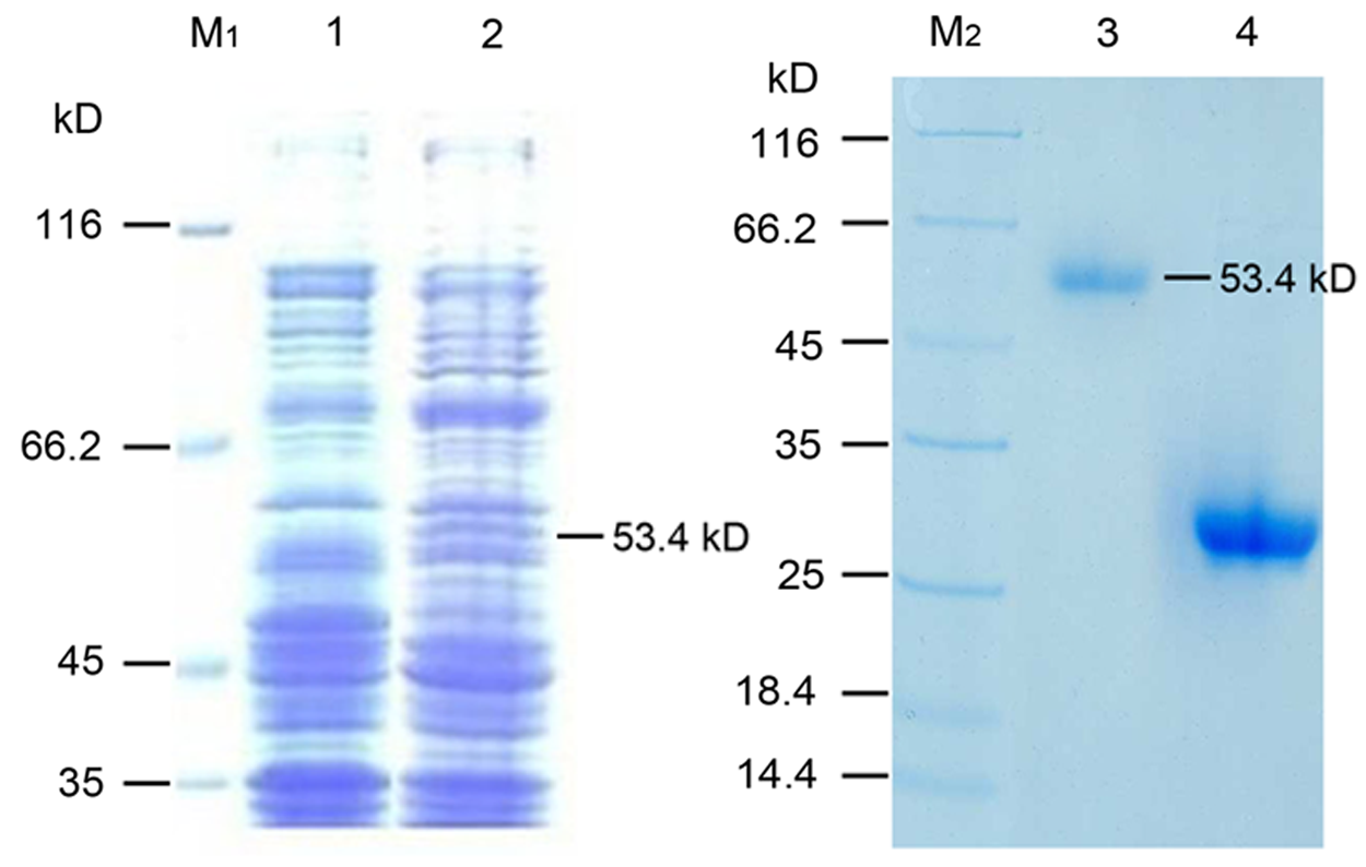
SDS-PAGE analysis of GST-VP7 protein. Lane M1 and lane M2, protein molecular weight marker. The protein extracts from uninduced recombinant bacteria (lane 1), 1.0 mM induced recombinant bacteria (lane 2), GST-VP7 protein purified from induced recombinant bacteria (lane 3), and GST protein purified from empty vector transformed bacteria (lane 4) separated on 12% sodium dodecyl sulfate-polyacrylamide gel and stained with Coomassie Brilliant Blue R250.
Optimal concentration determination of antigen
The ELISA assay (matrix titration) suggested that the titer of positive serum is 15625 at 1:50 diluted antigen. The purified antigen is diluted 50 times (final concentration, 3.6 μg/mL) for coating microtiter plates when screening for hybridoma cell lines.
ELISA screening for hybridoma cell lines
Cell fusion was performed as before and the fused proportion was 80%. After screening for positive clones and subcloning three times, one hybridoma cell line, which could stably secrete antibody, was obtained and designated as 2A10.
Purification of anti-GST-VP7 monoclonal antibody
MAb was produced by ascites revulsion and purified by octanoic acid/ammonium sulphate precipitation method. The SDS-PAGE analysis of purified MAb is shown in Figure 2. The bands correspond with the heavy chain (about 50 kDa) and light chain (about 25 kDa) of purified MAb, corresponding to the molecular weight of the heavy chain and light chain of immunoglobulin of BALB/c mice. The titer of purified antibody was 100,000 measured by indirect ELISA.
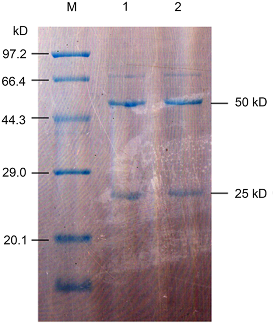
SDS-PAGE analysis of MAb 2A10. Lane M, protein molecular weight marker; lanes 1 and 2, purified ascites; MAb produced by ascites revulsion from a BALB/c mouse and purified by octanoic acid/ammonium sulphate precipitation method. The bands correspond with the heavy chain (∼50 kDa) and light chain (∼25 kDa) of purified MAb.
Western blot analysis of MAb
The results of Western blot suggested that MAb 2A10 binds VP7 specifically, while no band is observed with proteins of uninduced recombinant E. coli TOP10 (Fig. 3), demonstrating that 2A10 specifically bind the VP7 protein.
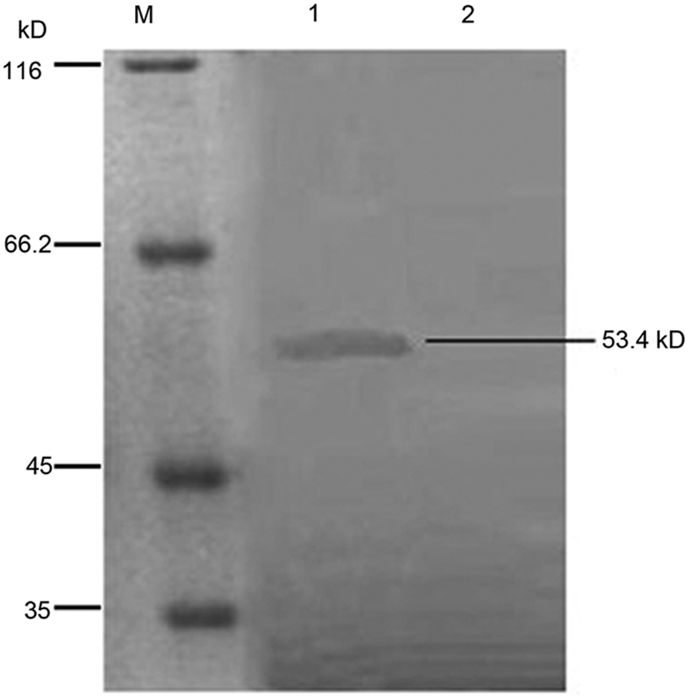
Western blot analysis of MAb 2A10. Lane M, protein molecular weight marker. GST-VP7 and GST protein separated on 12% SDS-PAGE, transferred to nitrocellulose membranes and probed with MAb 2A10. Lanes 1 and 2 show the GST-VP7 fusion protein and GST protein stained with DAB, respectively.
MAb stability
To test the MAb secreting stability of hybridoma cell line 2A10, the titers of hybridoma supernatants were determined by indirect ELISA. The titers of fifth, tenth, and twentieth subculturing of 2A10 are 128,000, 64,000, and 128,000, respectively. The serum samples of the immunized and unimmunized BALB/c mice are positive control and negative control for stability testing, respectively. The results indicated a stable secreting rate of MAb in hybridoma cell line 2A10.
Discussion
The coat proteins VP4, VP7 of rotavirus, which can be used for identifying serotypes of RV, are also protective antigens.(8) Therefore VP4 and VP7 can be used as antigens to develop monoclonal antibodies used for developing genetic engineering vaccines against RV. VP7 has higher antigenicity and immunogenicity than VP4, and the corresponding research is a popular focus both domestically and internationally. Therefore, in this study, the vp7 gene was chosen for prokaryotic expression. However several factors affect the expression of vp7 in E. coli: (1) Vp7 gene is not stable enough in E. coli cells. Wang and colleagues(9) reported that they encountered difficulties in cloning vp7 gene into E. coli, because the existence of underlying promoters in vp7 may affect the stability and expression of vp7 gene in host cells; (2) the expression product of vp7 is not stable and is toxic to E. coli cells.(10) Therefore fusion expression was used in prokaryotic expression of vp7 gene. The optimized expression conditions were E. coli TOP10 strain and 1.0 mM of IPTG, and 30°C of inducing temperature for 6 h. The results of SDS-PAGE showed an obvious band at 53.4 kDa. The gel was cut and the protein was recovered and used to immunize BALB/c mice. The Western blot showed a specific band, proving that gel-cutting immunization is a practicable immunization strategy. This strategy was easy to perform and the protein was highly purified and concentrated. The MAb could stably secrete antibody after being subcultured 20 times.
Overall, we successfully expressed the recombinant protein GST-VP7 and obtained one hybridoma cell line 2A10, which could stably secrete antibody. All of these works will be the basis for further study on proteomics and anthropic vaccines of rotavirus.
Footnotes
Acknowledgments
This research was supported by the Natural Science Foundation of Hubei Province of China (no. 2005ABA131), self-determined research funds of CCNU from the college's basic research and operation of MOE, and the project of Hubei Key Laboratory of Genetic Regulation and Integrative Biology (GRIB201204).
Author Disclosure Statement
The authors have no financial interests to disclose.