
Abstract
Pancreatic cancer remains a major challenge for research studies and clinical management. Monoclonal antibodies (MAbs) against pancreatic cancer biomarkers could be used in the early diagnosis of pancreatic cancer. The hippocalcin-like 1 protein (HPCAL1) is expressed in most pancreatic cancer cell lines. The goal of this work was to generate anti-HPCAL1 MAbs that have potential clinical applications. Recombinant HPCAL1 protein was successfully produced in an Escherichia coli expression system and was used as immunogen in mice. MAbs against HPCAL1 were generated using the standard hybridoma technique. Anti-HPCAL1 MAb 1E10 detects denatured HPCAL1 protein in Western blot analysis and immunoprecipitates native HPCAL1 protein from cell lysates. MAb 5A5 detects HPCAL1 in formalin-fixed, paraffin-embedded pancreatic cancer cell lines, making it a potentially useful reagent for pathologists. The MAb pair 1E10 and 5A5 had the optimal binding affinity for soluble HPCAL1 detection in a sandwich ELISA system, which is a potential clinical detection method for pancreatic cancer. In conclusion, multi-functional anti-HPCAL1 MAbs 1E10 and 5A5 have been generated and characterized, which may be used in early diagnosis of pancreatic cancer.
Introduction
P
PC is a group of heterogeneous diseases and includes cancer of the endocrine (islet cell carcinoma, neuroendocrine carcinoma, and carcinoma of carcinoid tumors) and exocrine (pancreatic ductal adenocarcinoma and acinar) pancreas. Among clinical cases, 74% of patients die within the first year of diagnosis and approximately 94% of patient deaths occur within 5 years of diagnosis, leading to the lowest relative survival rate among all cancers.(3) The major barriers against successful therapy are the complex pathophysiology, absence of early diagnostic and prognostic markers, and unresponsiveness to radiation and chemotherapies.(4) Currently, the only potentially curative approach to PC is surgical resection, which is often unsuccessful because the invasive and metastatic nature of the tumor masses makes complete removal difficult. Consequently, patients suffer relapses from remaining cancer stem cells or drug resistance that eventually lead to death. To improve the survival rate, early detection of PC is critical.(5) Since early detection is useful for improving survival and early diagnostic markers can positively impact the outcome of patients, biomarkers play an important role in the detection of pancreatic cancer at an early stage.
With the advent of post-genomic era, various microarray techniques have been used as detection biomarker of diseases. Several potential biomarkers of PC have been reported and used for early diagnosis of PC.(5) The protein hippocalcin-like 1 (HPCAL1) (
Materials and Methods
Cell lines
Human pancreatic cancer cell lines (CAPAN 2, Miapaca, Mpanc 96, NOR-P1, Panc 1, Panc 2.03, Panc 5.04, Panc 10.05, L33, PL45, BxPC3, and L36pl) were a kind gift from Drs. Martin McMahan and Stephan Gysin (University of California, San Francisco), and were cultured as described previously.(8) The 293 cells were grown in DMEM containing 10% fetal bovine serum (FBS). All cell lines were cultured at 37°C with 5% CO2.
Expression and purification of HPCAL1 protein
An HPCAL1 gene expression vector was constructed by GenScript (Piscataway, NJ). Origami (DE3) bacteria cells transformed with the pCold II-HPCAL1 plasmid were selected for expression of the pCold II-HPCAL1 target protein. Expression was optimized and analyzed by SDS-PAGE. The recombinant strains were grown overnight at 37°C in 20 mL of LB medium containing 100 μg/mL ampicillin, then were inoculated to 2 L of LB medium at a ratio of 1:100 for large-scale production and grown until an OD600 of 0.6 was reached. Recombinant HPCAL1 was expressed in Origami (DE3) cells by inducing with IPTG (0.5 mM final concentration) at 15°C over 16 h. The bacterial pellets were collected by centrifugation at 2000 g for 10 min, resuspended in 200 mL of lysis buffer, and then sonicated on ice until the bacteria were lysed. The expressed HPCAL1 was identified by SDS-PAGE.
Monoclonal antibody production
Mouse MAbs against HPCAL1 were produced by injecting BALB/c mice intraperitoneally with purified native and denatured (boiled in SDS sample buffer) HPCAL1 protein (20 μg/mouse) in Freund's complete adjuvant, followed by two additional injections at 3-week intervals in Freund's incomplete adjuvant. After 1 month, the final HPCAL1 injections were given intraperitoneally and intravenously without adjuvant. Spleen cells were isolated from the sacrificed mice and then were fused with OUR-1 myeloma cells using standard techniques 4 days after the final injections, and hybridomas were generated by the method described previously.(9)
To screen for positive hybridoma clones, 96-well plates were coated with 2.0 μg/mL HPCAL1 protein in coating buffer (0.2 M Na2CO3/NaHCO3 [pH 9.6]; 50 μL/well) at 4°C overnight. The plates were then blocked with PBS containing 1% BSA (200 μL/well) overnight at 4°C. Fifty μL of hybridoma supernatant were added to the coated wells and incubated for 1.5 h at room temperature (RT). Plates were washed twice in wash buffer (PBS with 0.05% Tween-20), and alkaline phosphatase-coupled goat anti-mouse IgG (Sigma, St. Louis, MO) was added (50 μL/well) at 1:2000 dilution for 1.5 h at RT. After washing four times in washing buffer, phosphatase substrate CP-nitrophenyl phosphate (Kirkegaard & Perry Laboratories, Gaithersburg, MD) was added for 30 min, and absorbance was measured at 405 nm. Hybridoma clones with strong reactivity with HPCAL1 (OD value >0.5, negative controls <0.02) were subcloned twice by limited dilution, and reactivity was again confirmed by ELISA. Subcloned hybridoma cells were cultured in the OPTIMEM medium (Gibco, Grand Island, NY) containing 10% FBS, weaned gradually to serum-free medium (Hybridoma-SFM medium, Gibco), and then transferred to a bioreactor (Integra Biosciences, Chur, Switzerland). The supernatant was harvested twice a week. Anti-HPCAL1 MAbs were purified from the culture supernatants by affinity chromatography using a protein-G column.
Western blot analysis and immunoprecipitation
To screen for antibodies that recognize denatured HPCAL protein in Western blot analysis, 293, Miapaca, Mpanc96, and Panc1 cells were lysed in RIPA buffer. Thirty-five μg of total protein from each lysate was resolved by 10% SDS-PAGE gel and transferred onto a nitrocellulose membrane. After the membrane was blocked with blocking buffer (Li-Cor Biosciences, Lincoln, NE) at 4°C overnight, it was incubated with hybridoma culture supernatants for 1.5 h at RT. The membrane was then washed in wash buffer and incubated with goat anti-mouse 680 at 1:15,000 dilution for an additional 1.5 h at RT. Following another wash using wash buffer, the blot was scanned and visualized using an Odyssey Infrared Imager (Li-Cor).
For immunopreciptation, MPanc 96, Panc 2.03, Nor-P1, and 293 cells were lysed in RIPA buffer. One mg of total protein (of either 293, MPanc 96, Nor-P1, or Panc 2.03 cell lysates) was mixed with 5 μg of anti-HPCAL1 1E10 MAb and 20 μL of protein-G Sepharose (Invitrogen, Carlsbad, CA) at 4°C overnight, followed by immunoblotting. Briefly, the immune complexes were washed twice, and the proteins were extracted and denatured in 2×SDS sample buffer before being separated on a 10% SDS-PAGE gel and transferred onto a nitrocellulose membrane (Bio-Rad, Hercules, CA). The membrane was blocked with blocking buffer (Li-Cor) at 4°C overnight, incubated with MAb 1E10 at 4 μg/mL for 1.5 h at RT, washed in PBS with 0.05% Tween-20, and reacted with goat anti-mouse 680 at 1:15,000 dilution for an additional 1.5 h at RT. Following the same washing, the proteins were detected with the Odyssey Infrared Imager.
Monoclonal antibody pair-search by Octet system
To screen for an antibody with the highest affinity to HPCAL1, protein A biosensors were dipped in and saturated with hybridoma cell culture supernatants (protein A–antibody binding) for 5 min. After washing for 2 min, the biosensors were dipped in HPCAL1 protein solution for 5 min (antibody–antigen association), then in eluting buffer (glycine/HCl [pH 2.0]) for 5 min (dissociation), and finally in Tris (pH 9.0) for neutralization. To search for a second antibody that binds a different epitope of HPCAL1, protein A biosensors were first saturated with one antibody (protein A–antibody binding); after washing in PBS, the biosensors were dipped in HPCAL1 protein (antibody–antigen binding). After another washing, the biosensors were moved to hybridoma cell culture supernatants (antibody–antigen–antibody association) and then to the eluting buffer (dissociation). The association curves show different affinities of the antibodies in culture supernatants.
Sandwich ELISA
Wells of a 96-well plate were coated with 50 μL of anti-HPCAL1 5A5 MAb at 10 μg/mL in 0.2 M Na2CO3/NaHCO3 (pH 9.6) overnight at 4°C. Wells were washed and blocked with 200 μL/well of PBS containing 1% BSA overnight at 4°C. The blocking solution was removed and purified HPCAL1 protein was added starting at 250 ng/mL, followed by serial dilutions. The protein was incubated at RT for 1.5 h. The plate was then washed and a biotinylated antibody against HPCAL1, 1E10-Biotin, was added at 8 μg/mL to each well and incubated at RT for 1.5 h. The plate was washed again and the secondary antibody streptavidin AP conjugate (Kirkegaard & Perry) was added at 1:2000 dilution and incubated at RT for an additional 1.5 h. The phosphatase substrate CP-nitrophenyl phosphate was added to the plate (50 μL/well) and incubated at RT for 30 min; absorbance was measured at 405 nm.
Immunofluorescent staining
To screen for antibodies that work for immunofluorescent (IF) and immunohistochemical (IHC) staining, Miapaca and 293 cells were plated into ten 96-well plates. The next day the cells were fixed with 10% neutral buffered formalin. Fifty μL of hybridoma supernatant were added to wells containing fixed cells for 1.5 h at 37°C. Plates were washed twice in washing buffer (PBS with 0.05% Tween-20), and Rhodamine Red-conjugated goat anti-mouse IgG (Jackson ImmunoResearch Lab, West Grove, PA) was added (30 μL/well) at 1:100 dilution for 1.5 h at 37°C. After washing two times in washing buffer, cells were examined under fluorescence microscope and positively stained clones were picked. 293 cells were transiently transfected with a GFP-HPCAL1 construct using Fugene 6 reagent (Roche, Mannheim, Germany). After 48 h, cells were seeded on chamber slides overnight and fixed with 10% neutral buffered formalin. After washing twice with PBS, the cells were blocked with PBS containing 5% dry milk and 0.05% Tween-20 overnight at 4°C, and then were incubated with purified antibody 5A5 (20 μg/mL) with or without HPCAL1 protein (30 μg/mL) at 37°C for 1.5 h. The chamber slides were then washed and incubated with sheep anti-mouse-TRITC (Jackson ImmunoResearch) at 1:50 dilution for an additional 1.5 h at 37°C. Nuclei were stained with DAPI for 10 min at RT. After washing twice in PBS, slides were coverslipped and photographs were taken using confocal microscope.
Immunohistochemical staining of pancreatic cancer cell array
A cell array was made using the HistoGel (Richard-Allan Scientific, Kalamazoo, MI). Pancreatic cancer cells were cultured in flasks, fixed by 10% formalin, and harvested into cell pellets. Liquified Histogel was mixed into the cell pellet and the mixture was solidified at 4°C. Cell pellets were then embedded in paraffin. A cell array block was made and cut into slide sections. Immunocytochemistry was done with standard procedures. Deparaffinized sections were immunostained with MAb 5A5 as primary antibody at 2 μg/mL by standard procedure of DAB staining on Discovery XT Immunostainer from Ventana Medical Systems (Tucson, AZ).
Results
Recombinant HPCAL1 protein was produced in Origami (DE3) bacteria and affinity-purified
Expression vector pCold II was shown in Figure 1A. According to its sequence, the molecular weight of HPCAL1 should be 21–23 kDa. HPCAL1 target protein expression in pCold II-HPCAL1-transformed Origami (DE3) bacteria was evaluated by SDS-PAGE analysis followed by Coomassie Brilliant Blue staining (Fig. 1B). The HPCAL1 protein was expressed successfully in Origami (DE3). The supernatant of the bacterial lysate was purified using HisTrap HP (GE Healthcare, Piscataway, NJ) affinity columns and an AKTA purification system according to the manufacturer's instructions. The fraction containing HPCAL1 was dialyzed against 5 mM Tris buffer (pH 8.0). About 10 mg of fusion protein were obtained from 1 L of culture, with purity above 90%.
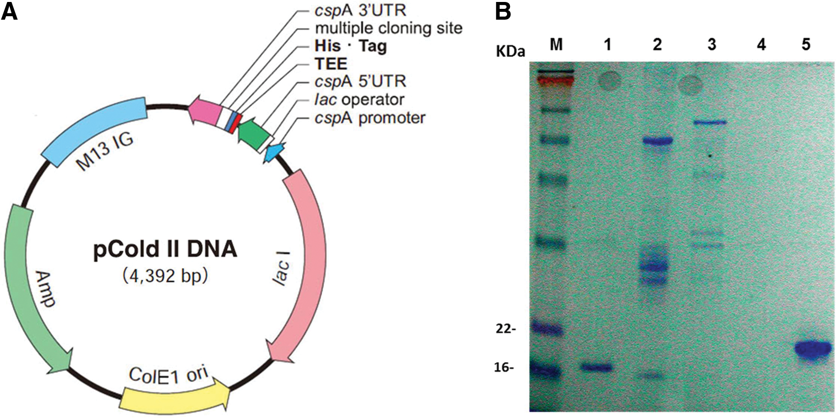
Expression and characterization of HPCAL1 protein. (
Production of monoclonal antibodies against HPCAL1
BALB/c mice were immunized with native and denatured HPCAL1 protein. After three immunization injections, the titer of the antibody against the antigen in mouse sera reached up to 1:160,000. Four days after the final injections, the mouse with the highest antibody titer was splenectomized and the spleen cells were fused with OUR-1 myeloma cells. Thirty-four clones with the highest OD values in capture ELISA were picked and expanded. Among them, clones 1E10 and 5A5 were subcloned as a result of their reactivities with HPCAL1 in different applications. The MAbs were purified through a protein-G column.
MAb 1E10 detects denatured HPCAL1 in Western blot analysis and immunoprecipitates native HPCAL1 from cell lysates
In Western blot analysis, purified MAb 1E10 was able to detect denatured HPCAL1 protein endogenously expressed in the pancreatic cell lines Miapaca, MPanc 96, and Panc 1. No HPCAL1 expression was detected in 293 cells (Fig. 2A). An immunocomplex was set up with 5 μg 1E10 and 20 μL of protein-G beads in pancreatic cancer cell lysates. 1E10 was also able to immunoprecipitate HPCAL1 from the cell lines MPanc 96, Panc 2.03, and Nor-P1 (Fig. 2B). The heavy and light chain bands were 1E10 IgG in the lysates recognized by anti-mouse secondary antibody.
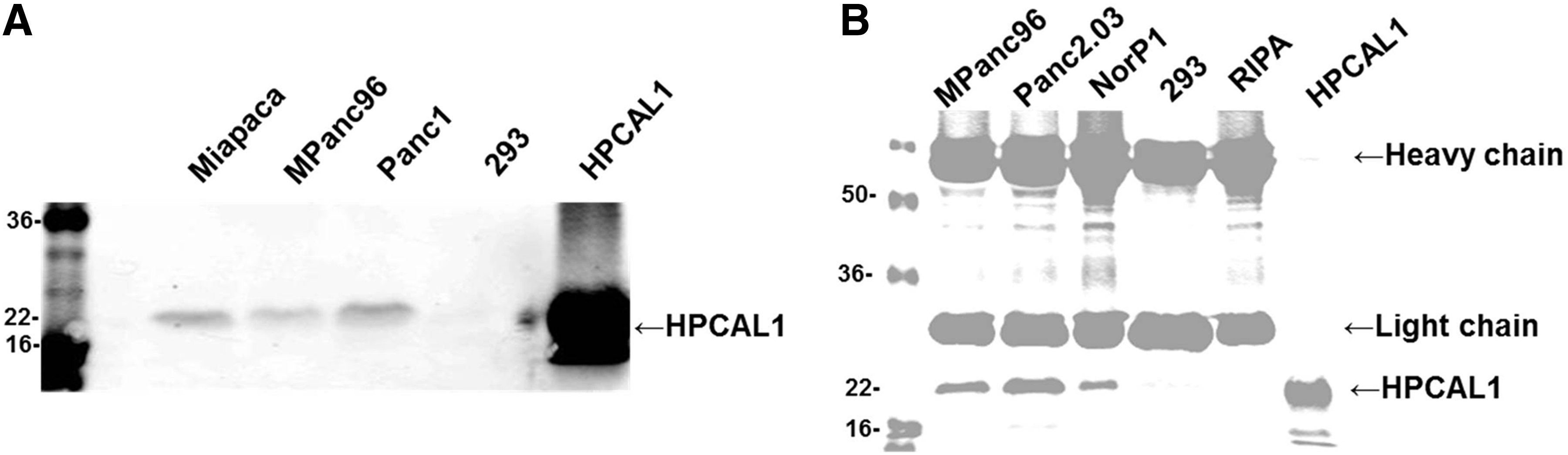
Western blot analysis and immunoprecipitation. (
MAb pair 1E10 and 5A5 has the optimal binding affinity for soluble HPCAL1 detection in sandwich ELISA
Each monoclonal antibody binds a specific epitope on an antigen. A pair of antibodies that recognize different epitopes can be used to establish a detection system such as sandwich ELISA. To screen for an antibody pair having optimal binding affinity towards soluble HPCAL1, ForteBio's Octet system was used. ELISA data showed that only MAb 1E10 was IgG2; the rest of the MAbs were all IgG1 (data not shown). We know that Protein A binds strongly to mouse IgG2, while it binds weakly to mouse IgG1 and does not bind mouse IgG3. In Figure 3A, MAb 1E10 shows stronger binding to HPCAL1 relative to the other IgG1 MAbs. 1E10 was therefore used as the capture antibody to screen for a second antibody (Fig. 3B, C). Of the antibodies screened, 5A5 was found to have the highest binding affinity to 1E10-captured HPCAL1 protein.

HPCAL1 monoclonal antibody pair search using the ForteBio Octet system. (
To set up a sandwich ELISA, MAb 5A5 was used as the capture antibody while 1E10 was biotinylated and used as the detecting antibody. The lowest concentration that the system could detect was less than 10 ng/mL. At a range of 7 to 70 ng/mL of HPCAL1, the absorbance and the protein concentration displayed a linear correlation (Fig. 4).

HPCAL1 sandwich ELISA. A 96-well plate was coated with the 5A5 MAb at 10 μg/mL overnight at 4°C. After blocking, HPCAL1 protein was added starting at 250 ng/mL, followed by serial dilutions. The protein was incubated at RT for 1.5 h. The plate was washed and incubated with a biotinylated antibody 1E10-biotin at 8 μg/mL at RT for 1.5 h. A secondary antibody, Streptavidin AP conjugate, and the phosphatase substrate CP-nitrophenyl phosphate were used to detect the reactivity. Absorbance was measured at 405 nm.
MAb 5A5 detects HPCAL1 in formalin-fixed 293 cells transfected with GFP-HPCAL1
In screening for IF- and IHC-positive clones, clone 5A5 displayed the strongest staining signal for HPCAL1 expression in Miapaca cells versus 293 cells (data not shown). To confirm the result, GFP-HPCAL1 was transiently transfected into 293 cells. After formalin fixation, the transfected cells were stained with purified MAb 5A5 followed by TRICT-labeled secondary antibody. MAb 5A5 demonstrated specific staining against HPCAL1 (i.e., red, HPCAL1 expression; green, GFP) stains co-localized in the cytoplasm and nuclei (Fig. 5). In the presence of HPCAL, the staining signal was abolished (data not shown).
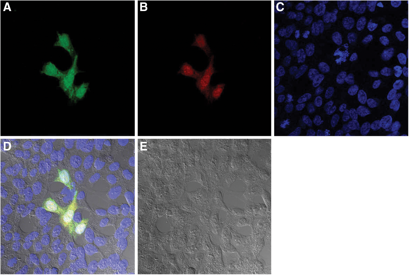
Immunofluorescent staining of GFP-HPCAL1-transfected 293 cells with the 5A5 MAb. The 293 cells were transiently transfected with a GFP-HPCAL1 construct using Fugene 6 reagent. After 48 h, cells were seeded on chamber slides overnight and fixed with 10% neutral buffered formalin. After blocking, the slides were incubated with 5A5 (20 μg/mL) with or without HPCAL1 protein (30 μg/mL) and detected with sheep anti-mouse TRITC. Nuclei were stained with DAPI. (
Immunohistochemical staining of pancreatic cancer cells with MAb 5A5
An array of formalin-fixed, paraffin-embedded pancreatic cancer cells was immunostained with MAb 5A5. HPCAL1 expression was detected in cell lines CAPAN-2, Panc 1, Panc 5.04, Panc 10.05, and PL45 cells, but the expression was low in BxPC-3, L33, and L36pl cells (Fig. 6).

HPCAL1 expression in pancreatic cancer cells. A formalin-fixed, paraffin-embedded pancreatic cancer cell array was immunostained with MAb 5A5 as described in the Materials and Methods section. HPCAL1 was highly expressed in CAPAN-2, Panc 1, Panc 5.04, Panc 10.05, and PL45 cells, but expression was low in BxPC-3, L33, and L36pl cells.
Discussion
HPCAL1 is a member of neuron-specific calcium-binding protein family found in the retina and brain.(10,11) It is highly similar to human hippocalcin protein and nearly identical to the rat and mouse hippocalcin like-1 proteins. Hippocalcin-like 1 (also known as visinin-like protein 3, VILIP-3) is used to study the calcium-dependent regulation of rhodopsin phosphorylation and is relevant to neuronal signaling in the central nervous system.(12,13) HPCAL1 is also a highly plastic epigenetic mark whose hypermethylation depends on both types of early-life exposure and adult-life events.(14–17) However, recently new discoveries indicate that these proteins also have key roles in pathological processes of disease, including cancer, as well as Alzheimer's disease.(14)
In this study, our goal was to generate a panel of murine MAbs against HPCAL1 that could be used to detect HPCAL1 expression in pre-clinical and clinical settings. Our purified HPCAL1 protein had strong immunogenicity. The choice of an appropriate hybridoma screening assay is one of the most important parts of hybridoma production. The assay is often selected according to the characteristics of the antigen and the laboratory conditions, as well as the intended use of the antibody. MAb 1E10 displayed the highest signal compared with background ratio and its reactivity to HPCAL1 was confirmed with a number of cell lines in Western blot analysis. To screen for antibodies that work for immunofluorescent and immunohistochemical staining, HPCAL1-expressing Miapaca cells and non HPCAL1-expressing 293 cells were cultured and fixed with formalin. Clone 5A5 demonstrated the strongest staining signal. MAb 5A5 was able to detect HPCAL1 expression in formalin-fixed, paraffin-embedded pancreatic cancer cell lines (Fig. 6); therefore it has potential applications in clinical settings. Further studies are needed to evaluate HPCAL1 expression in well-annotated clinical patient sample sets (pancreatic tumor tissues, normal tissues, and cancer-adjacent normal tissues).
Furthermore, ForteBio's Octet system was used to screen an antibody pair with the optimal binding affinity towards soluble HPCAL1. MAb pair 1E10 and 5A5 displayed the optimal binding affinity; with them we were able to detect soluble HPCAL1 in a sandwich ELISA at levels of nanograms per milliliter. Therefore MAbs 1E10 and 5A5 together are a candidate pair of reagents that can be used in a sandwich ELISA detection system for rapid and early detection of pancreatic cancer. For validation of this platform with established pancreatic cancer cell lines and clinical serum samples from pancreatic cancer patients, normal and pancreatitis samples are needed to evaluate sensitivity and specificity of the HPCAL1 ELISA method in clinical settings. We are currently gathering these cell lines and serum specimens through collaborators to optimize the detection system.
Footnotes
Acknowledgments
We thank Drs. McMahon and Gysin at UCSF for providing the pancreatic cancer cell lines used in this study.
Author Disclosure Statement
The authors have no financial interests to disclose.