
Abstract
B7-H4, a member of B7 family, is widely expressed in tumor tissues and plays an important role in the negative regulation of T cell immunity. In this study, we report on the establishment and characterization of a functional anti-human B7-H4 monoclonal antibody (MAb) 5G3 through hybridoma method. Flow cytometry analysis showed that MAb 5G3 specifically bound to B7-H4 molecule. Functional experiments indicated that MAb 5G3 could block the inhibitory role of B7-H4 molecule on A549 cells in and reduce the apoptosis of Jurkat cells, suggesting that MAb 5G3 is an antagonistic antibody and a useful tool for further studies of B7-H4 functions in cancers.
Introduction
I
In humans, the gene of B7-H4 is located on chromosome 1p11.1, spans 66.6 kb, and contains six exons and five introns. Alternative splicing in exon 6 forms two different transcripts with identical open reading frames. The cDNA of B7-H4 is about 1.8 kb, containing 849 bases that encode 282 amino acids. The extracellular region of B7-H4 molecule includes a pair of IgV and IgC domains, belonging to the immunoglobulin superfamily.(6) In addition, B7-H4 molecule contains a long hydrophobic transmembrane region, but its intracellular region has only two amino acids.(3–5)
B7-H4 mRNA is widely expressed in peripheral tissues. Reverse transcription polymerase chain reaction (RT-PCR) shows that B7-H4 mRNA is expressed in placenta, kidney, liver, lung, spleen, thymus, liver, testis, ovary, skeletal muscle, pancreas, and small intestine.(3,5,6) At the protein level, the B7-H4 molecule is almost undetectable in normal tissues,(7) including lung, liver, kidney, skeletal muscle, pancreas, and so on. In contrast, the upregulating expression of B7-H4 molecule has been reported in a number of tumors including gastric,(8) breast,(9) ovarian,(10–13) and lung cancers,(14) thus suggesting that the upregulation of B7-H4 expression on tumor cells is perhaps a mechanism by which these cells inhibit T cell activity, evade the immune cell attacks, and finally promote tumor progression.
The purpose of our current work is to generate a new mouse monoclonal antibody (MAb) against the human B7-H4 molecule. We used this mouse monoclonal antibody to investigate the effects of B7-H4 molecule in A549 cells (non-small cell lung cancer cell line) on Jurkat cells (an immortalized T cell leukemia cell line conserving functional characteristics of activated T cells).
Materials and Methods
Cell lines and reagents
The mouse fibroblasts L929, Jurkat cells, and A549 cells were obtained from American Type Culture Collection (Manassas, VA). L929/mock cells (L929 cells transfected with empty vector) and L929/B7-H3 and L929/B7-H4 cells (L929 cells transfected with human B7-H3 or B7-H4 genes) were from our laboratory (Medical Biotechnology Institute of Soochow University, Suzhou, China). RPMI-1640 and DMEM media were purchased from Gibco (Carlsbad, CA). All the cells were cultured in RPMI-1640 medium supplemented with 10% fetal calf serum (FCS, Sijiqing Company, Hangzhou, China). PE-conjugated mouse anti-human CD3, CD69 and B7-H4 (clone no. H74) antibodies were the products of eBioscience (San Diego, CA). Mouse IgG, PE-conjugated mouse IgG, and PE-conjugated goat anti-mouse IgG were also from eBioscience. HAT and HT products were purchased from Sigma (St. Louis, MO).
Immunization of mice and establishment of hybridoma
L929/B7-H4 cells were used as immunogens. BALB/c mice (female, 6–8 weeks old) were chosen and intraperitoneally injected with L929/B7-H4 cells pretreated by mitomycin. The booster injection was repeated every 3 weeks for three times. Three days after the final booster immunization, the splenocytes from the selected mice were harvested and fused with mouse myeloma cells (SP2/0 cell line) in the presence of 50% polyethylene glycol (PEG) according to the method described by Groth and Scheidegger.(15) The fusion cells were cultured with HAT-DMEM medium containing 15% FCS in 96-well plates to select hybrid clones. Two weeks later, the above medium was changed into HT-DMEM medium supplemented with 15% FCS. All hybrid clone supernatants were screened for the detection of antibody using flow cytometry. The hybrid clone secreting antibody that was reactive with L929/B7-H4 cells but not with L929 cells, L929/mock cells, or L929/B7-H3 cells was selected and subcloned by limiting dilution method three times to establish hybridoma cell line secreting monoclonal antibody.
MAb production, purification, and isotype determination
One hybridoma cell line (5G3) detected to secrete MAb against B7-H4 was selected and cultured for injection to produce the ascites. BALB/c mice pretreated with sterile paraffin oil 7 days ago were injected with hybridoma cells. Ten days later, ascites were collected and centrifuged to obtain the supernatant, which was purified by Protein G-Sepharose CL4B affinity column (Pharmacia, Uppsala, Sweden). The isotype of MAb was determined by mouse antibody isotyping kit (Sigma-Aldrich) following the manufacturer's protocol.
Competitive inhibition test and epitope analysis
L929/B7-H4 cells were incubated with 5G3 at 4°C for 30 min; then PE-conjugated MAb H74 was added for another 30 min of incubation. In the meantime, L929/B7-H4 cells with the addition of PE-conjugated mouse IgG was used as negative control and L929/B7-H4 cells with the addition of PE-conjugated MAb H74 were used as positive control. After all the cells were washed twice with PBS, flow cytometry was employed for epitope analysis.
Flow cytometric analysis of B7-H4 expression on A549 cells
B7-H4 expression on A549 cells was examined by 5G3 and PE-conjugated MAb H74. A549 cells were incubated with 5G3 at 4°C for 30 min. Cells were washed and resuspended in goat anti-mouse antibody conjugated with PE. Cells were incubated at 4°C for another 30 min, washed twice, and finally analyzed using flow cytometry. A549 cells labeled with PE-conjugated MAb H74 were also analyzed using flow cytometry after incubation for 30 min and twice washed with PBS.
Jurkat cells co-cultured with A549 cells
A549 cells were seeded into six-well plates at 2×10⁵ cells/mL, and total medium was added to 3 mL. When the cultured cells reached 60% confluency, the old medium was removed and 4 mL fresh media containing Jurkat cells (2×10⁵ cells/mL) were added. The co-cultured cells were divided into three groups: the first group with 5G3 antibody (20 μg/mL); the second group without 5G3 antibody; and the third group with mouse IgG. After 72 h of co-culture, the six-well plate was gently shaken to dislodge loosely attached Jurkat cells, which were collected and centrifuged at 1500 r/min for 5 min for the following experiments.
Examination of CD3 and CD69 expression on Jurkat cells
The co-cultured Jurkat cells were incubated with PE-labeled CD3 and CD69 antibodies at 4°C for 30 min and then washed twice for the examination of CD3 and CD69 expressions using flow cytometric analysis. The non-co-cultured Jurkat cells were used as the control group.
Cell proliferation tested by CCK8 assay
Cell proliferation was tested using CCK8 assay. The co-cultured Jurkat cells were seeded into 96-well bottom tissue culture plate at a concentration of 8×10⁴ cells/well containing 200 μL of RPMI-1640 medium supplemented with 10% FCS. The control group cells were non-co-cultured Jurkat cells. After the addition of 20 μL CCK8 to each well and incubation at 37°C for 4 h, the optical density (OD) of each well was read at 450nm by a microreader (Thermo Fisher, Cambridge, MA). Each group was measured in triplicate and three independent experiments were performed. Data were analyzed using Student's t-test. A value of p<0.05 was considered significant.
Analysis of apoptosis
The co-cultured Jurkat and non-co-cultured Jurkat cells were suspended in tubes at 1×10⁵ cells/mL with PBS, washed twice with PBS, centrifuged at 800 r/min for 5 min, and then resuspended in 50 μL of Annexin-V-binding buffer. After 5 μL of FITC-conjugated Annexin-V and 2 μL of propidium iodide (PI) were added, the cells were incubated in a dark room at room temperature for 20 min. Finally, 350 μL of Annexin-V binding buffer was added to each tube, and the cells were analyzed using flow cytometry.
Results
Generation of hybridoma cell line secreting MAb
After cell fusion, the supernatants of growing hybrid clone were screened for the secretion of antibody against B7-H4 by flow cytometry. One of the hybrid clones secreting antibody that was reactive with L929/B7-H4 cells but not with L929 cells, L929/mock cells, or L929/B7-H3 cells was obtained. Hybridoma cell line was finally established after subcloning three times by limiting dilution, and was named 5G3 (Fig. 1).
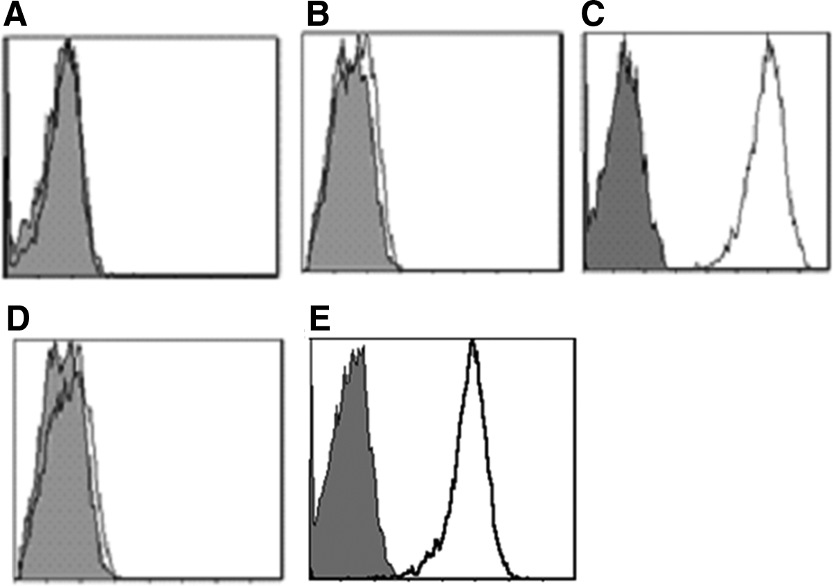
MAb 5G3 specifically recognized B7-H4 (
MAb ascites production, purification, and isotype identification
5G3 hybridoma cells were massively cultured and injected into BALB/c mice pre-treated with sterile paraffin oil. The collected ascites were centrifuged to obtain the supernatant that was purified by Protein G affinity column. The purified MAb 5G3 was tested by isotyping kit. The result of isotype identification showed that MAb 5G3 was IgG1 isotype with κ light chain.
Epitope analysis
Using the competitive inhibition test, results (Fig. 2) showed that, compared with anti-B7-H4 antibody H74 alone, MAb 5G3 could not block the binding of antibody H74 to L929/B7-H4 cells after the histograms of negative control, positive control, and experimental group were overlayed. This result illustrated that commercial antibody H74 and MAb 5G3 recognized the different antigenic sites on the B7-H4 molecule.
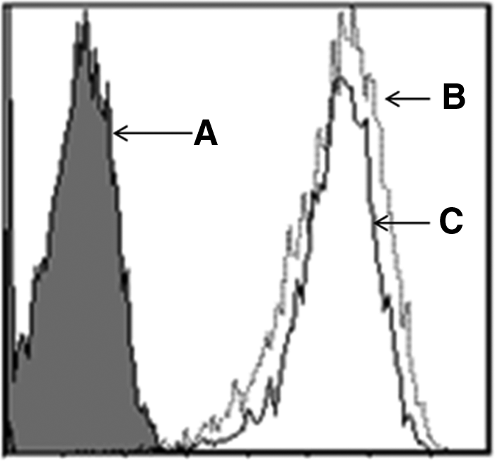
Flow cytometric analysis of B7-H4 epitopes recognized by H74 and MAb 5G3.
Biological activity of 5G3 MAb
A549 cells expressed B7-H4 that was recognized by MAb 5G3 and commercial antibody H74 (Fig. 3). After the co-culture of A549 and Jurkat cells for 72 h, we found that these A549 cells dramatically inhibited the expression of CD3 and CD69 on co-cultured Jurkat cells compared with non-co-cultured Jurkat cells. MAb 5G3 could block the inhibition role of A549 cells in CD3 and CD69 expressions on co-cultured Jurkat cells, while mouse isotype control IgG could not have such blocking effect (Fig. 4). A549 cells also caused co-cultured Jurkat cells to exhibit the obvious apoptosis compared with non-co-cultured Jurkat cells and MAb 5G3 significantly reduced the apoptosis of co-cultured Jurkat cells, whereas mouse isotype control IgG did not do this (Fig. 5). Finally, in comparison with non-co-cultured Jurkat cells, the proliferation of co-cultured Jurkat cells was inhibited by A549 cells and MAb 5G3 could abolish such inhibition effect, which was still displayed in the presence of mouse isotype control IgG (Fig. 6).

Expression of B7-H4 on A549 cells was recognized by MAb 5G3 and H74 using flow cytometric analysis. Mouse IgG was used as negative control (gray graph). (
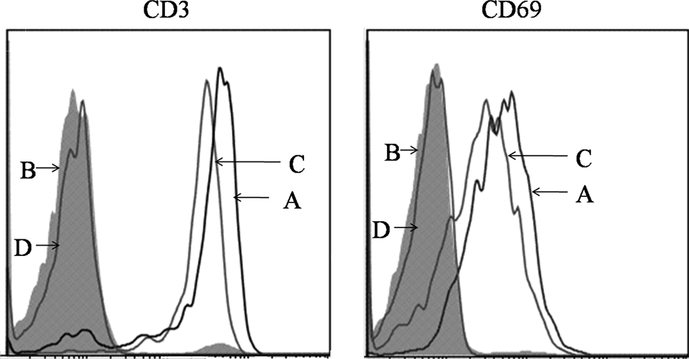
Flow cytometric analysis of expression of CD3 and CD69 on Jurkat cells. (
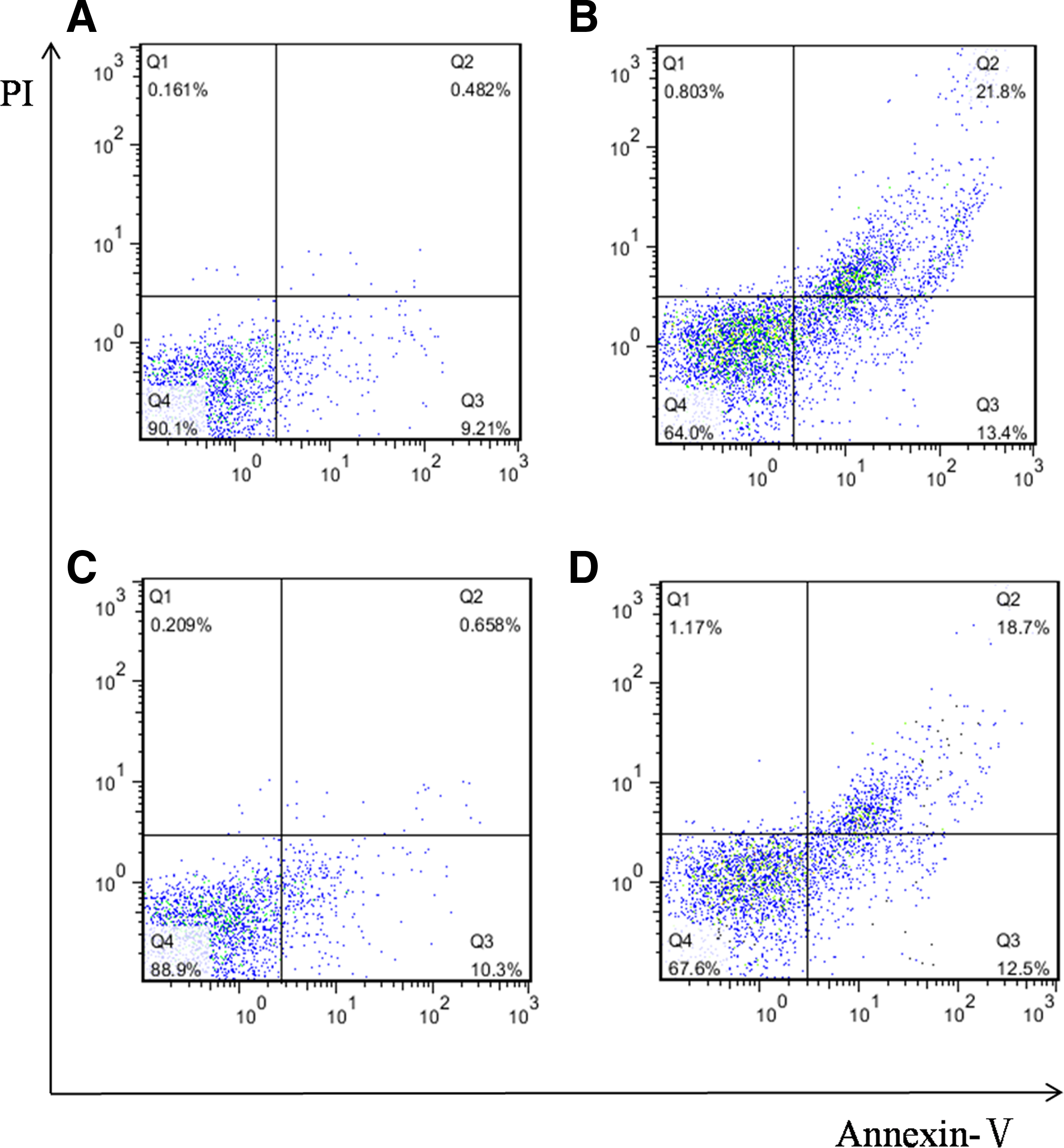
Flow cytometric analysis of B7-H4 induced apoptosis in Jurkat cells. (

Proliferation of Jurkat cells was determined by CCK8 assay. (
Discussion
In this study, we used the L929/B7-H4 transfectant as immunogen to immunize BALB/c mice and successfully established a hybridoma cell line secreting MAb against B7-H4 molecule through hybridoma technology. Flow cytometric analysis showed that MAb 5G3 specifically recognized L929/B7-H4 and did not react with L929, L929/mock, or L929/B7-H3 cells. Competitive inhibition experiment demonstrated that MAb 5G3 and commercial antibody H74, which is also MAb against B7-H4, bound to the different antigenic epitopes on B7-H4 molecule.
The B7-H4 molecule plays a role in the negative regulation of T cell response. Many studies have reported that it was not detected in normal tissues, but was observed in various cancers including gastric,(8) breast,(9) ovarian,(10–13) and lung cancers.(14) Expressing B7-H4 on tumor cells is thought to be one of the most important mechanisms by which tumor cells exert the immune escape of T cell attacks.(16,17) Although the B7-H4 receptor has not been cloned and identified as yet, it was proved that activated T expressed this unknown receptor through which B7-H4 signaling had inhibitory effects on activated T cells.(18,19) Such studies mainly adopted purified B7-H4-Fc fusion protein. In the present study, we used NSCLC cell line A549 cell expressing B7-H4 and Jurkat cell maintaining activated T cell characteristics to establish an in vitro co-cultured system to better mimic the pathological situation. The results showed that in such co-cultured systems of cross-talk between tumor cells and T lymphocytes, A549 cells could directly contact Jurkat cells, inhibit their activities (cell proliferation and membrane molecule expression), and facilitate apoptosis. More importantly, adding MAb 5G3 to this co-cultured system showed that it could block the inhibition role of A549 cells and reduce the apoptosis of Jurkat cells, illustrating that anti-B7-H4 MAb 5G3 was a functional antibody with antagonistic activity and that B7-H4 was a potential molecular target for tumor immunotherapy.
In summary, we generated MAb 5G3 directed against B7-H4 and demonstrated that it possesses antagonistic function in the co-cultured system of A549 cells and Jurkat cells. MAb 5G3 may prove a useful tool for the further investigation of the function of B7-H4.
Footnotes
Acknowledgment
This study was supported by the project of Jiangsu Province's Key Discipline and Laboratory of Medicine (XK201135).
Author Disclosure Statement
The authors have no financial interests to disclose.