
Abstract
Type I insulin-like growth factor receptor (IGF-1R) is a receptor tyrosine kinase that is involved in the transformation of cells, cancer proliferation, and metastatic events in various types of cancer. The present study reports on the generation of a mouse monoclonal antibody (MAb) to IGF-1R using the mouse lymph node method. MAb 1B1, which reacts specifically with the extracellular domain of IGF-1R, was obtained using flow cytometry (FCM) screening using MCF-7 cells. Using immunostaining, MAb 1B1 detected IGF-1R mainly on the plasma membrane of MCF-7 cells. MAb 1B1 would be useful in FCM and immunofluorescence assays to detect IGF-1R-expressing cells for basic and clinical research.
Introduction
T
IGF-1R and its ligands are reportedly involved in the transformation of cells, cancer proliferation, and metastatic events in various types of cancer.(4–6) Based on a large amount of previously reported evidence, IGF-1R is regarded as an attractive target molecule for cancer treatment, and several IGF-1R inhibitors have been developed and examined in clinical trials.(7,8) Some IGF-1R antibodies have been shown to have tolerability and to exhibit preliminary antitumor activity in early clinical trials.(9,10) However, a sufficient efficacy of IGF-1R antibody has not yet been demonstrated in late phase trials.(7,8) To further the development of anti-IGF-1R agents, predictive biomarkers of efficacy are needed.(8,11) The protein expression level and the gene copy number gain of IGF-1R are considered to be potential biomarkers, and technologies for quantifying these parameters are crucial.
In this present study, we report on the production and characterization of a monoclonal antibody (MAb) against the extracellular domain of IGF-1R. This MAb, 1B1, has the potential for use in flow cytometry (FCM) and immunostaining analyses.
Materials and Methods
Cell culture
MCF-7 cells were cultured in DMEM (Wako, Osaka, Japan) supplemented with 5% fetal bovine serum (FBS) in a humidified atmosphere of 5% CO2 at 37°C.
Immunization
Three 8-week-old female C57/BL6j mice were injected via the tail base with 100 μL of an emulsion containing 40 μg of IGF-1R recombinant protein (aa 1–932) and Freund's complete adjuvant. Seventeen days after the first immunization, an additional immunization was performed without adjuvant into the tail base of the mice. Immune sera collected from the mice were analyzed for antibody titers against IGF-1R using an enzyme-linked immunoadsorbent assay (ELISA).
ELISA
Recombinant IGF-1R protein (3 μg/mL) in a 10 mM sodium phosphate buffer was adsorbed on the surface of a 96-well plate by overnight incubation at 4°C. The plates were blocked with 1% bovine serum albumin (BSA) in phosphate buffered saline (PBS) to avoid non-specific binding. Mouse sera were serially diluted from 1:100 to 1:200,000 and were incubated for 1 h at room temperature. The plate was washed three times with TBS-T (20 mM Tris-HCl [pH 7.5], 150 mM NaCl, and 0.05% Tween-20) and then incubated for 30 min at room temperature with horseradish peroxidase-conjugated anti-mouse IgG antibody (Jackson Immune Research Laboratories, West Grove, PA). After washing with TBS-T three times, the immunoreactivity was visualized using the TMBZ system.
Production of mouse monoclonal antibody
Three weeks after the first immunization, the cells from the medial iliac lymph nodes of mice immunized with the antigen were fused with mouse myeloma SP2 cells at a ratio of 5:1 in a polyethylene glycol solution (PEG1500, Life Technologies, Grand Island, CA). The resulting hybridoma cells were plated onto 96-well plates and were cultured in HAT selection medium (Hybridoma-SFM [Life Technologies]; 10% FBS; 1 ng/mL mouse IL-6 [R&D Systems, Minneapolis, MN]; 100 mM hypoxanthine [Sigma, St. Louis, MO]; 0.4 mM aminopterin [Sigma]; and 16 mM thymidine [Wako]). At 8 days post-fusion, the hybridoma supernatants were screened using flow cytometry (FCM). Positive clones were subcloned and rescreened using FCM.
Flow cytometry
MCF-7, a breast cancer cell line expressing IGF-1R, was used for the hybridoma screening. The cells were incubated with the hybridoma supernatants for 30 min at 4°C. Subsequently, the cells were washed and incubated with phycoerythrin (PE)-conjugated anti-mouse Ig (BD Biosciences, Franklin Lakes, NJ) for 30 min at 4°C. Then the cells were stained with 7-amino-actinomycin D (7-AAD) solution (BD Biosciences) for 10 min at room temperature to exclude the non-viable cells. The stained cells were analyzed using FCM (FACSVerse, BD Biosciences). Anti-IGF-1R antibody (1H7) and isotype control were purchased from BioLegend (San Diego, CA) and were used as positive and negative control, respectively.
Immunofluorescence
Cells were grown on a multi-test slide (MP Biomedicals, Illkirch, France) in culture medium. The cells were incubated with anti-IGF-1R MAb 1B1 for 60 min at room temperature and then fixed with 3.7% formaldehyde in PBS for 15 min at room temperature, followed by detection with Alexa 488-conjugated anti-mouse IgG (Life Technologies). The samples were examined using an IX-71 fluorescent microscope (Olympus, Tokyo, Japan).
Results and Discussion
Recombinant human IGF-1R was used as an antigen. Three mice were immunized via the tail base with an injection. Following booster immunization, antisera against IGF-1R were evaluated using an ELISA. All the immunized mice had high titers (data not shown). At 3 weeks after the first immunization, the lymphocytes collected from the enlarged medial iliac lymph nodes of the mice were fused with mouse myeloma cells. To obtain hybridoma clones producing an antibody specific for the extracellular domain of IGF-1R, we conducted FCM screening using MCF-7, a breast cancer cell line that expressed IGF-1R.(12) Fourteen candidate clones were identified when supernatants derived from the hybridoma were initially screened using FCM. One, designated as MAb 1B1, was selected for further study because it yielded a strong signal using FCM (Fig. 1). MAb 1B1 also detected a band corresponding to the size of α[alpha]-subunit of IGF-1R using immunoblotting analysis of recombinant protein and MCF-7 cell extracts (data not shown). Antibody isotyping analysis indicated that MAb 1B1 is in fact mouse IgG2b (κ[kappa]).

FCM analysis of the expression of IGF-1R. MCF-7 cells were labeled with MAb 1B1 (red line), positive control MAb (1H7, green line), or isotype control MAb (blue line), and were then stained with PE-conjugated secondary antibody.
To further characterize MAb 1B1, immunofluorescence staining was performed using MCF-7. IGF-1R is reported to be primarily localized on the plasma membrane of MCF-7.(12) MAb 1B1 was applied to the cells prior to fixation to confirm that MAb 1B1 binds to the extracellular domain of IGF-1R. As shown in Figure 2, the signal was observed mainly on the plasma membrane, suggesting the presence of an epitope of MAb 1B1 in the extracellular domain of IGF-1R. The internalization and subsequent down-regulation of cell surface IGF-1R are reportedly caused by the binding of a specific antibody to the receptor.(12,13) MAb 1B1 was also detected weakly, but certainly, in the intracellular component (Fig. 2), suggesting that MAb 1B1 might trigger the internalization of the receptor. Furthermore, MAb 1B1 did not detect any signals using cells with no or very low levels of IGF-1R expression (data not shown).
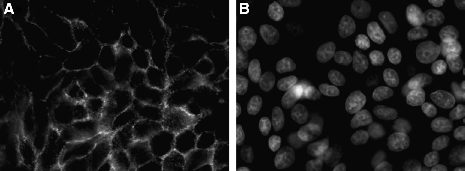
Indirect immunofluorescence using MAb 1B1. MCF-7 cells were incubated with MAb 1B1 prior to fixation. (
In summary, we have reported the production and characterization of a MAb that specifically recognizes the extracellular domain of IGF-1R. This MAb has potential for research and clinical applications in the fields of cancer diagnosis and therapy.
Footnotes
Author Disclosure Statement
Taro Tachibana is a founder of Cell Engineering Corporation (Osaka, Japan). [AU: Confirm statement, add other authors?]