
Abstract
ω-Conotoxins MVIIA (ω-CTX MVIIA) is a peptide with 25 amino acid residues. It is a selective and reversible N-type voltage-gated calcium channel blocker, which could be used as an analgesic for pain. To date, there are no monoclonal antibodies (MAb) for immunoassay against ω-conotoxin MVIIA. In this study, an MAb against ω-conotoxin MVIIA was prepared. The conotoxin-coding DNA sequence was chemically synthesized and cloned into expression vector pGEX-6p-1 and pET32a (+), respectively. The fusion protein GST-CTX was expressed and purified, and was used to immunize BALB/c mice for preparing the anti-CTX antibody. The spleen cells were fused with SP2/0 myeloma cells after the titer of antiserum was detected and qualified. After being screened by indirect ELISA and cloned by limiting dilution, a hybridoma named 4A12, which produces monoclonal antibody specifically against ω-CTX MVIIA, was successfully obtained. It was found that there are 102 chromosomes in the 4A12 cell, and the subclass for the MAb is IgM. The MAb affinity against ω-CTX MVIIA was 7.33×10⁹ L/mol, and the cross-reaction test showed that the MAb specifically bound ω-CTX MVIIA. The MAb could be used as a specific antagonist for ω-CTX MVIIA in the physiological study on the CaV channels in the nervous system.
Introduction
C
ω-conotoxins MVIIA (ω-CTX MVIIA) is a peptide with 25 amino acid residues originally isolated from Conus magus.(5) ω–CTX MVIIA has an amidated C-terminus and a four-loop Cys framework (C-C-CC-C-C) arranged by six Cys-residues.(6) ω-CTX MVIIA is a selective, reversible, and potent N-type CaV channel blocker; it has been reported to inhibit both neuronal excitability and neurotransmission.(6–8) ω-CTX MVIIA also shows analgesic and neuroprotective effects in humans.(9) The artificially synthetic ω-CTX MVIIA, ziconotide, has been recently approved by the FDA as an analgesic for the management of severe and chronic pain.(3)
The typical methods of analysis and detection of conotoxin are high performance liquid chromatography (HPLC), nuclear magnetic resonance (NMR), and so on. The MAbs against ω-conotoxin GVIA were developed to recognize native and synthetic toxin and are useful for the analysis of toxin in tissue extracts or biological fluids.(10) It was also carried out to detect ω-conotoxin MVIIA based on polyclonal antibody.(11) Thus far there still is not an immunoassay against ω-CTX MVIIA based on MAb. In this study, MAbs against ω-CTX MVIIA were successfully obtained and can be used as a specific antagonist to ω-CTX MVIIA for the study of the physiology of CaV channels in the nervous system.
Materials and Methods
Isopropyl thio-β-D-galactosidase (IPTG), β-mercaptoethanol, and ampicillin were purchased from Sangon (Shanghai, China). Ni2+-NTA column was purchased from Novagen (Darmstadt, Germany). Glutathione Sepharose 4B was from GE Healthcare Biosciences (Waukesha, MI). BCA protein assay kit and horseradish peroxidase (HRP)-labeled goat anti-mouse IgG were purchased from Beyotime (Shanghai, China). Complete and incomplete Freund's adjuvant, 50% polyethylene glycol (PEG) 1450, hypoxanthine-aminopterin-thymidine (HAT), hypoxanthine-thymidine (HT), and the mouse monoclonal antibody isotyping kit were purchased from Sigma (St. Louis, MO). RPMI 1640 medium was purchased from Gibco (Grand Island, NY). Fetal calf serum was from PAA (Cölbe, Germany). BALB/c mice were purchased from Slac Laboratory Animal (Shanghai, China). All animal work was performed according to relevant national and international guidelines. All animal experiments complied with the rules of the Animal Ethics Committee of the Fujian Agriculture and Forestry University.
Preparation of complete antigen
The artificial DNA sequence encoding ω-CTX MVIIA peptide was designed according to Escherichia coli favorable codons with a BamHI site at 5'-terminus and a XhoI site at 3'-terminus (the sequence is shown in Table 1). The expression vectors (pGEX-6p-1-ctx and pET-32 a(+)-ctx) for ω-CTX MVIIA were constructed by the insertion of the ω-CTX MVIIA peptide coding DNA sequence into pGEX-6p-1 and pET-32a(+). The fusion proteins (GST-CTX and Trx-CTX) were expressed and then purified by Glutathione-sepharose 4B Fast Flow column and Ni2+-NTA column, respectively. The protein concentrations were determined by BCA protein assay kit. The purified fusion protein GST-CTX was used as immunogen, and Trx-CTX was used as coating protein to detect the sensibility of the anti-CTX antibody in the study.
Immunization and anti-serum titer assays
Initially female BALB/c mice (6–8 weeks old) were immunized with 20 μg purified recombinant protein GST-CTX in complete Freund's adjuvant by subcutaneous injection as the first immunization. Then, three subsequent doses were injected with incomplete Freund's adjuvant at 14-day intervals. The titer of the anti-serum was determined by indirect non-competitive enzyme-linked immunosorbent assay (ELISA). Finally, a booster injection with 20 μg GST-CTX was given 3–4 days prior to cell fusion.
Indirect ELISA
The protocol used for indirect ELISA was similar to that described previously by Kim and colleagues, with some improvement.(12,13) In brief, the purified protein Trx-CTX was diluted to 2 μg/mL by coating buffer (0.05 mol/L carbonate buffers, pH 9.6), and microtiter plate was coated with the protein overnight at 4°C. The plates were washed three times with PBS and blocked with 200 μL of PBSM (0.01 mol/L PBS with 5% fat-free milk) at 37°C for 2 h. After washing with PBS, the plates were incubated with 100 μL of anti-serum or MAbs (diluted by doubling dilution method). Then the plates were washed four times with PBST (PBS containing 0.05% Tween-20) and incubated with 100 μL of HPR-labeled goat anti-mouse IgG (1:10,000) at 37°C for 1 h. Finally, the plates were washed six times with PBST and incubated with 100 μL of freshly prepared 3,3′,5,5′ tetramethylbenzidine with 30% hydrogen peroxide (TMB/H2O2) at 37°C for 10 min. The reaction was stopped by 50 μL of 2 mol/L H2SO4. The absorbance was measured by Multiskan (Thermo Scientific, Waltham, MA) at 450 nm.
Cell fusion and hybridoma clone screening
Hybridomas producing MAbs against CTX were prepared according to the protocol used by Köhler and Milstein, with some improvements.(13,14) The spleen was aseptically removed and the spleen cells were fused with the SP2/0 myeloma cells at a rate of 5 to 10: 1 in the presence of polythylene glycol (50%, w/v). After fusion, the cells were cultured in RPMI-1640 medium containing 15–20% fetal calf serum and HAT for 8–14 days. Then the positive hybridomas were screened by indirect ELISA as described above. The positive hybridomas were further cloned by limiting dilution in RPMI-1640 medium with 10% FCS and HT. After selection 3–4 times, the monoclonal hybridomas were expanded in RPMI-1640 with 10% FCS and cryopreserved.
MAb subclass identification
The subtype of MAbs was analyzed using the mouse monoclonal antibody isotyping kit (St. Louis, MO). Firstly, IgA, IgM, IgG1, IgG2a, IgG2b, and IgG3 reagents were diluted by coating buffer (1:1000) and coated overnight at 4°C. The cell culture medium containing MAb was added onto plates and incubated at 37°C for 1 h. The following procedure was the same as that described in the indirect ELISA.
Chromosome analysis of hybridoma cells
The number of chromosomes in hybridoma cells, myeloma cells, and spleen cells were analyzed respectively. The hybridoma cells and myeloma cells in logarithmic growth phase were treated with 0.4 μg/mL colchicine in 5% CO2 at 37°C for 4–6 h, respectively. Then cells were harvested by centrifugation and resuspended in 10 mL of 0.075 mol/L KCl hypotonic solution at 37°C for 20 min. Cells were then centrifuged and suspended with 10 mL of stationary liquid (methanol-acetic acid=3:1) for 10 min. After centrifugation, the cells were incubated once again with stationary liquid for 30 min. Finally, the cells were harvested and suspended by 5 mL of stationary liquid and then dropped onto glass slides. After giemsa staining, the number of chromosomes was counted under a microscope. To analyze the chromosomes of spleen cells, the mouse was first injected with 4 μg/g colchicine by intraperitoneal injection. After 3∼4 h, spleen cells were harvested and incubated in hypotonic solution at 37°C for 20 min. The following procedures were the same as that described above.
Production of monoclonal antibody
To obtain large numbers of MAbs, 1.0×10⁶ hybridoma cells were injected intraperitoneally into an 8-week-old BALB/c mouse, which was injected with 0.5 mL atoleine a week previously. When the abdomen of the mouse was swollen, the ascites could be collected by injector after about 7 days. The supernatant containing a high concentration of MAb was collected by centrifugation at a speed of 12000 r/min for 15 min. The MAbs were purified by caprylic acid-cold ethanol precipitation. Firstly, the ascites were diluted with double volumes of acetate buffer (pH 4.0), then caprylic acid was added slowly (1 mL ascites need 33 μL caprylic acid) and stirred for 30 min. Secondly, after 2 h standing at 4°C, the solution was centrifuged at 12000 r/min for 30 min. Then the supernatant was transferred into 0.03 M acetate buffer (pH 5.1). Finally, the concentrated solution was stirred for 30 min at 0°C, and alcohol was added to a final concentration of 10% alcohol. The solution was centrifuged after 30 min stirring and 1 h standing at 4°C. Lastly, the precipitate was dissolved in PBS (pH 7.0).
Cross-reactivity assay
To test the specificity of the MAbs produced by hybridoma clones, indirect ELISA was performed. The wells were coated with GST, GST-CTX, Trx-CTX, TTX, AFB1-OVA, DA, BSA, and FB1, respectively, and incubated at 4°C overnight. After blocking, 100 μL of positive, negative serums, or ascites were diluted to 1:1000 and added to the wells respectively at 37°C for 1 h. The following procedures were the same as that described above.
Affinity assay
The affinity of ascites was determined by indirect ELISA.(15) The wells were coated with GST-CTX antigen at concentrations of 0.5, 1.0, 2.0, and 4.0 μg/mL, respectively. Then the MAbs were doubly diluted and added to each well. After being washed, HPR-labeled goat anti-mouse IgG and TMB substrate were added in sequence. The reaction was stopped by 50 μL 2 mol/L H2SO4. The absorbance was measured at 450 nm by a microplate reader, and the affinity of the MAbs was calculated according to the results of ELISA.
Results
Preparation and purification of complete antigen
In order to obtain purified protein for immunization and detection, the constructed recombinant vectors of pGEX-6p-1-ctx and pET-32a(+)-ctx were expressed in E. coli BL21-DE3, and the expressed fusion proteins (GST-CTX and Trx-CTX) were purified by Glutathione-sepharose 4B Fast Flow column and Ni-NTA affinity chromatography, respectively. The results (Fig. 1) showed that GST-CTX (Fig. 1A) and Trx-CTX (Fig. 1B) were successfully expressed and purified. The concentration of GST-CTX (∼2 mL from 200 mL bacteria culture) and Trx-CTX (∼3 mL from 200 mL bacteria culture) were further detected (0.42 and 0.24 mg/mL, respectively) by BCA protein assay kit.
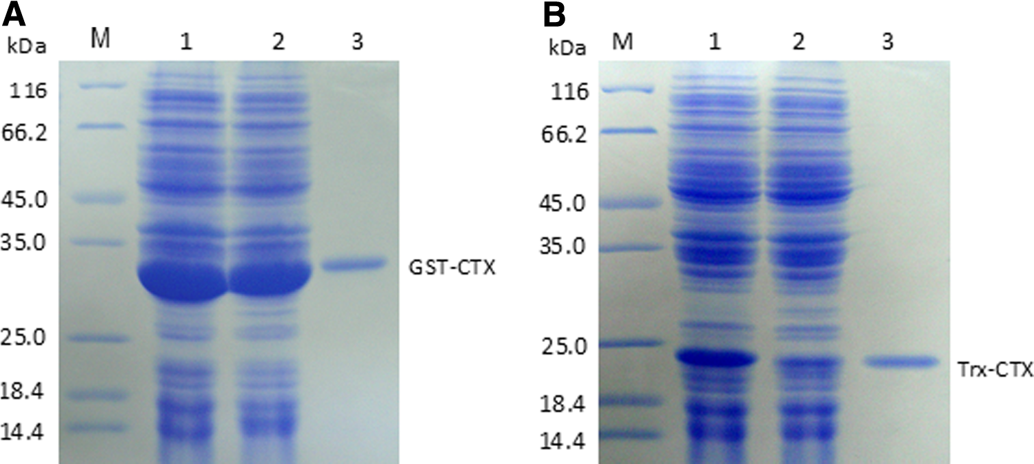
Expression and purification of fusion protein GST-CTX and Trx-CTX. All proteins were analyzed by 12% SDS-PAGE and stained with Coomassie brilliant blue. (
Immunization and anti-CTX serum titer assays
In this study, GST-CTX fusion proteins were used as antigen to immunize mice, and Trx-CTX was adopted to coat ELISA plates for immunoassay. A detection system containing an optimal concentration of coating antigen (Trx-CTX, 2 μg/mL) and an optimal concentration of HPR-labeled goat anti-mouse IgG (1:10,000) was set up. In the experiment, two mouse spleen cells were used to fuse with myeloma cells. Figure 2 shows that the titer of mouse 1 was 1:512,000, while mouse 2 was 1:32,000.
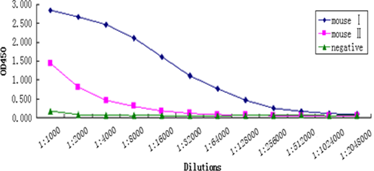
Titer of antiserum from mouse 1 was 1:512,000, and that of mouse 2 was 1:32,000.
Screening of hybridoma against CTX
The result of the successful fusions that yielded monoclonal antibodies is summarized in Table 2. The fusion rates for the two time fusions were 99.27% and 100%, respectively. In the initial ELISA screening assay, hybridoma supernatants were tested for antibodies that recognized Trx-CTX, and the positive rate were 2.52% and 5.03%, respectively. Finally, one stable hybridoma cell against CTX (named 4A12) was successfully screened out from mouse 2. The titer of 4A12 culture supernatant was determined to be 1:8192 by indirect ELISA (Table 2).
MAb subclass identification
The subclass of the MAb was analyzed by mouse monoclonal antibody isotype assay kit, and Figure 3 shows that 4A12 belonged to IgM subclass. Other MAb cell lines against CTX in the study were also tested, but all were classified as IgM (data not shown). In order to obtain an anti-CTX IgG MAb, several other fusions were carried out, but no IgG MAb was detected in our further studies (data not shown).
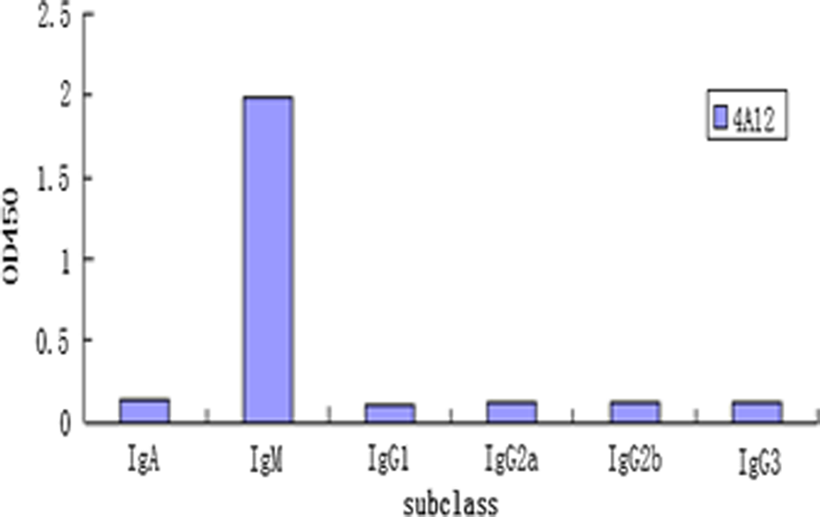
Subclass analysis for anti-CTX MAb.
Chromosome assay
The average chromosome number of spleen cells is 40 (Fig. 4A) and SP2/0 myeloma cells is 62∼70 (Fig. 4B). It was found from this study that the average chromosome number of hybridomas was 102 (Fig. 4C). The chromosome analysis results indicated that the chromosomes of hybridoma were obtained from spleen cells and SP2/0 myeloma cells.
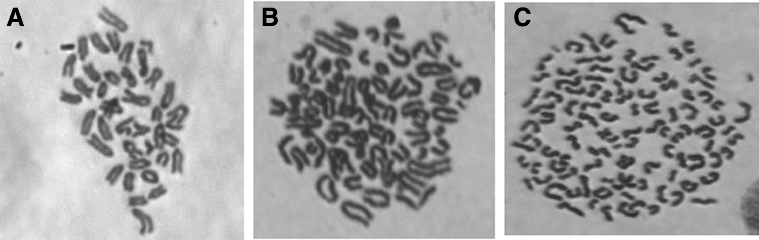
Chromosome analysis of cells. (
Production and characterization of anti-CTX MAb
The anti-CTX MAb was prepared in large scale by intraperitoneal injection. The MAb concentration in ascites was found to be 33.6 mg/mL, and the titer of the ascites was 1:128,000. After purification, the concentration of the MAbs was detected to be 0.310 mg/mL, while the titer was 1:8000. The results showed that the molecular weight of the light and heavy chains of the anti-CTX MAb were about 27 kDa and 55 kDa, respectively (Fig. 5, lane 5).
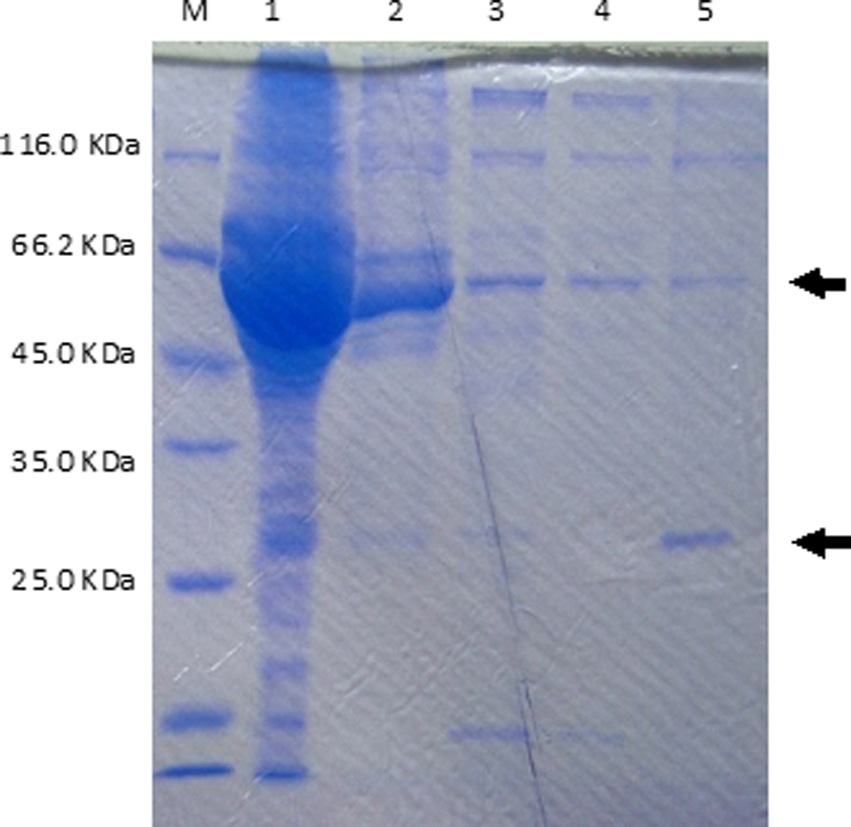
Purification of ascites. All proteins were analyzed by 12% SDS-PAGE and stained with Coomassie brilliant blue. Lane M, protein molecular weight markers; lane 1, ascites; lane 2, precipitate after caprylic acid treated; lane 3, supernatant after caprylic acid treated; lane 4, supernatant after ethanol treated; lane 5, precipitate after ethanol treated (MAb).
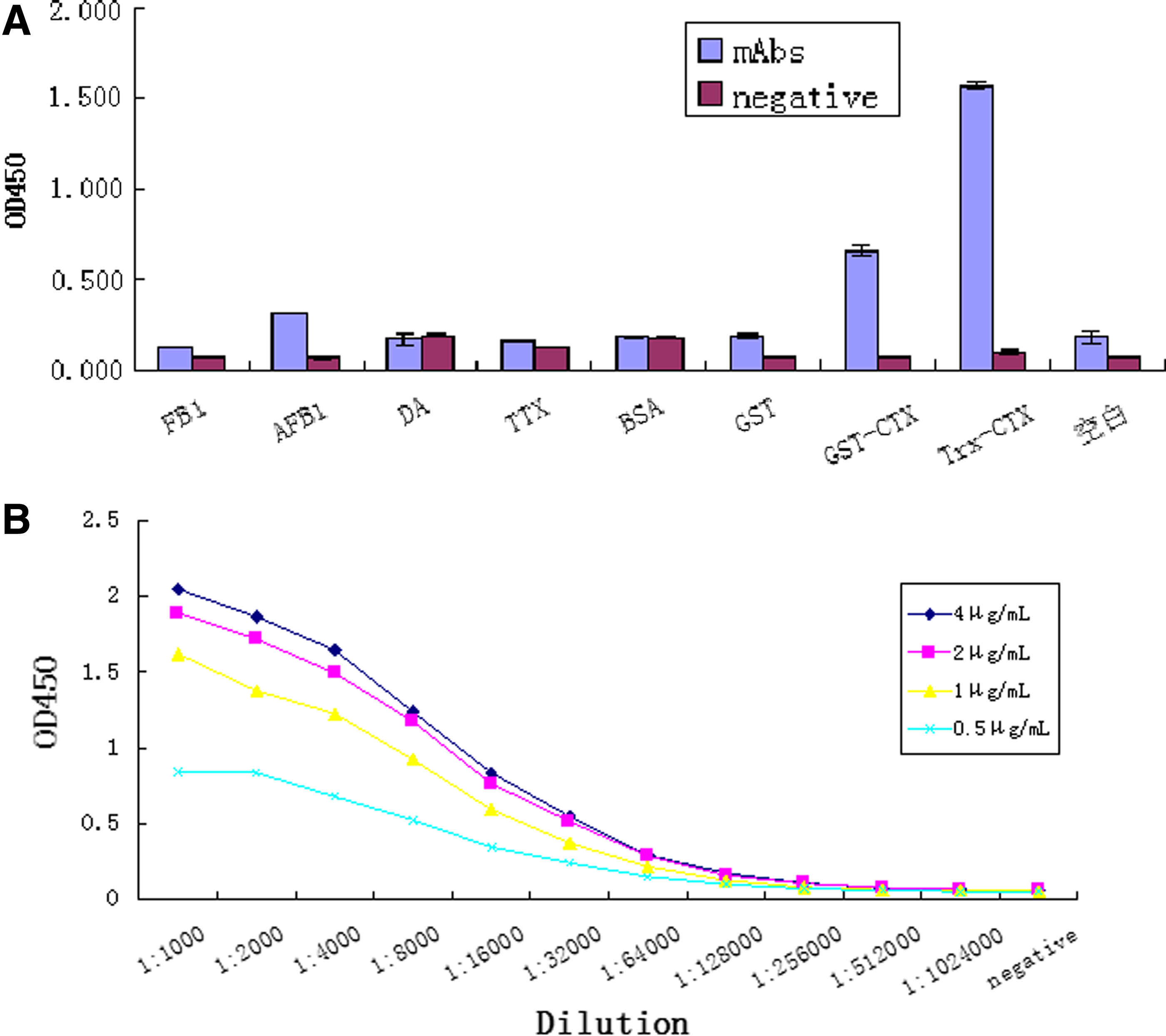
To test the specificity of the hybridoma clone, indirect ELISA was performed. AFB1 (aflatoxin B1), FB1 (fumonisin B1), DA (domoic acid), TTX (tetrodotoxin), BSA (bovine serum albumin), GST, GST-CTX, and Trx-CTX were coated onto microtiter plates and incubated at 4°C overnight. After blocking, 100 μL of positive and negative ascites were diluted and added to the microtiter plates at 37°C for 1 h. The following procedures were the same as that described above. The results show that the MAb was specific to CTX (Fig. 6). The affinity of ascites to CTX was further determined by indirect ELISA, which was found to be 7.33×10⁹ L/mol (Fig. 6B).
Detection of specificity and affinity of anti-CTX MAb by indirect ELISA. (
Discussion
Since CTX MVIIA is a small and disulfide-rich peptide, it is difficult to analyze the product.(16) Currently, the chemical synthetic CTX MVIIA has been analyzed by RP-HPLC and NMR, but the immunological assay based on monoclonal antibody for CTX MVIIA was unavailable prior to our study.(11) In fact, the immunological assay is a quicker, easier, and more specific way to detect conotoxin than is HPLC, especially in cases of detecting unknown samples.(17) However, due to its low antigenicity, it is difficult to prepare antibody by using ω-CTX MVIIA as antigen directly.(11) In the study, to make ω-CTX MVIIA a complete antigen, a prokaryotic plasmid pGEX-6p-1-CTX was used to obtain fusion protein GST-CTX MVIIA. After purification, the fusion protein was directly used to immunize the mice to produce anti-CTX MVIIA antibody. Because anti-GST antibodies would be produced at the same time, another prokaryotic plasmid pET32a(+)-CTX was used to produce fusion protein Trx-CIX as coating protein for anti-CTX MVIIA antibody screening. This strategy is commonly used in producing polyclonal antibody against small peptides, such as conotoxins.(11) As the fusion tags, Trx is not homologous to GST, the antibodies produced against them have no cross-reaction characteristics, so Trx-CTX MVIIA could be used to cross-test (screen and measure the titer of) the specific anti-CTX MVIIA MAb. The specificity of the hybridoma clone (4A12) was tested by indirect ELISA; the results showed it did not react with BSA, GST, and other toxins (AFB1, FB1, DA, TTX), and it reacted specially with CTX (Fig. 6A). It was also found that the affinity of 4A12 to Trx-CTX is higher than that to GST-CTX (Fig. 6A), which may suggested that there is some difference lying in the spatial conformation between GST-CTX and Trx-CTX.
To test the specificity of the hybridoma clone, indirect ELISA was performed. AFB1, FB1, DA, TTX, BSA, GST, GST-CTX, and Trx-CTX were coated onto microtiter plates and incubated at 4°C overnight. After blocking, 100 μL of positive and negative ascites were diluted and added to the microtiter plates respectively at 37°C for 1 h. The following procedures were the same as that described above. The results in Figure 6A shows that the MAb was specific to CTX. The affinity of ascites to CTX was further determined by indirect ELISA, and it was found to be 7.33×10⁹ L/mol (Fig. 6B).
Conclusion
In this study, six hybridoma cell clones were separated, but only 4A12 hybridoma was selected, and it was confirmed to secrete antibody persistently in the study. All six hybridoma cell clones were detected to be IgM subclass. IgG and IgM are the most frequently generated classes in the preparation of MAbs.(18,19) IgM antibody was produced from the primary response (the first immunization). After the second immunization, the main antibodies are IgG.(20) Usually the interval time of the first and second immunizations was 2–3 weeks. If the interval time between them was too short, the concentration of IgM would be greater. In this report, the interval time between the first and second immunization was 2 weeks; in our further study on anti-CTX MVIIA MAb preparation, the interval time was elongated to 3 weeks so there would be more opportunity to isolate an IgG subclass anti-CTX MVIIA MAb.
In this study, a hybridoma clone (4A12) that could persistently secrete anti-ω-CTX MVIIA MAb was obtained. The MAb specifically binds ω-CTX MVIIA with high affinity. It could be used as a specific antagonist for ω-CTX MVIIA on the CaV channels in the nervous system, or to detect native or synthetic ω-CTX MVIIA in extracts from food, fodder, or biological tissues.
Footnotes
Acknowledgments
This work was supported by the Key Scientific and Technology Project of Fujian Province of China (no. 2012Y002), Natural Sciences Foundation of Fujian Province (no. 2011J05049), State Key Laboratory of Freshwater Ecology and Biotechnology (grant no. 2009FB13), and Agricultural Five-new Engineering Projects of Fujian Development and Reform Commission.
Author Disclosure Statement
The authors have no financial interests to disclose.