
Abstract
The sterile alpha motif and HD domain 1 (SAMHD1) protein has been identified as a novel innate immunity restriction factor that participates in processes crucial to the viral life cycle. In the present study, we describe a procedure to generate monoclonal antibodies (MAbs) against porcine SAMHD1 and investigate its characteristics to analyze the expression of endogenous SAMHD1. The open reading frame of porcine SAMHD1 was cloned into the prokaryotic expression vector pCold-TF DNA to construct a recombinant plasmid pcold-pSAMHD1 and induce expression of recombinant porcine SAMHD1 protein by IPTG in Escherichia coli Rosetta. The purified recombinant porcine SAMHD1 protein was used to prepare MAbs of SAMHD1. After subcloning five times hybridoma cell clones expressing SAMHD1, MAbs were generated. Western blot analysis and indirect immunofluorescence assay showed that the overexpressed porcine SAMHD1 in 293T cells and endogenous SAMHD1 protein in porcine cell lines could be specifically recognized by the MAbs produced in this study. In conclusion, specific MAbs of porcine SAMHD1 are reported, and these MAbs provide a valuable tool for further studies of SAMHD1-mediated signaling in virus-infected cells to elucidate the underlying antiviral mechanism.
Introduction
W
SAMHD1 is expressed in nearly all tissues, but it is highly expressed in myeloid lineage cells, such as dendritic cells and macrophages.(4–6) There are no relative data, however, on whether SAMHD1 is expressed in porcine cells lines. Although there are various monoclonal or polyclonal antibodies against human SAMHD1 that have cross-reactivity with other species, such as monkey, mouse, and dog, there are no antibodies that specifically recognize porcine SAMHD1. Here, we prepared monoclonal antibodies against porcine SAMHD1, which are the preferred antibodies for investigating the functions of SAMHD1 and monitoring the behavior of endogenous SAMHD1.
Materials and Methods
Cell lines and animals
Peripheral blood lymphocyte (PBMC) and porcine alveolar macrophage (PAM) isolations were performed as previously described.(7,8) SP2/0, HeLa, BHK-21, A549, U251, and 293T cells were purchased from the Shanghai Cell Biology Institutes, Chinese Academy of Sciences (Shanghai, China). MARC-145 and PK-15 cells were preserved in our laboratory. Cells were maintained in Dulbecco modified Eagle medium (DMEM, Gibco, Grand Island, NY) or minimum essential medium (MEM, Gibco) supplemented with 10% fetal bovine serum (FBS, Gibco) at 37°C in a 5% CO2 atmosphere. The 6-week-old female BALB/c mice (SPF grade) were purchased from the Center of SLAC Laboratory Animals of Shanghai (China) and bred in independent ventilation incubators. All animal protocols used in our study were approved by the Shanghai Veterinary Research Institutional Animal Care Committee.
Molecular cloning and expression vector construction
The porcine-SAMHD1 primers, pcold-pig SAMHD1-forward: 5′-CG
Expression and purification of recombinant porcine SAMHD1
The recombinant expression plasmid pcold-pSAMHD1 was transformed into E. coli Rosetta and was cultured at 37°C for 2 h; then protein expression was induced with isopropyl-β-D-thiogalactoside (IPTG) at a final concentration of 0.5 mM for 5 h at 16°C. The induced bacteria were harvested by centrifugation at 6000 rpm for 10 min at 4°C. The pellet was resuspended and boiled in 5× SDS-PAGE loading buffer for 10 min. The samples were then subjected to sodium dodecyl sulfate-polyacrylamide gel electrophoresis (SDS-PAGE) to validate expression of the recombinant porcine SAMHD1 protein. The PAGE gels were stained with Coomassie brilliant blue dyes. The expression of recombinant porcine SAMHD1 was also confirmed by anti-His antibody (Sigma-Aldrich, St. Louis, MO) by Western blot analysis. Purification of the His-tagged recombinant porcine SAMHD1 was performed using the Ni-NTA His•Bind Resin (Novagen, Madison, WI) according to the manufacturer's instructions. The eluted recombinant protein was purified by ultrafiltration using the tubular ultrafiltration modules (Millipore, Billerica, MA) to remove the imidazole and to obtain the highly purified protein, named rpSAMHD1. The rpSAMHD1 was stored at −80°C until used.
Procedure for immunization
The purified rpSAMHD1 was quantified by RC DC Protein Assay kit (Bio-Rad, Hercules, CA) and used as the immunogen to prepare the anti-porcine SAMHD1 monoclonal antibody. At the first immunization, purified rpSAMHD1 (100 μg) was emulsified with an equal amount of Freund's complete adjuvant and subcutaneously injected into the mice. The mice were immunized with purified rpSAMHD1 (50 μg) emulsified with equal amount of Freund's incomplete adjuvant by subcutaneous injection in 2 weeks. Four weeks after the initial injection, the mice were injected intraperitoneally twice with 50 μg of purified rpSAMHD1 at 2-week intervals. After 2 weeks, the mice were orbital-bled to obtain sera. The sera titers for anti-SAMHD1 antibody were confirmed by indirect ELISA.
Preparation of porcine SAMHD1 monoclonal antibody
Cell fusion was performed when the antibody titers reached a certain level. The procedure was performed as previously described with modifications.(9) Briefly, spleen cells obtained from rpSAMHD1-immunized BALB/c mice were fused with SP2/0 cells using polyethylene glycol (Sigma-Aldrich). The hybridoma cells were screened for antibody production against SAMHD1 using an indirect enzyme-linked immunosorbent assay (indirect ELISA). After subcloning five times, the positive hybridoma cells were inoculated intraperitoneally into pristane-primed BALB/c mice, with a density of 1×10⁶ cells for MAb production.
Indirect enzyme-linked immunosorbent assay
The screen of positive hybridoma cells and antibody titer determination were analyzed by indirect ELISA assay. The ELISA plates were plated with 100 μL purified rpSAMHD1 (1 μg/well) in carbonate bicarbonate buffer (15 mM Na2CO3, 35 mM NaHCO3 [pH 9.6]) and coated at 4°C overnight. The plates were then blocked with 5% skimmed milk in phosphate buffer with 0.05% Tween-20 (PBST) at 37°C for 2 h. After washing three times, the plates were incubated with 50 μL diluted antibodies or cell culture supernatant at 37°C for 1 h. The plates were incubated with horseradish peroxidase-conjugated goat anti-mouse IgG (Sigma-Aldrich) with 1:10,000 dilution in PBST at 37°C for 30 min after washing three times in PBST. Following this, the plates were incubated with 50 μL/well TMB liquid (Amresco, Solon, OH) for 10 min at room temperature with protection from light. After being stopped by 2 M H2SO4, the results were read with an OD450 value. The wells coated with purified His-tag protein served as negative control for screening the positive hybridoma cells.
Eukaryotic expression vector construction and transfection
The sequence of the complete CDS of porcine SAMHD1 were amplified using the pcold-pSAMHD1 plasmid as template, primed by pFLAG-pig SAMHD1 primers: pFLAG-pig SAMHD1 forward: 5′-CC
For transfection, 293T cells were seeded in 6-well plates and transfected at 80% confluency with pFLAG-pSAMHD1 plasmids DNA by FuGENE HD transfection reagent (Promega, Madison, WI) in Gibco OPti-MEM cell culture medium (Life Technologies), according to the manufacturer's instructions. Empty vector transfection served as control. After 48 h transfection, the cells were harvested and overexpression of porcine SAMHD1 was detected by Western blot analysis.
Western blot analysis
Specificity of the MAbs was confirmed by Western blot analysis. The 293T cells overexpressing porcine SAMHD1, PAMs, PK-15 cells, MARC-145 cells, BHK-21 cells, HeLa cells, A549 cells, and U251 cells were harvested as previously described.(8) Briefly, cell suspension was collected and incubated on ice for 15 min, followed by centrifugation at 13,000 g at 4°C for 15 min. The concentrations of total protein were quantified. Equal amounts of cell lysates were separated by 12% sodium dodecyl sulfate–polyacrylamide gel electrophoresis (SDS-PAGE), then transferred to the nitrocellulose (NC) membrane. The membrane was probed with anti-FLAG antibody, β-actin antibody (Sigma-Aldrich), or anti-SAMHD1 MAbs. Detection of protein bands was visualized using an enhanced chemiluminescence detection system (Thermo Fisher Scientific, Waltham, MA).
Indirect immunofluorescence assay
PK-15 cells were plated in a 6-well plate, then washed with PBS twice and fixed by paraformaldehyde for 30 min at room temperature. After washing twice in PBS, cells were permeabilized by incubation with 0.25% Triton-100 in PBS for 5 min. The cells were blocked with 10% bovine serum albumin (BSA) in PBS for 30 min at 37°C after washing twice in PBS. The cells were then incubated with the anti-SAMHD1 MAb or normal mouse antibody diluted at 1:300 in PBS at 37°C for 1 h. This was followed by washing three times in PBS; the cells were then incubated with Alexa Fluor 488 donkey anti-mouse IgG (H+L) antibody (Life Technologies) at 37°C for 30 min. The cells were stained with DAPI for 5 min at 37°C, followed by washing. The cells were washed three times in PBS and then observed on Nikon inverted fluorescence microscope.
Results
Expression of recombinant porcine SAMHD1 protein
Total RNA was extracted from PBMCs and was used for cDNA synthesis. The CDS of porcine SAMHD1 gene was amplified by PCR with the porcine-SAMHD1 primers, using the PBMCs cDNA as template. The results showed that a gene fragment about 1884 nt was generated (Fig. 1A). The PCR product was purified and cloned into pCold-TF DNA vector, resulting in a recombinant plasmid that was named pcold-pSAMHD1. The recombinant plasmid was identified by sequencing and restriction enzyme digestion (Fig. 1B).
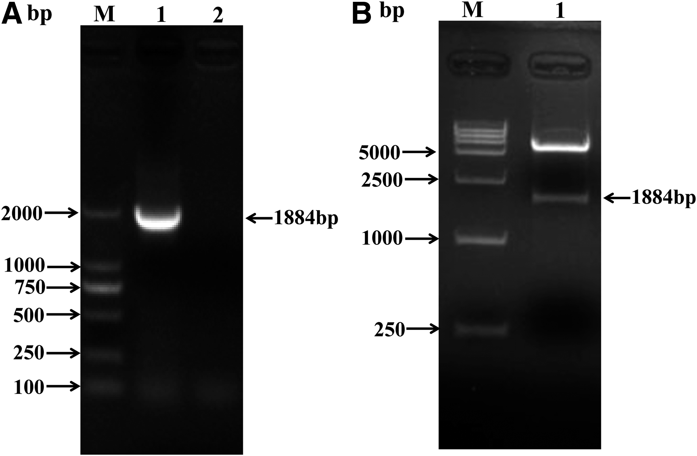
Amplification of CDS of SAMHD1 and prokaryotic expression vector construction. (
For the expression of the recombinant SAMHD1 protein, the recombinant plasmid pcold-pSAMHD1 was transformed into E. coli Rosetta. The host bacteria were induced by 0.5 mM IPTG for 5 h at 16°C. The results showed that the high-level expression of rpSAMHD1 was achieved under this condition and the rpSAMHD1 was expressed in a soluble form (Fig. 2A). To obtain highly purified rpSAMHD1, the tubular ultrafiltration modules were utilized to remove the imidazole. As shown in Figure 2A, the purified rpSAMHD1 was of high purity. The rpSAMHD1 expression was also confirmed by Western blot analysis. The results showed that rpSAMHD1 was recognized by anti-His-tag antibody in the predicted size; the empty vector transformed bacteria served as control (Fig. 2B).
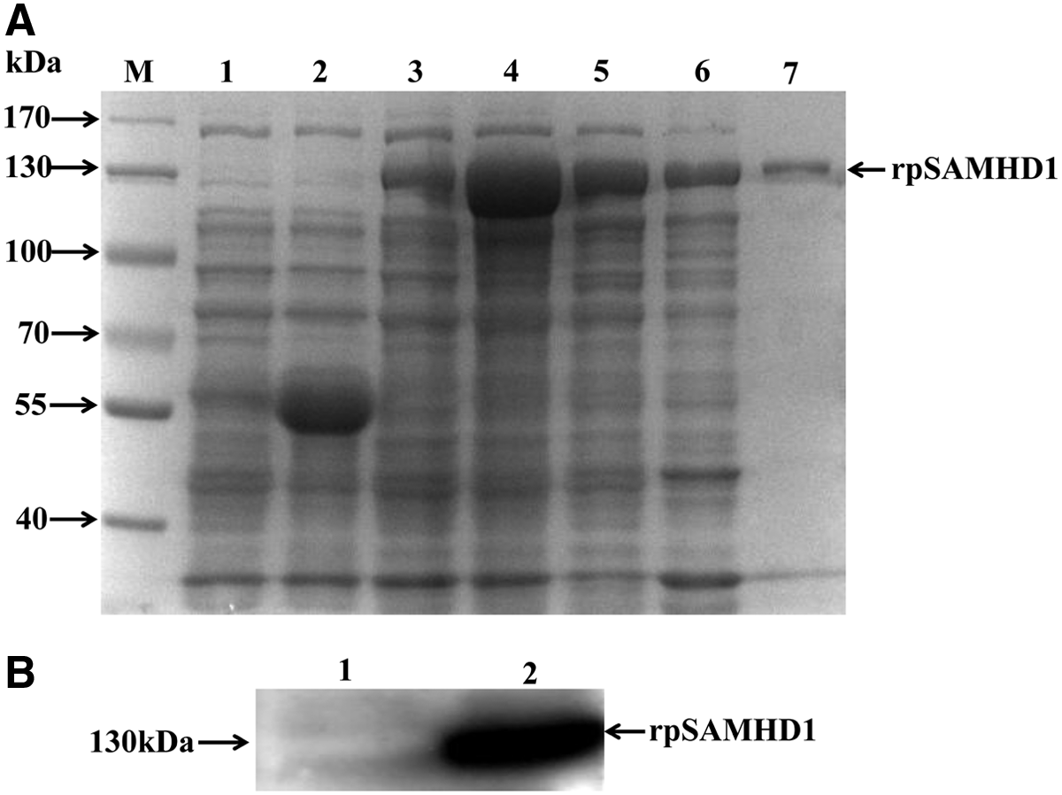
Expression, purification, and identification of rpSAMHD1. (
Characterization and specificity of porcine SAMHD1 monoclonal antibodies
The rpSAMHD1 was expressed and purified. After protein quantification, rpSAMHD1 was used to immunize BALB/c mice to prepare MAbs. After four immunizations, the antisera titers were determined by indirect ELISA. The mice with the antibody titers up to 1:106 were used in cell fusion. After subcloning five times by limiting dilution and screening, three clones of hybridoma cell lines were acquired and named clones 5M1, 5M2, and 5M5. The three hybridoma cell clones were used to prepare the ascites containing monoclonal antibodies. The antibodies titers of the ascites were up to 1:1.2×10⁶, 1:7.5×10⁵, and 1:5.8×10⁵, respectively. Purification of the ascites was performed by NAb™ protein G Spin Columns (Thermo Fisher Scientific). To explore the specificity of the MAbs, overexpressed porcine SAMHD1 and endogenous expression of porcine SAMHD1 were confirmed by the MAbs. As shown in Figure 3A, the overexpressed porcine SAMHD1 was detected both by anti-FLAG antibody and anti-SAMHD1 MAb 5M1. MAbs could specifically recognize the endogenous expression of SAMHD1 in PK-15 cells (Fig. 3B). Furthermore, the endogenous expression of SAMHD1 in porcine cell lines could also be specifically recognized by the MAb compared to other cells (Fig. 3C).
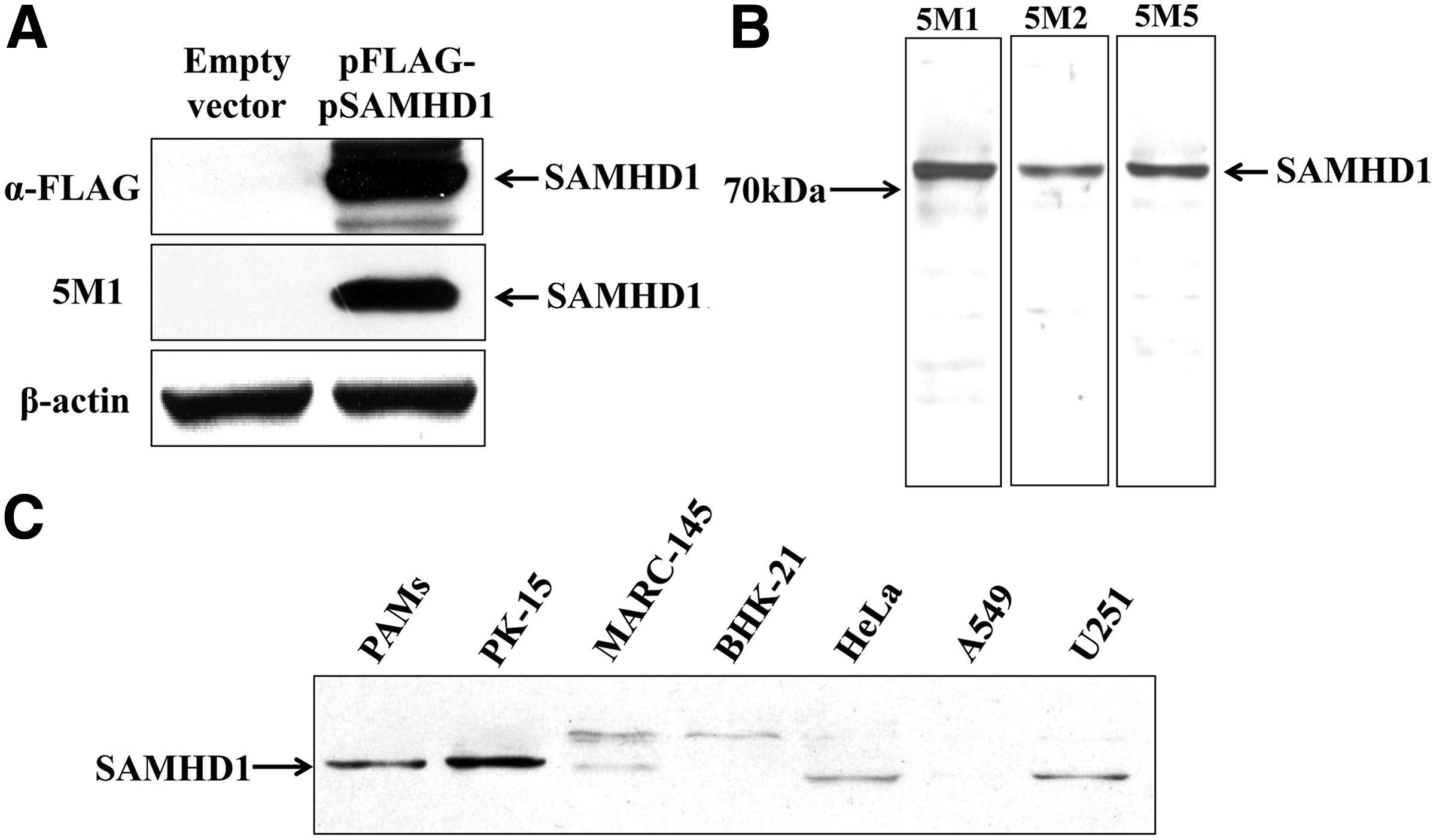
Specificity of prepared MAbs against porcine SAMHD1. (
Detection of endogenous SAMHD1 expression by immunofluorescence assay
Immunofluorescence assay (IFA) plays important roles in the study of cell signaling pathways. IFA was used to confirm whether the MAb would also work successfully in an immunofluorescence assay. PK-15 cells, a porcine kidney cell line, were plated in 6-well plates and used for IFA. Endogenous SAMHD1 expression was detected by the clone 5M1. The results showed that the MAb exhibited positive immunoreactivity with the SAMHD1 and SAMHD1 was detected in the nucleus, demonstrating that the MAb could be used for IFA analysis (Fig. 4).
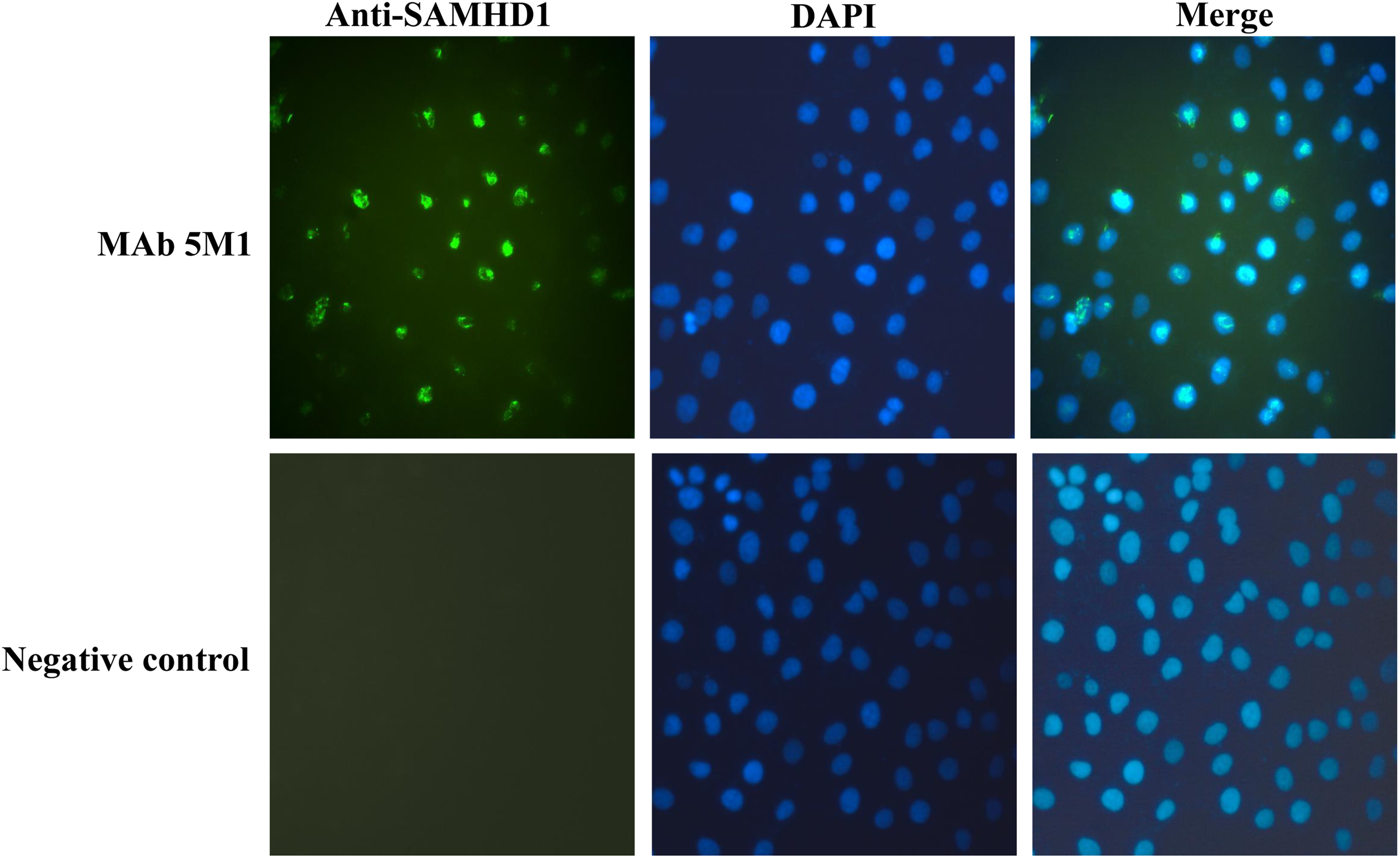
Detection of endogenous SAMHD1 expression by IFA. PK-15 cells were plated in 6-well plate and fixed. The fixed cells were incubated with MAb 5M1 or normal mouse antibody (negative control), followed by Alexa Fluor 488 donkey anti-mouse IgG (H+L) antibody.
Discussion
Monoclonal antibodies are necessary for the study of cell signaling pathways, and the specificity of the MAbs is crucial. SAMHD1 is a nuclear-localized protein with a protein molecular weight of 72 kDa; the full-length SAMHD1 protein is hard to express in soluble form with the conventional prokaryotic expression vector in E. coli expression systems, such as pET-28a (+), pET-32a (+), and pGEX-6P-1. pCold-TF DNA vector is a novel protein expression vector that is incorporated into the cold shock protein A (cspA) promoter and related elements to upregulate target protein production at lowered incubation temperatures (16°C). This process allows expression of target proteins at high yield and increased solubility. Here, the whole recombinant porcine SAMHD1 protein was acquired by prokaryotic expression. Specific MAbs against porcine SAMHD1 were prepared using the purified rpSAMHD1 as immunogen. Western blot analysis and IFA showed that the MAbs could recognize the endogenous SAMHD1, and indicated that the MAbs could be used in the study of cell signaling pathways of SAMHD1.
The innate immunity plays important roles in host defense against infections. As an innate immunity restriction factor, SAMHD1 could block the replication of human immunodeficiency virus-1 (HIV-1) in myeloid lineage cells by depleting cells of deoxynucleotide triphosphates (dNTPs), inhibiting lentiviral complementary DNA (cDNA) synthesis, and replication.(4,10) Viruses have developed many adoption strategies to evade the antiviral responses of host cells by regulating relative cell signaling pathway activation. Subsequent studies showed that the Vpx proteins encoded by human immunodeficiency virus (HIV)-2 and the simian immunodeficiency virus-sm could utilize the CRL4DCAF1 and E3 ubiquitin ligase complex to recruit SAMHD1 for proteasome-dependent degradation.(11–13) Highly pathogenic porcine reproductive and respiratory syndrome virus (HP-PRRSV), classic swine fever virus (CSFV), porcine epidemic diarrhea virus (PEDV), porcine pseudorabies virus (PRV), swine Japanese encephalitis virus (JEV), and other viruses pose a serious threat to the swine industry worldwide. The relationship between SAMHD1 and virus infection is not well determined and there are no specific MAbs against porcine SAMHD1 that could be used.
In summary, MAbs against porcine SAMHD1 were successfully prepared, and they are suitable for Western blot analysis, IFA, and ELISA assay. The MAbs can specifically recognize the overexpressed and endogenous porcine SAMHD1. Endogenous expression of porcine SAMHD1 in PK-15 cells and PAMs was confirmed by the cloned MAbs, which provide a valuable tool for further investigation of the regulation of SAMHD1 and expand the fields of SAMHD1 research.
Footnotes
Acknowledgments
This study was supported by grants from the National Basic Research Program of China (973 Plan, grant no. 2014CB542702); the National Natural Science Foundation of China (grant no. 31100121); and the Natural Science Foundation of Shanghai (no. 11JC1415200).
Author Disclosure Statement
The authors have no financial interests to disclose.