
Abstract
Female, 8-week-old BALB/c mice were immunized with purified recombinant proteins of the predicted immunodominant region of bovine haptoglobin (pirBoHp). Two monoclonal antibodies (MAbs), named 1B3 and 6D6, were prepared by conventional B lymphocyte hybridoma technique. Titers of ascitic fluid and cell culture supernatant of MAb 1B3 were 1:9.6×10⁸ and 1:8.2×10⁴, respectively, and that of MAb 6D6 were 1:4.4×10⁵ and 1:1.0×10⁴, respectively. The subtype of MAbs 1B3 and 6D6 was IgG1κ. In Western blot analysis, MAbs 1B3 and 6D6 could recognize the α-chain of native BoHp from plasma of dairy cows. These data indicated that MAbs 1B3 and 6D6 have a potential use for developing diagnostic reagents of BoHp.
Introduction
H
Previously the nucleotide sequence of the predicted immunodominant region of bovine haptoglobin (pirBoHp), removing the signal peptide sequence, was synthesized based on the codon usage bias of Escherichia coli.(9) The synthesized pirBoHp gene was successfully expressed in E. coli BL21 (DE3) cells. The polyclonal antibody against the recombinant pirBoHp protein could recognize α- and β-chains of the native bovine haptoglobin. These data provide evidence that the recombinant pirBoHp protein is similar to native BoHp in terms of immunogenicity. In the current study, monoclonal antibodies against the recombinant pirBoHp protein were prepared by using conventional B lymphocyte hybridoma technique. Our aim was to provide some basis for the development of rapid diagnostic reagents of BoHp.
Materials and Methods
Antigen, animal, and reagent
The recombinant protein of the predicted immunodominant region of bovine haptoglobin (pirBoHp) was expressed in a previous study.(9) The purified pirBoHp recombinant protein was stored at the Department of Veterinary Clinical Medicine, College of Animal Science and Veterinary Medicine, Heilongjiang Bayi Agricultural University. Female, 8-week-old BALB/c mice were purchased from Experimental Animal Center of Harbin Veterinary Research Institute (Chinese Academy of Agricultural Sciences, Harbin, China). Horseradish peroxidase (HRP)-conjugated sheep anti-mouse IgG (H+L), HAT supplements, HT supplements, 50% polyethylene glycol-1450 (PEG1450), and Freund's adjuvant were all purchased from Sigma (St. Louis, MO). Dulbecco's Modified Eagle's Medium (DMEM) and fetal calf serum (FCS) were obtained from Gibco BRL (Grand Island, NY). Rapid ELISA Mouse MAb Isotyping Kit were purchased from Thermo Pierce (Rockford, IL).
Development of monoclonal antibodies
Development of monoclonal antibodies (MAbs) against BoHp was carried out according to the report described by Sun and colleagues.(10) Briefly, female, 8-week-old BALB/c mice were immunized with 60 μg of the purified pirBoHp recombinant protein emulsified in complete Freund's adjuvant. At 2-week intervals, two boosters of 60 μg of the purified pirBoHp recombinant protein emulsified in incomplete Freund's adjuvant were administered, and mice were sacrificed 3 days after the last booster inoculation using 100 μg of the purified pirBoHp recombinant protein. Spleen cells from immunized mice were fused with SP2/0 myeloma cells using 50% (v/v) of PEG1450, and the fused cells were cultured in DMEM supplemented with 20% FCS, HAT medium. Positive hybridoma clones were selected by indirect ELISA using the purified pirBoHp recombinant protein with a His tag as coating antigen. In the ELISA, the purified His tag protein was used as control. After preparation of the ascitic fluid of MAbs, titers of the ascitic fluid and cell culture supernatant of the MAbs were tested by ELISA, respectively. Moreover, the subtype of MAbs secreted by the final hybridoma clones was identified using the Rapid ELISA Mouse MAb Isotyping Kit (Thermo).
Western blot analysis of MAbs 1B3 and 6D6
Two pooled plasma samples from unaffected and foot-affected dairy cattle were used to evaluate MAbs 1B3 and 6D6 by Western blotting. In the footrot-affected plasma sample, the presence of BoHp was confirmed by Sun and colleagues.(8) The two pooled plasma samples were subjected to separation by 12% SDS-PAGE, and then transferred to a nitrocellulose (NC) membrane using a semi-dry transfer apparatus. After blocking using 5% (w/v) non-fat dried milk in phosphate-buffered saline (PBS) at 37°C for 1 h, the NC membrane was incubated with MAbs 1B3 and 6D6 (1:200 dilution in PBS) at 37°C for 1 h, respectively. The NC membrane was washed five times using PBS and then incubated with horseradish peroxidase-conjugated goat anti-mouse IgG (1:4000 dilution in PBS) at 37°C for 1 h. After washing five times with PBS, the NC membrane was incubated with enhanced chemiluminescence detection reagents (Biotopped, Beijing, China) at room temperature for 3 min. The captured image of the signal was processed using the Molecular Imager ChemiDoc™ XRS+ Systems with Image Lab ™ Software. In the Western blot, the recombinant pirBoHp protein and His tag were used as control for evaluation of specificity of MAbs 1B3 and 6D6.
Results
Preparation of MAbs against BoHp
Using the recombinant pirBoHp protein as immunogen, a total of two monoclonal antibodies were obtained by the B lymphocyte hybridoma technique. For MAb 1B3, the titers of cell culture supernatant before and after revivification of hybridoma cells were 1:8.2×10⁴ and 1:1.3×10⁴, respectively; for MAb 6D6, the titers of cell culture supernatant before and after revivification of hybridoma cells were 1:1.0×10⁴ and 1:6.4×10³, respectively (Table 1). Titers of the ascitic fluid of MAbs 1B3 and 6D6 were 1:9.6×10⁸ and 1:4.4×10⁵, respectively. Moreover, the identification of subtypes of the MAbs 1B3 and 6D6 was performed by using the Rapid ELISA Mouse MAb Isotyping Kit. Results revealed that the MAbs 1B3 and 6D6 were classified into the IgG1κ subtype (Fig. 1).
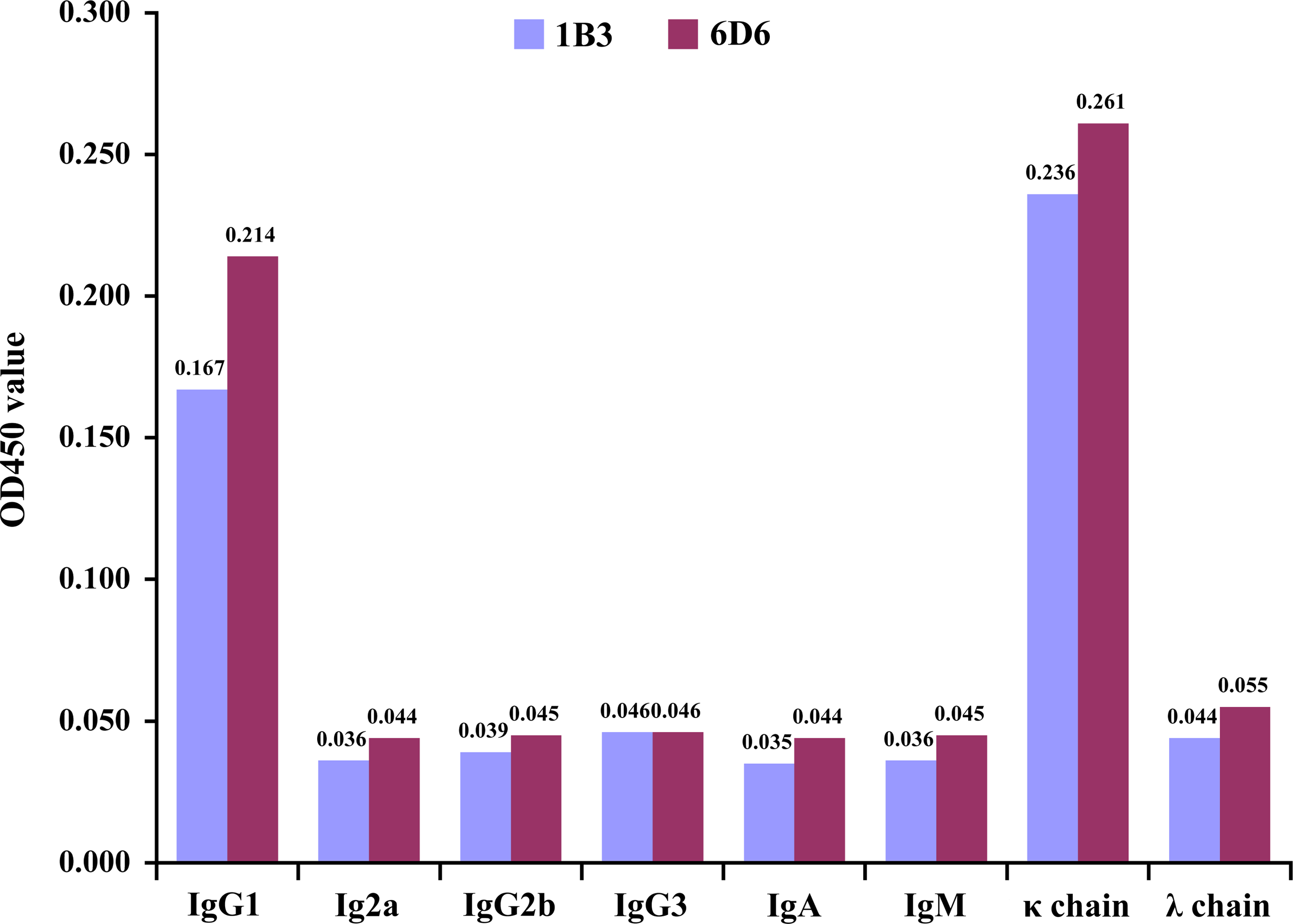
Identification of subtypes of MAbs 1B3 and 6D6.
Western blot analysis of MAbs 1B3 and 6D6
To confirm specificity of MAbs 1B3 and 6D6, Western blot analysis was carried out using plasma sample of footrot-affected dairy cattle as the detected antigen, in which presence of the native BoHp was validated. Results indicated that MAbs 1B3 and 6D6 could recognized α-chain (about 20 kDa) of the native BoHp and the recombinant pirBoHp protein, and no specific bands were observed in lanes of the His tag and footrot-unaffected plasma sample (Fig. 2).
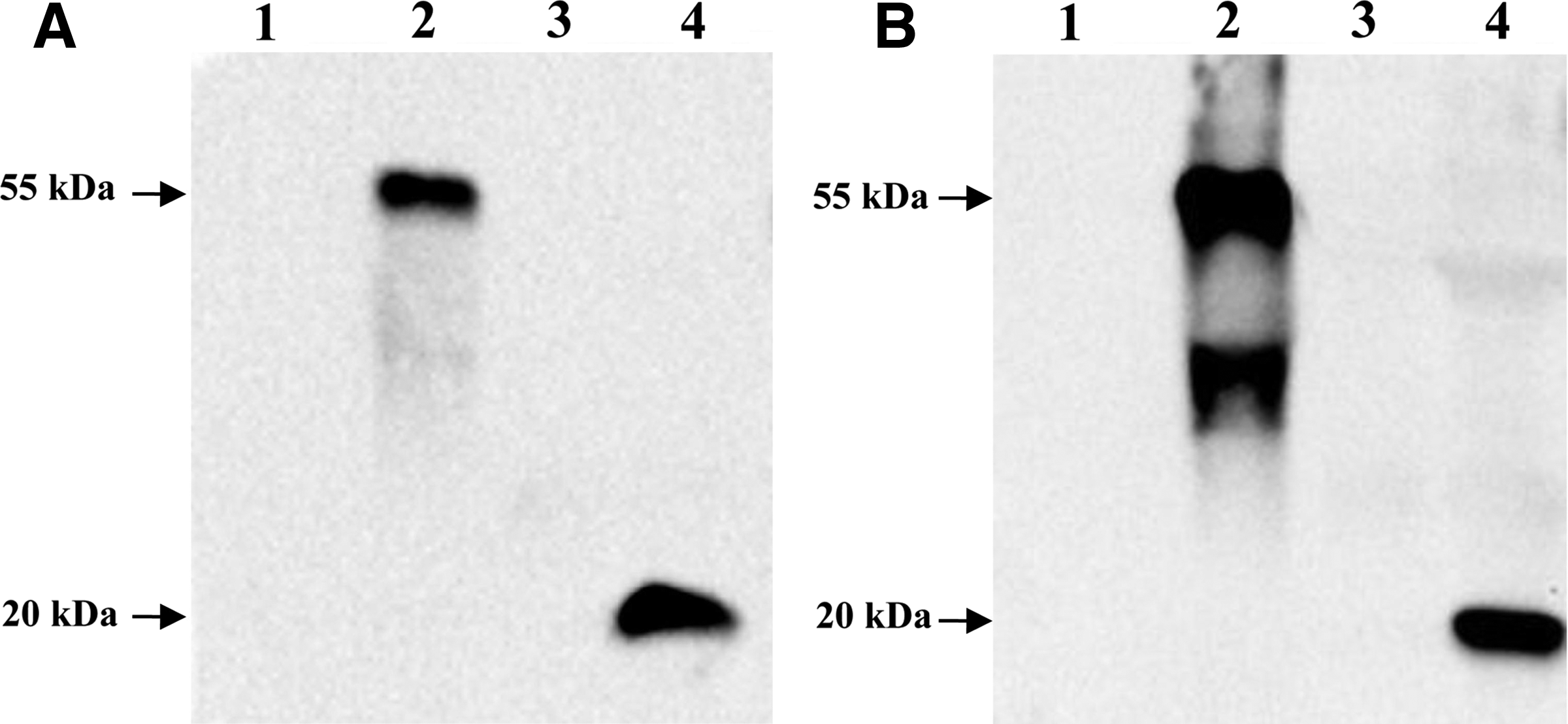
Western blot analysis of MAbs 6D6 and 1B3. (
Discussion
Subclinical inflammatory diseases in dairy cows have a serious impact on animal production performance, involving mastitis, metritis, and liver abscess, among others.(11–17) It is a focusing field where early diagnosis and monitoring techniques are developed for subclinical inflammatory diseases of dairy cows. At present, many small molecules are reported as diagnostic reagents, including monoclonal antibody, single-chain fragment variable (scFv), aptamer, and peptide ligand.(18–21) Of those diagnostic reagents, monoclonal antibody is still the most effective diagnostic tool in the biomedical field.
In our study, two monoclonal antibodies against BoHp were obtained using the recombinant pirBoHp protein as antigen. The titers of cell culture supernatant of MAbs 1B3 and 6D6 were 1:1.3×10⁴ and 1:6.4×10³ after revivification of hybridoma cells, respectively. This data demonstrated that the MAbs 1B3 and 6D6 were stably capable of secreting specific antibodies after revivification from liquid nitrogen. Compared to cell culture supernatants, the ascitic fluid of MAbs 1B3 and 6D6 showed higher antibody titers, facilitating subsequent antibody labeling for diagnosis.
Specificity of the monoclonal antibody determines whether the prepared MAbs can be used in development of the subsequent diagnostic reagents. In our study, the His tag protein was used as a screening antigen for elimination of false-positive hybridoma cells. In a validation test of MAbs 1B3 and 6D6, the native BoHp present in footrot-affected dairy cattle was choosen as targeted antigen for the evaluation of specificity of the prepared MAbs. Results indicated that two MAbs, both 1B3 and 6D6, could recognize the α-chain of the native BoHp and had no cross-reaction with other proteins in plasma from dairy cows. However, a non-specific band was found in the Western blot lane of the recombinant pirBoHp protein of MAb 1B3. It is speculated that degradation of the recombinant pirBoHp protein may occur, resulting in the appearance of the non-specific band in the Western blot. In a previous study, polyclonal antibody against the recombinant pirBoHp protein could recognize α- and β-chains of native bovine haptoglobin.(9) In the current study, the MAbs 1B3 and 6D6 only showed specific reaction with α-chain of the native BoHp. This result suggests that in the BoHp molecule, the α-chain may have better antigenicity than the β-chain does.
In conclusion, we obtained two specific monoclonal antibodies recognizing the native BoHp using the recombinant pirBoHp protein as immunogen. Combination of the prepared monoclonal antibodies with polyclonal antibody against BoHp may have potential use for early diagnosis of many inflammatory diseases in dairy cattle.
Footnotes
Acknowledgments
This work was supported by the National Key Technologies R&D Program of China during the 12th Five-Year Plan Period (grant no. 2012BAD12B05-2), the Young Academic Backbone Support Program of Education Department of Heilongjiang Province (grant no. 1253G003), and the Postgraduate Innovation Program of Heilongjiang Bayi Agricultural University (grant no. YJSCX2013-10BYND/Y27).
Author Disclosure Statement
The authors have no financial interests to disclose.