
Abstract
CXCL4 plays important roles in numerous disease processes, which makes the CXCL4 signaling pathway a potential therapeutic target. In this study, we aimed to develop a neutralizing antibody against both human and mouse CXCL4. Rats were immunized with recombinant human CXCL4 (rhCXCL4). Hybridoma clones were created by fusion of the immunized rat spleen cells with mouse myeloma SP2/0 cells and screened using recombinant mouse CXCL4 (rmCXCL4) and rhCXCL4. The CXCL4 monoclonal antibody (CXCL4 MAb) produced by the 16D6-3 hybridoma clone was sequenced and characterized by Western blot and Biacore assays. It recognized both human and mouse CXCL4 with high affinity and neutralized the effect of rhCXCL4 in vitro. Thus, the antibody may be used in the studies of CXCL4 in murine disease models and as a template in the antibody humanization for clinical developments.
Introduction
CXCL4/PF4
As one of the most abundant proteins released during platelet activation, CXCL4 has been shown to modulate the inflammatory response in many disease models such as inflammatory bowel disease,(16) malaria,(17) rheumatoid arthritis,(18) and, in particular, early tumor growth of human liposarcoma, mammary adenocarcinoma, and osteosarcoma.(19) Recently, CXCL4 mRNA levels are reported significantly upregulated in Henoch-Schönlein purpura (HSP) peripheral blood mononuclear cells (PBMCs) of patients with kidney damage.(20) The low basal expression of CXCL4 mRNA and protein in intestinal epithelial cells (IEC) are also dramatically increased after 120 min of reperfusion of mice.(21) The mRNA and protein expression of CXCL4 in kidney and gut may be important for mediating tissue homeostasis. Upregulation of CXCL4 in the vessel wall within minutes of platelet attachment to the site of endothelial injury(22) and introduction of a CXCL4 null locus into the ApoE-/- mouse(23) suggest that CXCL4 promotes the development of atherosclerotic lesion. In summary, identification of CXCL4 in different pathological conditions suggests that it may become an interesting target for disease diagnosis or therapy.
Here we describe the development of the CXCL4-specific neutralizing antibody. We have successfully produced and characterized a rat monoclonal anti-mouse and human CXCL4 antibody based on Western blot analysis, Biacore, and functional in vitro assays. It provides a useful tool to evaluate the effect and to measure the level of CXCL4 in numerous biological processes.
Materials and Methods
Materials
Pathogen-free, male BALB/c, nude mice and Sprague-Dawley (SD) rats (8–10 weeks old, SLACCAS, Shanghai, China) were maintained in air-filtered units at 23°C±5°C with 50±15% relative humidity throughout the experimental period. SP2/0 myeloma cell line was purchased from Chinese Academy of Sciences (Shanghai, China).
Establishment of monoclonal antibodies against CXCL4
Rat monoclonal antibodies against human CXCL4 were generated by immunizing SD rats at five sites with 200 μg recombinant human CXCL4 (rhCXCL4) in Freund's complete adjuvant at the ratio (1:1). Reimmunization was accomplished using the same protocol but with the antigen in Freund's incomplete adjuvant once a week for 3 weeks. Testing bleed was performed until serum became positive to the antigen in enzyme-linked immunosorbent assays (ELISAs) against rhCXCL4. Three days after the last injection of the antigen, lymphocytes were isolated from the spleen of the immunized rat and fused with the mouse myeloma cell line SP2/0 in tissue culture. Several hybridoma clones were isolated and established with ELISA against both human and mouse recombinant CXCL4 (4 μg/well). The positive clones were subcloned at least three times using the limiting dilution method. Furthermore, we excluded the His-tag provoked immunogenicity by re-screening the clones that were not recognizing recombinant mouse CXCL14 protein (rmCXCL14) with His-tag. rmCXCL4 also shares 39% amino acid identity with rmCXCL14, which provided additional high specificity to the positive clones. We calculated the ratio of the absorbance of samples and the negative control (P/N), and chose the P/N value of 2 for our cutoff base line.
Antibody production
To produce ascitic fluid, hybridoma cells were injected into the peritoneum of paraffin liquid-primed nude mice. Ascitic fluid was then drained from the peritoneum by using an 18-gauge needle, and the monoclonal antibody (MAb) was purified by protein G affinity chromatography (HiTrap protein G HPcolumn, GE Healthcare, Buckinghamshire, United Kingdom). The MAb concentration was detected according to BCA kit (Beyotime Biotechnology, Haimen, China). The properties of the antibody were analyzed by sodium dodecylsulfate-polyacrylamide gel electrophoresis (SDS-PAGE) and stained with Coomassie brilliant blue.
Western blot analysis
rhCXCL4 and rmCXCL4 was loaded in equal amounts and separated by SDS-PAGE, followed by immunoblotting with MAb produced by hybridoma clones for CXCL4. Briefly, samples were mixed with Laemmli buffer, boiled at 95°C for 10 min and loaded onto SDS-PAGE. Proteins were separated by electrophoresis and blotted onto nitrocellulose (Pierce, Rockford, IL). Non-specific binding was reduced by blocking the membrane in 5% non-fat dry milk. The purified antibody (diluted 1:100 in TBS) was applied at 4°C overnight. After washing, the membranes were incubated in peroxidase-coupled goat anti-rat IgG (Beyotime Biotechnology) and were diluted 1:1000 in 5% non-fat dry milk for 1 h at room temperature. After four washes, enhanced chemiluminescence (ECL, Pierce) was applied to the membranes, which were then exposed to an X-ray film (Kodak, Rochester, NY).
Amplification of VL and VH gene fragments and nucleotide sequencing
The total RNA was extracted from 107 cells of hybridoma 16D6-3 with TRIzol reagent (Invitrogen, Carlsbad, CA) and retro-transcripted into cDNA with a retro-transcriptase kit (Toyobo, Osaka, Japan) according to the manufacturer's protocol. The resulting cDNA was split into six tubes (3 for VH and 3 for VL PCR) in equal amount and subjected to amplification: one step of denaturation (95°C, 5 min), 30 cycles (95°C, 30 s; 60°C, 30 s; 72°C, 30 s), and a finishing step (72°C, 10 min). PCR reactions were performed by ExTaq DNA polymerase (Takara Biotechnology, Dalian, China) using the degenerated primers at a concentration of 1 μM each. All forward primers were used separately with a mix of the corresponding backward primers as described previously.(24) The amplified VH and VL genes were cloned into pMD19-T Vectors (Takara Biotechnology), and sequenced using M13 primers (Jie Li Bio., Shanghai, China).
Measurement of affinity and binding kinetics
The Kd of CXCL4 MAb was determined using Biacore 3000, and the data were analyzed using Biaevaluation software, v. 4.1 (Biacore, Piscataway, NJ). Standard EDC/NHS coupling was used to covalently immobilize CXCL4 MAb to CM5 sensor chips. Flow cell 1 was left blank as a negative control. Association rates were measured under continuous flow of 10 μL/min using rhCXCL4 concentrations ranging from 3×10⁻⁷ M to 2×10⁻⁶ M, plus zero concentration, and the data were fitted using a 1:1 Langmuir binding with no bulk refractive shift.
Cell culture
Human renal cell carcinoma cell line (ACHN) were routinely grown in culture flaks at 37°C in a 95% humidified atmosphere of 5% v/v CO2 in MEM medium supplemented with 5% fetal bovine serum (Hyclone, Logan, UT), 100 IU penicillin, and 100 μg/mL streptomycin (Invitrogen).
Cell inhibition assay
About 3000 ACHN cells were plated per well of a 96-well plate and left overnight for attachment. Cells were incubated with both rhCXCL4 and CXCL4 MAbs at indicated series concentrations. Cells were subjected to MTT survival assay 48 h later.
Statistical analysis
All experiments were performed at least in triplicates. A two-tailed Student's t test was used to establish statistically significant differences between the treatment groups; where applicable, mean±SD of multiple measurements were reported, as indicated.
Results
Production and analysis of monoclonal antibodies against CXCL4
The monoclonal antibody against CXCL4 was obtained by the fusion of SP2/0 myeloma cells and the spleen cells from rhCXCL4 immunized SD rat. From the fusion experiments, hybridomas positive in ELISA against both human and mouse CXCL4 were identified; these mostly produced IgG. Thirty-five clones with the highest OD values in capture ELISA were picked and expanded. Futhermore, we identified several candidate clones that exhibited rmCXCL4 positive but rmCXCL14 negative for excluding the His-tag provoked immunogenicity (Fig. 1). Thus, hybridoma clone 16D6 (i.e., its subclone 16D6-3) and the corresponding antibody 16D6-3 were assigned. The antibody produced in the ascitic fluid was purified by the chromatography on HiTrap protein G HP column and analyzed with SDS-PAGE (Fig. 2).

Analysis of anti-CXCL4 antibodies by ELISA. Hybridomas were tested in ELISA using rmCXCL4 (His-tag) and rmCXCL14 (His-tag). Clones exhibiting P/N values (ratio of OD450 of a sample to that of the negative control) >2 were considered positive. Data are from a representative experiment repeated two times independently with similar results.
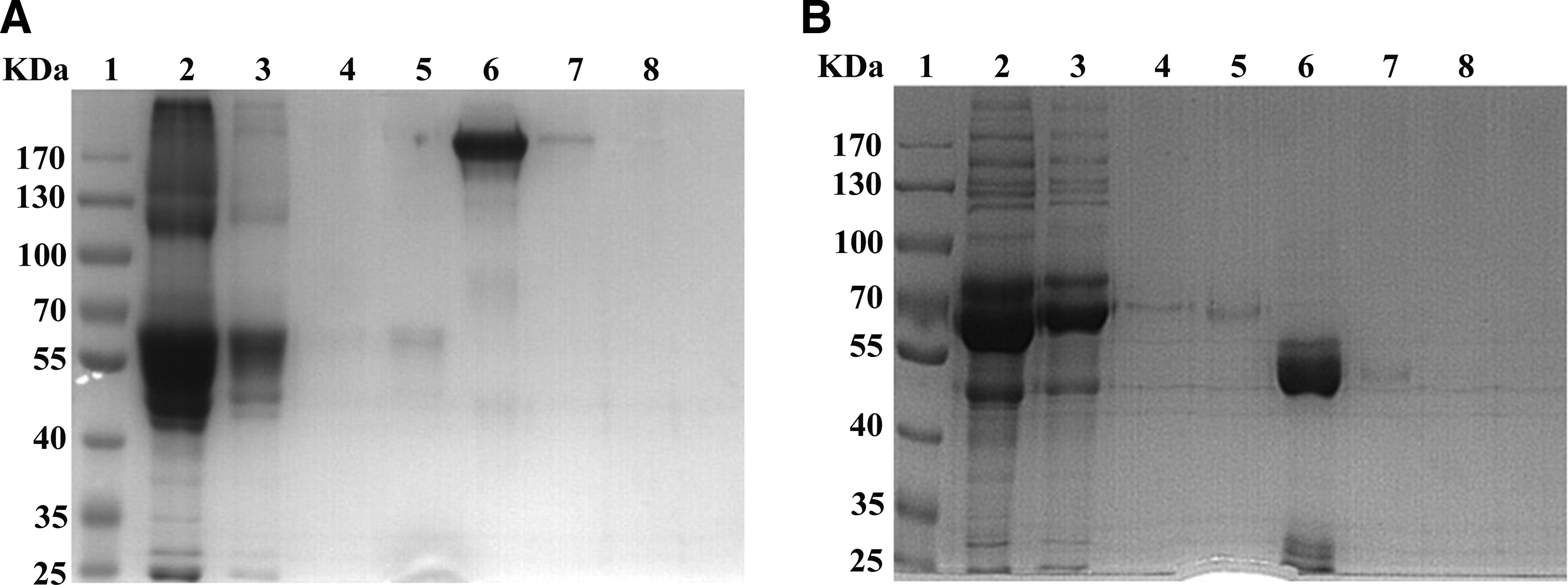
SDS-PAGE analysis of purified anti-CXCL4 monoclonal antibody. Eluted solutions from the protein G affinity column were estimated by Coomassie staining after 8% SDS-PAGE. (
Cloning VH and VL gene fragments of 16D6-3 antibody
Hybridoma 16D6-3, which secretes a CXCL4 antibody, was established and maintained in our laboratory. The gene encoding 16D6-3 antibody was sequenced. Electrophoresis of the total RNA from hybridoma 16D6-3 showed that there were three bands demonstrating the integrity of the extracted RNA (Fig. 3A). The results of electrophoresis of the PCR products, obtained using the primers VH backward and VH forward to amplify the VH gene fragment from retrotranscripted cDNA and using primers VL backward and VL forward to amplify the VL gene fragment, are shown in Figure 3. The VH and VL gene fragments had the expected molecular sizes of 360 bp and 320 bp, respectively (Fig. 3B). Alignment of the amino acid and DNA sequences with the IMGT databank revealed that the cloned VH and VL genes closely matched the sequence of the variable regions of the rat antibody (Fig. 3C).
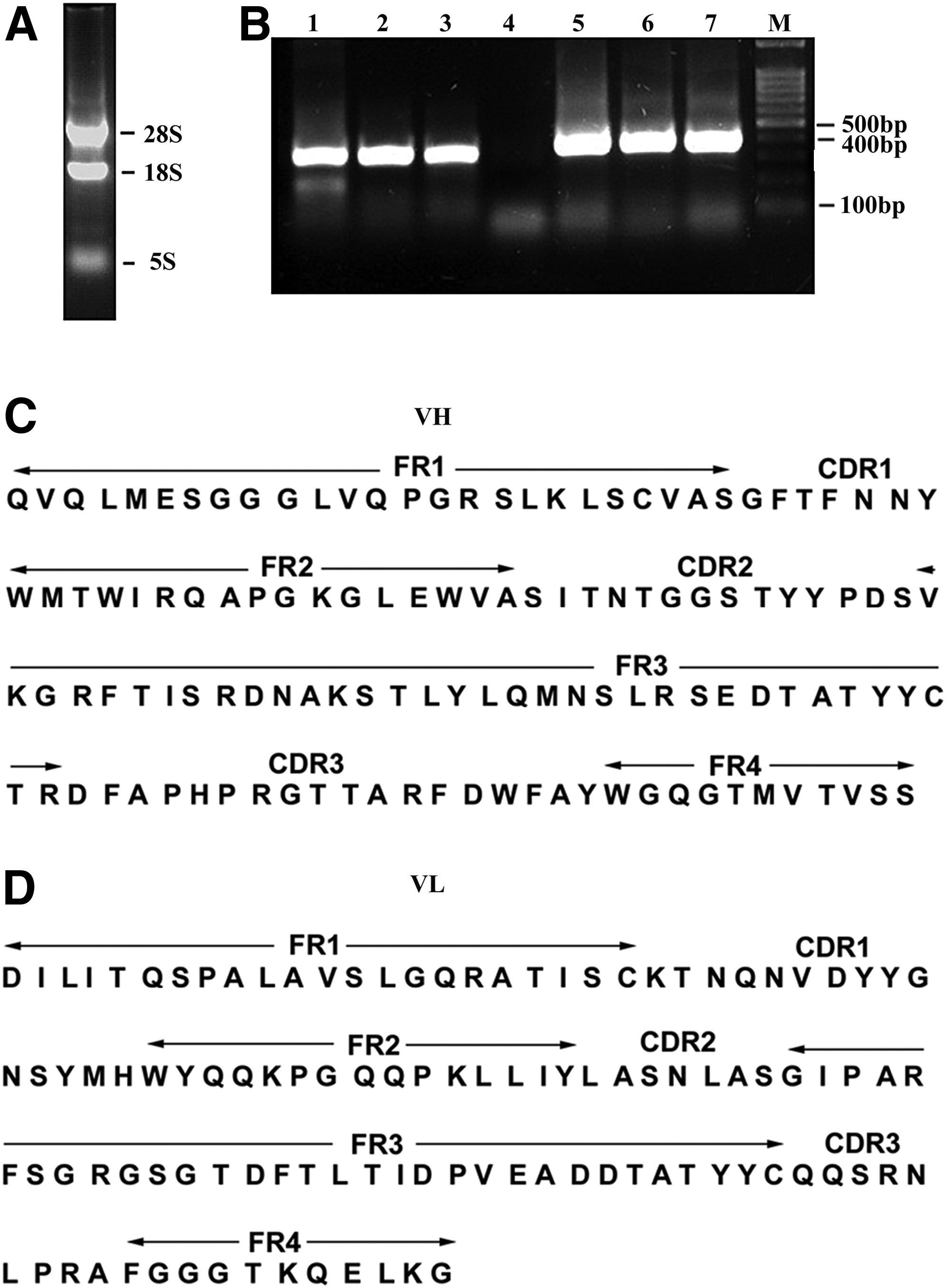
Cloning and sequence analysis of 16D6-3 antibody. (
Characterization of CXCL4 monoclonal antibody
In Western blot analysis, protein G purified 16D6-3 antibody (5.2 mg/mL) strongly reacted against both mouse and human CXCL4 proteins resulting in bands corresponding to a relative molecular weight between 10 and 15 kDa. Furthermore, the binding affinity was analyzed by Biacore. The values for Ka and Kd were calculated from sensorgrams using five concentrations. Fitting the data to a 1:1 binding model yielded an apparent binding affinity of Kd=1.83×10⁻⁸ M, obtained from the ratio of the rate constants koff/kon (Fig. 4B).

Characterization of CXCL4 MAb. (
Because CXCL4 reportedly inhibits the proliferation of ACHN cells by binding CXCR3,(25) CXCL4 MAb was tested for its role in neutralizing rhCXCL4 in the culture of ACHN cell line. As shown in Fig. 4C, CXCL4 (IC50=72.6 μg/mL) induced growth inhibition of the ACHN cell line with a peak response occurring at 72 h at concentrations of 10–100 μg/mL, which was in agreement with the previous report.(25) The inhibition effect of CXCL4 (72.6 μg/mL) was significantly blocked (p<0.01) by CXCL4 MAb on a dose-dependent manner.
Discussion
Our study describes the generation and characterization of a highly specific rat monoclonal antibody against both human and mouse CXCL4. This development is important for the further study of the role of CXCL4 in many biological processes.
In this work, we produced an antibody against CXCL4 by fusing the rat spleen cells with the mouse SP2/0 cells. The 16D6-3 antibody was produced in the ascites and purified by protein-G affinity chromatography. Next, we sequenced the 16D6-3 antibody gene. Blast analysis of the Genebank showed that it matched with a rat immunoglobin protein domain. The complementarity determining regions (CDRs) were identified by examining the sequence in IMGT database. In typical characterizations of rat CDRs, variable fragments containing four frame regions (FRs), three CDRs, and two cysteine residues were identified. The binding affinity of CXCL4 MAb to rhCXCL4 with Kd of 1.83×10⁻⁸ M was determined.
CXCL4 is a growth inhibitor of the tumor cell line ACHN.(25) We have chosen this activity as a functional assay to test the antibody for its neutralization property. The activity is believed to be mediated by the chemokine receptor activation. Neutralizing antibodies against CXCR3 blocked CXCL4-induced growth inhibition of the ACHN cell.(25) Our CXCL4 antibody was also shown to be effective in the inhibition of CXCL4-induced growth arrest of ACHN cells.
Increasing data support a central role for platelets and their products in the pathogenesis of many inflammatory diseases.(26–29) Platelet-derived PF4 plays important roles in endothelial cell proliferation and apoptosis, angiogenesis,(11) tumor development,(19) and atherosclerosis.(23,30–33) Recently, it has been reported that PF4 participates in tissue damage, especially as an important mediator of intestine tissue damage.(34) Thus, the monoclonal antibody should be a useful tool for further studies of CXCL4 in murine disease models.
In conclusion, we demonstrated that the antibody produced from the monoclonal hybridoma line 16D6-3 recognized both human and mouse CXCL4. CXCL4 MAb may be used for studies of CXCL4 in vitro and in vivo. In particular, it will be helpful for efficacy studies using murine disease models.
Footnotes
Acknowledgments
This work was supported by the National Natural Science Foundation of China (grants 81373467/H3108, 81173113/H3109, and 81273573/H3108).
Author Disclosure Statement
The authors have no financial interests to disclose.