
Abstract
Angiogenesis, a neovascularization process, is the most important occurrence in developmental and pathological conditions. Key factors involve belonging to the vascular endothelial growth factor family. Recently, placental growth factor (PlGF) has been considered in medicine because of its pathological importance in solid tumor invasion and metastasis. Therefore, PlGF targeting plays a promising role in the hindrance of tumor progress. In this study, murine PlGF was cloned, expressed in an Escherichia coli system, and used for development of polyclonal camel antibody. Codon-optimized mouse PlGF (mPlGF) cDNA was synthesized and cloned in to pET-28a. The expression was performed in BL21 (DE3) E. coli strain and purified by immobilized metal affinity column chromatography. A camel was subcutaneously immunized six times over a one-week interval using purified protein. IgGs were purified by applying the serum on two sequential column affinity chromatography using proteins A and G. Then, anti-PlGF IgG was identified by Western bolt analysis and ELISA using commercial and expressed PlGF. Synthesized mPlGF was cloned successfully into the pET-28a expression vector, and the accuracy of construct was confirmed by map restriction analysis and sequencing. The expression was induced by 1 mM IPTG and confirmed by SDS-PAGE and Western blot using anti-His monoclonal antibody. The camel was immunized using recombinant mPlGF, and IgG was purified by two-step affinity chromatography. Identification of specific IgG against mPlGF was confirmed by ELISA assay.
Introduction
A
The mouse form (mPlGF, 158 amino acids) is expressed as a unique form, unlike the human forms, which have several isotypes produced by seven exons using an RNA splicing process.(4) Studies on inactivation gene illuminate that mice with PlGF gene ablation are fully viable and healthy. The studies indicate the redundant role of PlGF during the evaluation. Moreover, it is demonstrated that PlGF is not detectable at transcriptional or translational levels in evolution and physiological conditions. However, in pathological conditions, the expression of PlGF and its receptor significantly increases at transcriptional and translational levels, which are detectable by common molecular methods.(5) Other investigations demonstrated that defection of protein in tumor tissues leads to tumor growth inhibition.(5,6) According to recent experiments, PlGF targeting by antibody or small molecules could reduce tumor growth and metastasis, both in vitro and in vivo. Therefore, PlGF has been recently considered a promising target for drug development in cancer treatment.(7,8)
In addition to the common four-chain antibodies, about half of immunoglobulin of camel and llama serum involves special antibodies, called heavy chain antibodies (HcAbs). They consist of only two heavy chains attached to the antigen via antigen-binding domain known as VHH or nanobody. In addition, CH1 domain is deleted from the constant regions of HcAbs naturally. Substitution of amino acid residues in critical points of HcAbs, rather than conventional antibodies, creates a stable and hydrophilic structure without light chain fragments.(9,10) Because of the small size and polar charge, nanobodies have advantages such as high yield expression in bacteria and yeast, high stability, good affinity to antigen, easy manipulation, and close homology to human antibodies.
The prokaryotic expression vectors like pET series are routinely used for expression of recombinant proteins. Low-cost, time-saving, and easy production are advantages for this type of expression system. Optimizing strategies like codon optimization, in the case of rare codons, could be performed on coding sequences of eukaryotic genes for better expression in prokaryotic systems.
In the current study, mPlGF coding sequence was expressed in BL21 (DE3) Escherichia coli strain. The resulting recombinant protein was purified using Ni-NTA affinity chromatography. Finally, the preparation and characterization of HcAbs against purified mPlGF were investigated by camel immunization and ELISA.
Material and Methods
Cloning of pET28-mPLGF construct
The mPlGF construct was synthetically produced by Biomatik (Cambridge, Canada). The construct was sub-cloned into the pET-28a expression vector at NcoI and XhoI restriction sites and then transformed into the TOP10F′ E. coli strain. The accuracy of the construct was confirmed by restriction map analysis and sequencing. The confirmed recombinant vector was transformed to BL21 (DE3) competent cells for protein expression.
Expression and purification of mPlGF protein
A single transformed BL21 colony harboring the recombinant construct (pET28-mPlGF) was grown in 5 mL Luria-Bertani (LB) broth containing 50 mg/L kanamycin at 37°C, 220 rpm overnight. Two mL of the culture was transferred into 1 L of fresh LB medium containing kanamycin and was grown at 37°C. Isopropyl-β-d-thiogalactopyranoside (IPTG) was added to the culture at a final concentration of 1 mM to induce expression of the mPlGF. After an overnight incubation at 37°C, the bacterial cells were harvested and homogenized by sonication at 200 W for 7 min in an ice bath and centrifuged at 8000 g for 30 min in lysis buffer (8 M urea, 500 mM NaCl, 50 mM Tris-HCl, and 30 mM imidazole) at 4°C. The clarified supernatant was applied on a Ni-NTA column, and irrelevant proteins were washed with washing buffer containing 4 M urea, 500 mM NaCl, 50 mM Tris-HCl, 60 mM imidazole, and protease inhibitor. Finally, mPlGF protein was eluted with 500 mM imidazole in PBS.
SDS-PAGE and Western blot analysis
For assessment of the expression of mPlGF, the pellets were collected after 2 h, 4 h, and an overnight induction. Samples were analyzed on 15% poly-acrylamide gel electrophoresis (Mini-Protean Tetra cell®, Bio-Rad, Hercules, CA) using the Coomassie Brilliant Blue staining method.(11) Verification of target protein, was performed by Western blotting (Mini Trans-Blot®, Bio-Rad) using an anti-His monoclonal antibody.(12) Briefly, the protein contents of samples were transferred to a nitrocellulose membrane and blocked with BSA. The membrane was subsequently incubated with anti-His antibody (1:3000) and HRP-conjugated anti-mouse antibody (1:3000) as primary and secondary antibodies, respectively. Finally, the band was visualized using 3,3′-diaminobenzidine (DAB).
Preparation of polyclonal HcAb against rmPlGF
In this study, Camelus deromedaricus was used for animal immunization. Six weekly subcutaneous injections at neck and flank were done with purified recombinant mPlGF. At each injection, 100 μg of recombinant mPlGF were mixed with complete Freund's adjuvant at first injection and incomplete Freund's adjuvant for the balance of immunizations. Before the first injection and after the sixth, blood samples were collected and serums were separated and stored at −70°C. The isolation of heavy chain antibodies was carried out by G and A affinity chromatography columns using standard methods.(13)
mPlGF reactivity assessment
For assessment of HcAb binding capacity, the ELISA test was employed. The 96-well ELISA plate was coated with 5 μg/mL of recombinant mPlGF. The blocking process was done with 2% skimmed milk. After washing, 1:200 to 1:12,000 dilutions of HcAb fraction was added to positive and negative wells and incubated for 1 h at room temperature (RT). 100 μL of 1:3000 diluted rabbit anti-camel monoclonal antibody were added to each well and incubated for 1 h at RT. After washing with PBS contains 0.05% Tween-20, 1:5000 diluted HRP conjugate anti-rabbit polyclonal antibody was applied for 1 h at RT as a secondary antibody. Finally, colorimetric reaction was done using 3,3,5,5-tetramethylbenzidine and analyzed by a spectrophotometer at 450 nm wavelength.
Results
Construction of pET28-mPlGF
The integrity of pET28-mPlGF was confirmed by colony PCR with universal T7 promoter and T7 terminator primers, restriction enzyme (NcoI and XhoI) analysis, and sequencing (Fig. 1).

(
Expression and purification of mPlGF
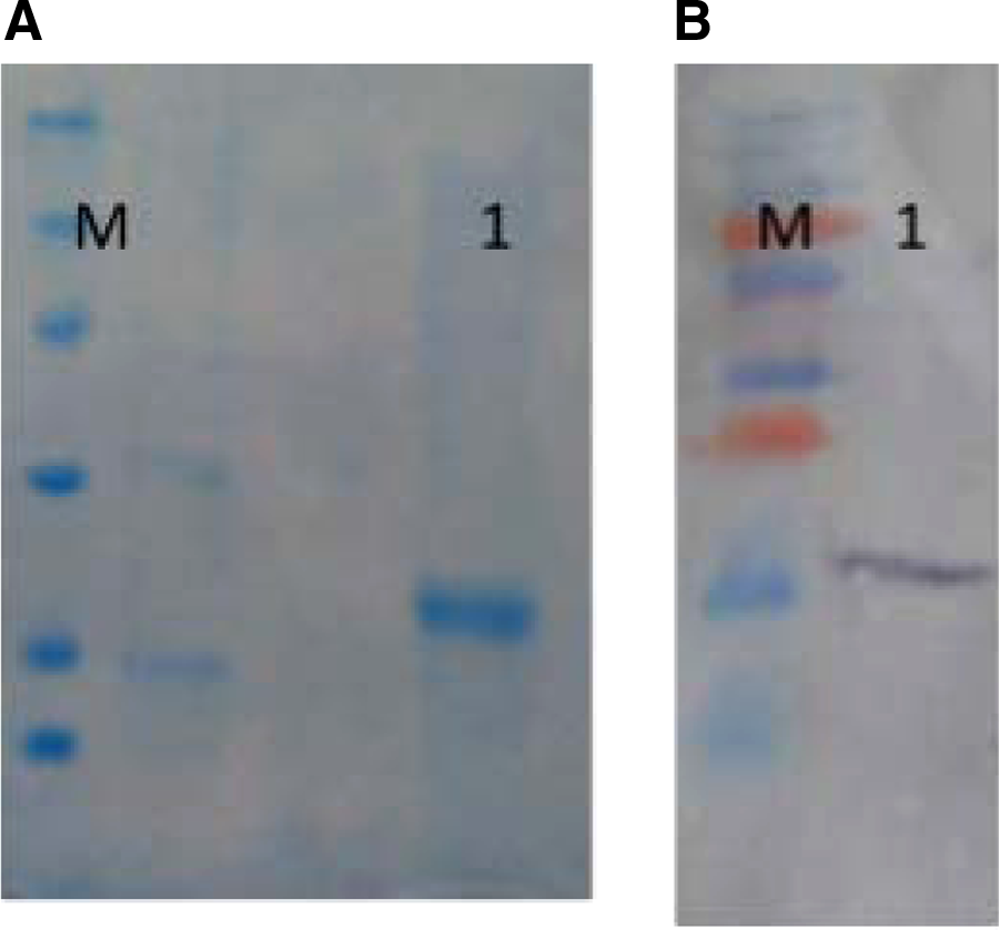
The prokaryotic expression vector pET28-mPlGF was introduced into the E. coli BL21 according to the standard method. The expression of the recombinant protein was induced with 1 mM IPTG. As shown in Figure 2A, mPlGF was expressed efficiently in the host with a molecular mass of about 18 kDa. Moreover, the level of protein expression at overnight culture was found to be more than 2 h or 5 h after induction with IPTG (Fig. 2B). The Western blot result confirmed the identity of recombinant mPlGF (Fig. 3).

Expression and characterization of rmPlGF. (
(
mPlGF-specific HcAb preparation
Camel immunization was done six times in weekly intervals. HcAbs purification was carried out by standard protocol, and specific reactivity of purified HcAb was monitored by ELISA test. As illustrated in Figure 4, the camelid HcAbs antibody at different dilutions (1:200–1:12,000) reacted with the recombinant mPlGF protein.

Reactivity analysis of HcAbs by ELISA. Camelid HcAbs antibody at different dilutions (1:200 to 1:12,000) reacted with the recombinant mPlGF protein.
Discussion
PlGF directly induces growth, proliferation, migration, and survival of endothelial cells. It also has regulatory effects on other types of cells involved in the vascularization process, especially in pathological angiogenesis. In this condition, PlGF plays a major role in angiogenesis, which may lead to either tumor development or metastasis.(14)
Therefore, PlGF targeting in pathological conditions (such as in solid tumors like breast cancer) could be considered as a new approach in cancer therapy. In this manner, Fischer and colleagues developed a neutralizing anti-PlGF monoclonal antibody against mPlGF that could simultaneously inhibit growth and metastasis of tumor cells, leading to improvements in the effect of chemotherapeutics agents as well as reducing side effects.(6) TB-403, introduced as a humanized monoclonal antibody against PLGF, completed phase I clinical trial, demonstrated anti-angiogenic and anti-neoplastic effects. Preclinical studies demonstrated TB-403 binds to PLGF, inhibiting the binding of PLGF to the VEGFR1, which results in the inhibition of tumor angiogenesis and tumor cell proliferation.(15) In one investigation, Taylor and colleagues selected BP1-peptid against PlGF by phage display technology and subsequently employed it for treatment of breast cancer xenograft in vitro and in vivo. The treatment caused a decrease in the number of spontaneous metastatic lung nodules.(16) In another study, Bower and colleagues evaluated the potent and stable PlGF-1 targeting covX bodies (antibody that conjugated with a peptide antagonist) in phage display peptide discovery. The complex successfully inhibited tumor growth and metastasis in vitro and in animal models.(17)
Interestingly, about 50% of camelid immunoglobulin lack light chain and the 15 kDa antigen binding domain of the heavy chain antibody (VHH, obtained by protease cleavage) is considerably smaller than papain cleaved Fab fragments (60 kDa) of conventional IgG.(18) Manipulation of VHH is practically simply for various purposes like enhanced in vivo half-life, using attachment to compounds like Poly (ethylene glycol) (PEG), PASyaltion, HESylation, and other types of protein modifications. Moreover, the small size of nanobodies allows them to penetrate deeply into the solid tumors even when armed with chemical drugs. Nanobodies were also employed as tumor imaging agents, either in diagnosis or monitoring during therapeutic treatment due to rapid clearance from the circulation.
Bacterial systems are cost-effective, time-saving, and high-level producers for recombinant protein manufacturing at industrial scale. A problem that could have a negative effect on pET expression systems is the presence of rare codons for some amino acids in such systems. In the current study, we used a codon usage bioinformatics tool for codon optimization of mPlGF coding sequence. Codon-optimized mPlGF was cloned in pET-28a and successfully expressed at the level of 6 mg/L in the culture media.
Camel immunization was done successfully and the monitoring of immune responses illustrated that the purified mPlGF protein has good antigenicity and was able to be recognized by immune camel serum in the ELISA assay. In our previous study, we performed cloning and expression of human PlGF (hPlGF) in pET-26b expression system. Purified hPlGF could also induce immune responses so we successfully constructed an immune library for phage display.(19) The lymphocyte repertoire of the immunized camel can be further utilized for the construction of an immune phage library for obtaining an anti-mPlGF nanobody.
Footnotes
Acknowledgment
This project was financially supported by the Institute Pasteur of Iran. The authors wish to thank everyone who supported this current investigation.
Author Disclosure Statement
The authors have no financial interests to disclose.