
Abstract
SPLUNC1 (Short palate, lung and nasal epithelium clone1) protein is an abundant secretory product of epithelia present throughout the conducting airways. Although its function is still not fully known, most studies have focused on its defensive effect in the infection of human airways and its potential to serve as a molecular marker for lung cancer. In this study, we further evaluated the SPLUNC1 expression in patients with lung disease to explore its role in cancer or tuberculosis at the protein level. We generated a panel of antibodies by using protein from a eukaryotic expression system as the immunogen to mice. It was the panel of SPLUNC1 monoclonal antibodies that allowed us to comparatively determine SPLUNC1 protein in lung cancer and tuberculosis infection by detecting sera and pleural effusion other than airway surface. The results showed that the SPLUNC1 level was not significantly changed either from sera of lung cancer or control. There was a significant increase in pleural effusion from lung cancer when compared to tuberculosis. These results indicate that SPLUNC1 may be a useful marker for tracing lung cancer cells, based on its epithelial origin property in pleural effusion.
Introduction
S
The SPLUNC1 level was significantly lower in the persistent allergy group; in smokers the SPLUNC1 levels were much lower than in non-smokers. A correlation was found between SPLUNC1 and Vol 2 (volume 2) in non-smokers.(7) Protein profiling showed significantly higher levels of S100-A9 and lower levels of SPLUNC1, cystatin SN, IgJ, and β2-microglobulin among workers exposed to MWF (metal working fluids) with airway symptoms.(8) In wild-type mice, P. aeruginosa infection could significantly reduce SPLUNC1 protein and increase neutrophil elastase (NE) activity in bronchoalveolar lavage fluid (BAL).(9) SPLUNC1 may also serve as a potential molecular marker for detection of tumors, such as non-small-cell lung cancer,(10) gastric hepatoid adenocarcinoma,(11) salivary gland mucoepidermoid carcinomas,(12) and NPC.(6) Earlier research has shown that mRNA expression level and positive rate of SPLUNC1 in peripheral blood correlated with the pathologic stage of NSCLC.(10) However, SPLUNC1 protein level investigations from lung cancer patients were lacking. Furthermore, the role of SPLUNC1 in lung cancer is still not clearly defined. Qualified antibody is an essential tool for analyzing the expression of SPLUNC1 at the protein level under various pathological and physiological conditions. In this research, we generated a panel of antibodies and established immunoenzymatic techniques for detection of the protein. Then we probed the SPLUNC1 expression in lung cancer and respiratory infection with tuberculosis.
Materials and Methods
Production of fusion protein
Full-length SPLUNC1 cDNA was amplified by PCR and cloned into PCDM-L and PCDH-L,(13) which included the fragment of mouse IgG-Fc (mFc) and human IgG-Fc (hFc), respectively. The primers for the construction of PCDH-L-SPLUNC1 were: 5′-CGC
Both expression vectors were respectively transfected into COS-7 cells using DEAE- dextran (Sigma, St. Louis, MO). Expression supernatants were collected and fusion proteins were purified by rProteinA sepharose (GE Healthcare, Uppsala, Sweden). The purity of the fusion protein was confirmed by spectrophotometer (UV-2550, Shimadzu, Kyoto, Japan) and SDS-PAGE gel electrophoresis. A control fusion protein mE3-hFc, which was mouse CD137 cysteine-rich domains with human IgG-Fc, was produced by our laboratory,(13) and another control of human IgG1-Fc protein was purchased from Sino Biological (Beijing, China).
Production of hybridomas
Hybridoma production was performed according to standard protocol. All studies performed with laboratory animals were approved by the institution's review boards for the care and use of experimental animals. The fusion protein SPLUNC1-hFc from the eukaryotic expression system was used as the immunogen to mice. After fusions, wells containing hybridoma cells were screened for binding to SPLUNC1-hFc and not to mE3-hFc. Positive clones were subcloned by limiting dilution assay three times and expanded. Culture supernatants of positive clones were collected and antibodies were purified by Protein G Sepharose.
Characteristics of monoclonal antibodies
Specificity of monoclonal antibodies (MAbs) to recombinant proteins was further identified by ELISA.(14) Briefly, microtiter polystyrene plates were separately coated with 1 μg/mL of purified recombined proteins SPLUNC1-hFc, mE3-hFc,(13) and human IgG1-Fc. 100 μL cultured supernatant or diluted purified antibody and horseradish peroxidase (HRP)-conjugated goat anti-mouse IgG-Fc antibody were successively added. Tetramethylbenzidine (TMB) substrate was used to reveal the positive reaction. The reaction was stopped with 12.5% H2SO4, and optical densities were measured by a multiscan ELISA reader at 450nm. Antibodies were further identified by Western blot and Dot-blot analyses. Briefly, purified SPLUNC1-hFc (1 μg) was treated using loading buffer without β-mercaptoethanol and directly electrophoresed in 10% polyacrylamide gel and transferred onto polyvinylidene difluoride membranes, or 2 μL SPLUNC1-hFc (1 μg/mL) and mE3-hFc (1 μg/mL) were spotted onto nitrocellulose membrane. The membrane was then blocked, separately incubated with affinity-purified antibodies. The blots were detected with enhanced chemiluminescence (ECL).
The subclass of purified MAbs was analyzed with mouse monoclonal antibody isotyping reagents (Sigma, St Louis, MO, USA), according to the manufacturer's instructions, and relative affinity of MAbs was analyzed using indirect ELISA.(15) The optimal concentration of coating antigen and HRP-goat anti-mouse antibody was achieved via checkerboard titration. 4 μg/mL SPLUNC1-hFc were coated. As in the previous method, diluted purified MAbs and second antibody were added in turn. The absorbance was detected at 450 nm. The antibody diluted ratio corresponding to the half-maximum OD (OD-50) was considered as relative affinity when the binding curve reached the saturation level.(15) To test whether the prepared MAbs could recognize different epitopes, a competitive ELISA was applied.(16) 4 μg/mL SPLUNC1-hFc were coated; four strains of purified MAbs (100 μL/well) and the mixture from 50 μL of any two strains of MAbs was separately added. As in the previous method, the absorbance was detected at 450 nm. Additive index (AI) was calculated using the formula AI=[2A1+2 / (A1+A2) – 1]×100%. A1 and A2 respectively represented absorbance (A) of each monoclonal antibody. A1+2 represented absorbance of the mixture from 50 μL of any two strains.
Identification of MAbs binding to natural SPLUNC1
SPLUNC1 was mainly expressed in the trachea and bronchial epithelium.(1) To analyze the binding ability of MAbs for the natural protein, bronchoalveolar lavage fluid (BALF) was obtained from tuberculosis patients, according to the approval of the Ethics Committee. The participant enrolled received an intramuscular injection of atropine sulfate and diazepam, followed by local anesthesia with lidocaine. A flexible bronchofiber scope (Olympus, Tokyo, Japan) was inserted orally to pour 40 mL of 0.9% saline (37°C) into the bronchus of the right middle lobe, and the BALF was recovered. This process was repeated one time and the fractions pooled together without presence of blood contamination. BALF was filtered through double sterile gauze and 0.45 μm filter, then centrifuged at 4500 rpm for 40 min at 4°C by using protein ultrafiltration tubes (Merck, Darmstadt, Germany) with protease inhibitor cocktail (Roche, Mannheim, Germany). Total protein concentration was measured using a spectrophotometer. The SPLUNC1 protein in BALF was ascertained by Western blot analysis. The binding ability of MAbs from different clones to natural SPLUNC1 protein was evaluated. Briefly, 0.2 μg/mL rabbit anti-human SPLUNC1 polyclonal antibody, which have proved binding to the natural protein, was coated; then SPLUNC1 from BALF protein (0.2 μg/mL) was added. Thereafter MAbs from clones 9, 130, 160 were used as second antibody. SPLUNC1 MAb (Clone 251512, R&D, Minneapolis, MN) was used as positive control.
Establishment of SPLUNC1 detection method for clinical samples
The rabbit anti-human SPLUNC1 polyclonal antibody (0.2 μg/mL) (sc-133917 Santa Cruz, Dallas, TX, USA) was used as capture antibody and SPLUNC1 monoclonal antibodies (clone 130) prepared in this study were used as detective antibody; then a sandwich ELISA was established to test clinical samples. Series of 10–10,000 pg/mL SPLUNC1-hFc protein were separately added. The value of absorbance 450 nm was detected and a standard curve was established.
Test of SPLUNC1 in sera and pleural effusion
Lung cancer and tuberculosis patients were from Beijing Chest Hospital and all patients were clinically diagnosed. Health control group was from Center for Disease Control of Tongzhou District (Beijing) with a strict exclusion of cancer and tuberculosis. All samples were centrifuged and supernatants were sub-packaged and preserved in −70°C. The subjects included 29 healthy individuals and 57 lung cancer patients. Among the lung cancer patients, there were 26 adenocarcinoma, 17 squamous cell carcinoma, and 14 small cell carcinoma. Pleural effusion was inspired from 24 lung cancer patients and 16 tuberculosis patients. Ten mL pleural effusion was centrifuged at 3500 rpm for 10 min, and then supernatant was collected. As previously, rabbit anti-human SPLUNC1 polyclonal antibody was coated, while 100 μL serum or pleural effusion from subjects was added. Then 1 μg/mL SPLUNC1 monoclonal antibody (clone 130) was added, followed by HRP-conjugated goat anti-mouse antibody. The absorbance was also determined at 450 nm.
Statistical analysis
The data was analyzed using SPSS, v. 17.0. Results were expressed as means±standard deviation. Student's 2-sample t-test was used between the groups for normally distributed data. The no normal distribution data was analyzed by nonparametric tests. The differences among the three groups were tested using one-way ANOVA. The correlations between two variables were determined by Pearson's correlation. p<0.05 was considered statistically significant.
Results
Fusion protein expression and identification
Expression plasmids of PCDM-L-SPLUNC1 and PCDH-L-SPLUNC1 were identified by sequencing. SPLUNC1 mRNA could be detected in the COS-7 cells transfected separately with PCDM-L-SPLUNC1 and PCDH-L-SPLUNC1 compared to the control COS-7 cells without SPLUNC1 transfected by reverse transcription-polymerase chain reaction (RT-PCR) (Fig. 1A). SPLUNC1 fusion proteins in COS-7 supernatants were purified and verified using Western blot analysis. The SPLUNC1-mFc and SPLUNC1-hFc proteins were approximately 54.54 kDa and 52.50 kDa, and Western blot demonstrated that the proteins were respectively located at corresponding positions that were consistent with the predictive molecular weight (Fig. 1B).
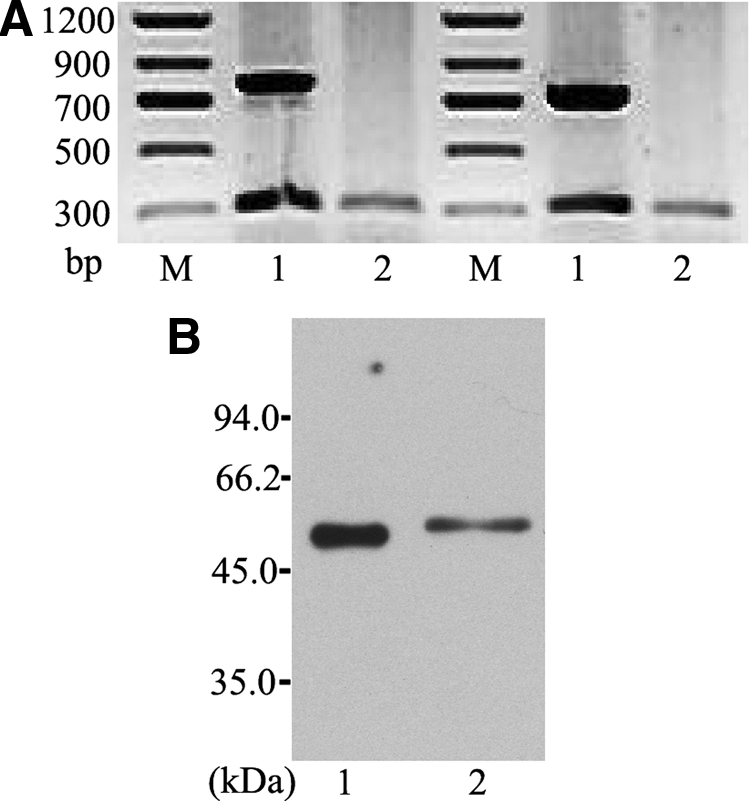
Identification of SPLUNC1 transfection and SPLUNC1 protein expression. (
Generation of anti-human SPLUNC1 hybridomas
Culture supernatants from growing hybridomas were screened by ELISA in three rounds, and four clones were selected with constant genetic stability and high production. Of the four, clones 9, 130, and 160 were determined strictly against SPLUNC1-hFc but not mE3-hFc, while clone 104 had cross-reactivity against both mE3-hFc and SPLUNC1-hFc (Fig. 2A). We also tested the ability of the MAbs to bind to SPLUNC1-hFc in Western blot and dot-blot under oxidized and undenatured states. The blots could be developed when the membrane was incubated with antibodies from the selected clones, including 9, 130, and 160, as with the positive control (Fig. 2B, C). The results further strengthened that the antibodies from clones 9,130, and 160 could bind to SPLUNC1-hFc but not to mE3-hFc.

Specificity of prepared MAbs against SPLUNC1. (
MAbs of clones 9, 104, 130, and 160 were all IgG1 subclass, and relative affinities tested by 50% of maximal binding to SPLUNC1-hFc were separately 1.95×10⁻⁴, 7.81×10⁻⁶, 9.77×10⁻⁵, and 1.33×10⁻⁴ (Table 1).
–, no binding; +, binding; ++ , stronger binding; ND, Not Done.
According to the value of AI, when it was more than 50%, it was suggested that the epitopes of MAbs from two strains were different. While the value of AI is from 10–50%, the epitopes of two antibodies were possibly regarded as different. When the value of AI is less than 10%, the epitopes were possibly the same between two antibodies. Compete ELISA indicated that the different antigenic site was recognized between clone 9 and other clones (e.g., 104, 130, and 160). The epitope that was recognized by clone 104 was different compared to clones 9, 130, and 160. Adversely, the same epitope was possibly recognized between 130 and 160 (Table 2).
–, epitopes were different between antibodies from two clones; ±, the epitopes were possibly different between antibodies from two clones; +, epitopes were the same between antibodies from two clones.
Binding capacity of MAbs against natural SPLUNC1
The ability of SPLUNC1 polyclonal antibody (Santa Cruz Biotechnology, Dallas, TX) binding to natural SPLUNC1 was verified by Western blot analysis (Fig. 3A). Thus, the polyclonal antibody was used as capture antibody. Detective MAb clones 9, 130, and 160 and a MAb from R&D were used in the sandwich ELISA. As shown in Table 1 and Figure 3B, clones 9, 130, and 160 and positive control antibody all can bind to natural and eukaryotic expressed SPLUNC1.

Establishment of ELISA for SPLUNC1 detection of clinical samples. (
SPLUNC1 expression in serum of patients and healthy group
A standard curve was established using sandwich ELISA. In the beginning, we detected the expression of SPLUNC1 in serum from lung cancer patients and healthy individuals. The outliers were excluded. However, SPLUNC1 expression was not significantly different between lung cancer patients and control (97.11±42.29 pg/mL vs 95.53±27.38 pg/mL, Z=8.07, p>0.05) (Fig. 4A) using non-parametric tests. Furthermore, we tested whether the SPLUNC1 expression was significantly different among adenocarcinoma (95.47±18.52 pg/mL), squamous cell carcinoma (93.34±43.91 pg/mL), and small cell carcinoma (104.62±33.93 pg/mL). The results suggested that there was no statistical difference among these lung cancer patients (F=0.229, p>0.05) (Fig. 4B).

Scatter distribution of SPLUNC1 expression in clinical samples. (
SPLUNC1 expression in pleura of lung cancer and tuberculosis patients
The SPLUNC1 protein levels were significantly increased in malignant pleural effusion from lung cancer (108.31±34.45 pg/mL) compared to pleural effusion from tuberculosis patients (47.97±15.68 pg/mL) (t’=7.50, p<0.01) (Fig. 4C). These results indicated that SPLUNC1 was more related to the lung cancer environment than the tuberculosis infection in the disease site.
Discussion
SPLUNC1 has been identified as having an important role in lung innate defense by different means.(17) SPLUNC1 has also been shown to have an anti-inflammatory function in exogenous microbe-induced respiratory infection, such as K. pneumoniae,(18) Mycoplasma pneumoniae,(19) P. aeruginosa, and Epstein-Barr virus.(20) Additionally, SPLUNC1 could also regulate ion/mucus transport and hydration levels in the lung,(21) lower surface tension by its surfactant-like properties,(22) and act as an inflammatory mediator. The earlier studies demonstrated that SPLUNC1 was associated with tumorigenesis. Absence of SPLUNC1 expression was found in 93.85% of newly diagnosed NPC tissue specimens. With the progression of NPC, SPLUNC1 expression was gradually decreased. SPLUNC1 regulates NPC cell proliferation, differentiation, and apoptosis by MAPK and p27 pathway.(6)
As an important addition to these, SPLUNC1 was also known as a potential marker for the detection of non-small lung cell cancer.(23,24) However, SPLUNC1 protein level investigations were lacking from NSCLC patients in the studies. In order to further explore the role of SPLUNC1 in lung cancer, a panel of specific MAbs was produced in our lab. As for the structure of protein from the eukaryotic expression system close to the natural protein, with these expressed proteins, we succeeded in preparing a group of hybridomas. From the results of specificity determination of obtained MAbs, it was shown that the selected three MAbs not only could bind SPLUNC1 fragment of recombinant protein instead of hIgG-Fc fragment, but also combined specifically to the natural SPLUNC1 protein derived from human BALF, which was proven better than commercial antibody. Furthermore, in contrast to commercial antibody, the prepared MAbs could not efficiently bind to the SPLUNC1-hFc under denatured state (data not shown) by using Western blot. However, under undenatured state, the reactivity of the prepared MAbs against SPLUNC1-hFc and natural SPLUNC1 in BALF was more sufficient than commercial antibody. Thus, it is most likely that the epitopes recognized by the prepared MAbs from our lab were not linear but conformational in structure. In general, the MAbs from our lab are more suitable to detect SPLUNC1 protein in any clinical samples by using ELISA.
SPLUNC1 expression in the serum and pleural effusion was detected by an ELISA reaction. Our results showed that there was no significant difference between lung cancer patients and healthy controls for the expression of SPLUNC1 in sera. In addition, the expression of SPLUNC1 was no different among adenocarcinoma, squamous cell carcinoma, and small cell carcinoma. This is the inverse of studies of mRNA expression of SPLUNC1 in the peripheral blood for the diagnosis of micrometastasis and the prognosis of lung cancer.(23,24) The reason may be related to the sensitivity of different methodology for protein and nucleic acid detection, though the higher protein levels of SPLUNC1 was determined in serum from lung cancer patients by our ELISA. A more sensitive method may need development for the detection from serum based on the MAbs. The pleural space normally contained a very thin layer of fluid. The pleural effusion was produced when the pleural fluid is excessively formed and/or the fluid removal was decreased by the lymphatics.(25) Malignant pleural effusion was mainly caused by lung cancers and other carcinomas. The prognosis of lung cancer patients with malignant pleural effusion was usually poor. SPLUNC1 mRNA expression was highly increased in malignant pleural effusion caused by lung cancer(26); however, the change of SPLUNC1 protein level in pleural effusion of lung cancer was unknown. Our results demonstrated that SPLUNC1 protein levels were also increased in the malignant pleural effusion from lung carcinoma.
Although these results provided initial evidence of the increased SPLUNC1 expression in pleura from pulmonary carcinoma, the limited sample used in this study needs to be expanded to further clarify the correlation between SPLUNC1 protein level and lung cancer in pleural effusion, especially in blood. In the meantime, an improved sandwich ELISA established with our MAbs, such as 130 and 9, will be more helpful for the intensive study. Furthermore, the function of the anti-SPLUNC1 MAbs from our laboratory is also worthwhile to be evaluated for potential inhibitory effects on non-small cell lung cancer as showed recently in xenograft model both in vitro and in vivo.(27)
Conclusion
Hybridoma-secreted antibodies against human SPLUNC1 were produced and characterized in this study. To our knowledge, it is the first time that SPLUNC1 protein levels were detected in the serum and pleural effusion using the ELISA method that is based on eukaryotic expression protein prepared hybridomas. The specific antibodies provide valuable tools for further experimental investigation of pulmonary carcinoma, as well as the research and development of the correlation between SPLUNC1 protein and other pulmonary diseases.
Footnotes
Acknowledgments
This work was supported by the National Basic Research Program of China (the 973 program, no. 2014CB744403), Beijing Science and Technology Foundation of China (grant no. Z121107001012146), and a National 12.5 grant (2013ZX10003003).
JLX, LY, XJW, XL, PJW, and JEZ performed most of the experiments. HTZ designed the study. ZHY and YLZ performed statistical analysis. HTZ supervised the study, and JLX and LY wrote the manuscript. All authors read and approved the final manuscript.
Author Disclosure Statement
The authors have no financial interests to disclose.