
Abstract
1-(5-fluoropentyl)-3-(2-iodobenzoyl)indole (AM694) is one of the synthetic cannabinoids and an illegal drug in Japan. It is important to generate a monoclonal antibody (MAb) against AM694 for use in the rapid and sensitive detection of the drug. Two monoclonal antibodies, named HN0124 (IgG1) and NK0504 (IgG1), were obtained, which were possibly effective for detecting AM694 and its derivatives. The cross-reactive ability of these MAbs was evaluated using a competitive enzyme-linked immunosorbent assay. In the results, both of these antibodies recognize 1-(5-fluoropentyl)-3-(2-iodobenzoyl)indole, 1-(5-fluoropentyl)-3-(3-iodobenzoyl)indole, 1-(5-fluoropentyl)-3-(4-iodobenzoyl)indole. Forty nmol/L AM694 can be detected using HN0124 MAb. Thus, MAbs produced in this study could be considered a useful tool for the detection of AM694.
Introduction
S
It has been necessary to develop sensitive and useful methods for detection of AM694. Several methods such as gas chromatography/mass spectrometry (GC/MS) and liquid chromatography/mass spectrometry (LC/MS) are possible to apply for the detection of AM694. But these detection methods are difficult to use and generally time consuming.
In addition, there are various types of immunological assays used as qualitative and quantitative methods for the detection of other drugs except AM694.(3–10) Development of an immunological detection method for AM694 requires a monoclonal antibody specific for AM694. Here we describe the production and characterization of new monoclonal antibodies specific for AM694.
Materials and Methods
Chemicals and instruments
Horseradish peroxidase (HRP)-conjugated anti-mouse IgG antibody, bovine serum albumin (BSA; agarose gel electrophoresis grade, no. A3675), keyhole limpet hemocyanin (KLH), complete Freund's adjuvants (CFA), incomplete Freund's adjuvants (IFA), and RPMI1640 were purchased from Sigma-Aldrich (St. Louis, MO).
Fetal calf serum (FBS) was purchased from (Hyclone, Thermo Scientific, Cramlington, United Kingdom). 1-(5-fluoropentyl)-3-(2-iodobenzoyl)indole (AM694), 1-(5-fluoropentyl)-3-(3-iodobenzoyl)indole, and 1-(5-fluoropentyl)-3-(4-iodobenzoyl)indole were purchased from Cayman Chemicals (Ann Arbor, MI). Indole, 2-iodobenzoic acid, 3-iodobenzoic acid, 4-iodobenzoic acid, 1-(5-fluoropentyl)indole, n-hydroxysuccinimide (NHS), N, N-dicyclohexylcarbodiimide (DCC), sodium bicarbonate, sodium dodecylsulfate (SDS), 1-(heptyl-7-carboxylate)-3-(2-iodobenzoyl)indole, dichloromethane, oxalyl chloride, 2,4,6-trinitrobenzene sulfonic acid (TNBSA), hydrochloric acid, ethylmagnesium bromide, ether, ammonium chloride, methanol, sodium hydroxide, N,N-dimethylformamide, dimethyl sulfoxide, sulfuric acid, 3,3',5,5'-tetramethylbenzidine (TMB), sodium hydrate, 1-bromo-7-heptanic acid, and ethylacetate were purchased from Tokyo Chemical Industry Co. (Tokyo, Japan). Unless otherwise stated, all other inorganic chemicals and organic solvents were of analytical-reagent grade or better.
Synthesis of 1-(heptyl-7-carboxylate)-3-(2-iodobenzoyl)indole (AM694 derivative)
4.00 g (31.6 mmol) of oxalyl chloride were added dropwise to a suspension of 1.00 g (5.37 mmol) of 2-iodobenzoic acid in 30 mL of dichloromethane at 0°C. The reaction mixture was warmed to room temperature and stirred for 2 h. After cooling, the solvent and excess oxalyl chloride were removed in vacuo to give a brown residue (0.90 g; 4.40 mmol), which was used in the next step without further purification.
1.40 g (6.98 mmol) of indole in 5.0 mL of ether was added dropwise to a stirred solution of 3.17 mL (8.01 mmol) of 2.5 M ethylmagnesium bromide in ether, diluted with 1.1 mL of ether, at 0°C. The solution was stirred for 0.5 h at room temperature and a solution of 0.90 g of 2-iodobenzoyl chloride in 5 mL of ether was added dropwise. The reaction mixture was stirred for 1.5 h, quenched with saturated aqueous ammonium chloride, and stirred until the solid was broken up into a fine suspension. The residue was washed with water and ether, then suspended in 20 mL of methanol, to which was added 4 g of sodium hydroxide and 10 mL of water. The mixture was stirred at room temperature for 18 h, the solid was filtered off and washed with successive portions of methanol, water, and ether. Drying in vacuo at 100°C turned 1.06 g (70%) of 3-(2-iodobenzoyl)indole a viscous oil, which was used in the next step without further purification.
0.5 g of sodium hydrate was added to a solution of 1.06 g (3.06 mmol) of 3-(2-iodobenzoyl)indole in 10.0 mL of N,N-dimethylformamide. The reaction mixture was stirred at room temperature and 1.35 mL (6.49 mmol) of 1-bromo-7-heptanic acid were added slowly. The solution was stirred at 120°C for 1 h. After cooling, the reaction mixture was diluted with water and extracted with three portions of ethylacetate. The extracts were washed with brine and dried, and the solvent was removed in vacuo. Chromatography (silica gel [32–63 μm], petroleum ether/ethyl acetate, 7:1) gave 0.60 g (30%) of 1-(heptyl-7-carboxylate)-3-(2-iodobenzoyl)indole as a yellow solid. The synthetic route for the AM694 derivative is shown in Figure 1: 1H NMR (400 MHz, CDCl3) d 1.44 (m, 2H), 1.52 (m, 2H), 1.68 (m, 2H), 1.89 (quint, 2H), 2.18 (m, 2H), 4.12 (t, 2H), 4.45 (m, 2H), 7.14 (td, 1H), 7.29 (s, H), 7.34 (m, 2H), 7.35 (m, 1H), 7.45 (m, 1H), 7.39 (m, 1H), 7.91 (brd, 1H), 8.34 (m, 1H); 13C NMR (75.5 MHz, CDCl3) d 22.7, 25.4, 29.4, 29.8, 30.5, 47.0, 92.5, 109.9, 115.4, 122.8, 122.9, 123.7, 126.7, 127.7, 128.0, 130.5, 137.0, 137.9, 139.6, 146.3, 173.1, 191.2.

Synthesis procedure for AM694 derivative and artificial antigen.
Synthesis of immunogen (AM694 derivative-protein conjugate)
Antigen was prepared following the method described by Cervino and associates with some modifications.(11) Briefly, 1-(heptyl-7-carboxylate)-3-(2-iodobenzoyl)indole was suspended in 0.4 mL dimethyl sulfoxide, and 8 mg N-hydroxysuccinimide (NHS) was added and reacted for 1 h at room temperature. Fifteen mg dicyclohexylcarbodiimide (DCC) was added to the solution and reacted for 1 h at 4°C. Then the mixture was stirred for 8 h at room temperature. After being centrifuged at 8000 r/min for 5 min, the supernatant was added dropwise to 9.6 mg of the KLH dissolved in 5 mL PBS (50 mM, pH 7.4) and kept for 8 h at room temperature. After being centrifuged (3000 r/min, 60 min), the obtained supernatant was dialyzed with 0.1 M sodium bicarbonate buffer (pH 8.5) for 10 h. The dialyzed protein solution was diluted with supplied 5% TNBSA solution 500-fold in 0.1 M sodium bicarbonate buffer (pH 8.5). To the protein solution, TNBSA solution was added, and then was incubated at 37°C for 2 h. Mixture solutions were added in 0.5 mL of 10% SDS and 0.25 mL of 1 N hydrochloric acid to each sample to stop and stabilize the reaction. Then absorbance of the solution at 335 nm was measured, and the concentration of primary amines determined by calculation from the extinction coefficient or by comparison to amino acid standards.
The synthetic route for the AM694 derivative-protein conjugate is shown in Figure 1.
Cell line
The mouse myeloma cells (P3X63-Ag8.653) were cultured in RPMI1640 medium supplemented with 15% fetal calf serum in a 37°C humidified incubator.
Hybridoma preparation
Every 100 μL of the prepared adjuvant emulsion (a 1:1 emulsion of AM694 derivative-KLH conjugate [100 μg per mouse] and complete Freund's adjuvant was injected intraperitoneally into each of female A/J mice (8 weeks of age). The mice received the same injections 2 weeks later. After another 2-week interval, the mice were injected intraperitoneally with 100 μL booster solution containing 100 μg AM694 derivative-KLH conjugate for the third immunization. Immunity was assessed by non-competitive enzyme-linked immunosorbent assay (ncELISA).
The fusion of myeloma cells was carried out using standard methodology. The hybridoma cells of positive wells were cloned by limiting dilution method.
Monoclonal antibody preparation
The cloned hybridoma cells were injected into A/J mice to produce ascites. The mice were sacrificed 2 weeks after the injection. Ascites were extracted and centrifuged at 5000 rpm for 30 min at room temperature. The supernatants were purified by affinity chromatography using a protein A sepharose CL-4B.
Non-competitive ELISA assays
96-well polystyrene plates were incubated with 100 μg/well of AM694 derivative-BSA conjugate in PBS (pH 7.4) overnight at room temperature. The plates were washed and the unbound sites were incubated with 1% BSA in PBS (pH 7.4). After washing three times with 200 μL PBS buffer (pH 7.4), 200 μL of 1.0% BSA in the PBS solution was added to each well and incubated for 1 h at room temperature. After another three washing steps, 100 μL/well MAbs with an appropriate dilution were added into each well of the plates. After 1 h incubation at room temperature, the plates were rewashed three times, the wells were incubated with horseradish peroxidase (HRP)-conjugated anti-mouse IgG antibody (0.2 μg/mL) for 45 min at room temperature. After washing three times, the color was developed by adding 100 μL/well freshly prepared substrate solution (composed of 9.5 mL [pH 5.0] phosphate-citrate buffer, 0.5 mL 2 mg/mL TMB [dissolved by ethanol], and 32 mL 3% [w/v] urea-hydrogen peroxide), and the mixture was incubated for 15 min at room temperature. Then, 25 μL/well of the stop solution (1 M sulfuric acid) were added to each well. Finally, the absorbance of each well was determined at 492 nm with a microplate reader.
Competitive ELISA
Competitive ELISA was carried out to determine antibody sensitivity of mouse sera, cell culture supernatants, and monoclonal antibodies purified from ascites. The procedure was identical to that of non-competitive ELISA except that 50 μL/well of MAb diluted in PBS, and 50 μL/well of analytes dissolved in 10% DMSO-PBS were added after blocking. Sigmoidal curves were fitted to a logistic equation from which IC50 values (concentrations at which binding of the antibody to the coating antigen are inhibited by 50%) were determined.
Results and Discussion
1-(pentyl-7-carboxylate)-3-(2-iodobenzoyl)indole (AM694 derivative) and AM694-KLH conjugate were synthesized for use as an immunogen to prepare an antibody specific for AM694 (Fig. 1). Approximately four molecules of AM694 derivative were introduced per 1 mol of KLH. Mouse splenocytes produced a high-titer antibody immune response to the immunogen and were fused with myeloma cells resulting in hybridomas that were screened for the secretion of anti-AM694-specific antibodies by non-competitive ELISA. Hybridoma supernatants from two wells (HN0124; IgG1, NK0504; IgG1) were found to be specific for AM694 using competitive ELISA (Table 1). As shown in Table 1, both of the two antibodies recognize 1-(5-fluoropentyl)-3-(2-iodobenzoyl)indole, 1-(5-fluoropentyl)-3-(3-iodobenzoyl)indole, 1-(5-fluoropentyl)-3-(4-iodobenzoyl)indole. They fail to bind indole, 2-iodobenzoic acid, 3-iodobenzoic acid, 4-iodobenzoic acid, or 1-(5-fluoropentyl)-indole.
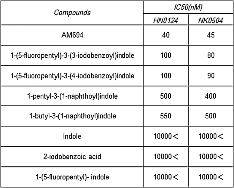
3-(n-iodobenzoyl)indole (n=2, 3, or 4) structure appears necessary for antibody recognition. In addition, the MAb that was produced from the HN0124 hybridoma has the highest affinity for AM694 (IC50=40 nM).
In future studies, the anti-AM694 MAbs that were obtained in this study will be used as an immunosensor for AM694 and evaluated on real samples.