
Abstract
Nanobodies (or VHHs) are the smallest antigen-binding domain of heavy chain antibodies, which naturally occur in camelidae. The small size, monomeric nature, low immunogenicity, high solubility and stability, as well as high affinity to target in nanomolar range, makes nanobodies a promising tool for diagnostic and therapeutic application. In the present study, we developed and identified the nanobody against human IgG from an immune library using phage display technique. For this goal, we performed four rounds of selection procedures on immobilized human IgG through biopanning. A clone named R1Nb was selected and expressed as His-tagged protein, and purified by nickel affinity chromatography. In addition, R1Nb was further characterized for binding specificity and affinity. Results demonstrated that R1Nb was highly specific for human IgG and its constant affinity was about 7.5 nM. Taken together, our achieved results indicate the potential of R1Nb as a promising tool in research and may be used for therapeutic purposes as a fusion part of target-specific nanobody.
Introduction
R
In this study, we generated, characterized, and purified the first example of human IgG-specific nanobody by phage display. In addition, we evaluated the specificity and affinity of the selected nanobody, named R1Nb, to bind to human IgG.
Materials and Methods
Materials
Human IgG was purified from human serum by protein A affinity chromatography and the purity was checked by SDS-PAGE (data not shown). Anti-M13HRP conjugated monoclonal antibody and anti-mouse HRP-conjugated were purchased from Amersham-Pharmacia-Biotech (Vienna, Austria). Anti-hemagglutinin A (anti-HA) was from Roche (Mannheim, Germany). 4-choloro-1-naftol (4-CN) substrate and goat anti-human IgG-HRP antibody were purchased from Sigma (St. Louis, MO). The nickel nitrilotriacetic acid (Ni+-NTA) resin was from Qiagen (Hilden, Germany). PHEN-4 and PHEN-6c vectors were generously provided by Serge Muyldermans (Laboratory of Cellular and Molecular Immunology, Vrije University Brussel, Brussels, Belgium).
Library construction
A young male camel (Camelus dromedarius) was injected six times every week with about 1 mg of human IgG. Antigen resuspended in PBS and mixed with equal volume of complete Freund's adjuvant for the first injection, and incomplete Freund's adjuvant for the booster immunization. 100 mL of blood sample were collected 1 week after the last immunization. Peripheral blood lymphocytes were isolated using density-gradient medium (Histopaque®-1077-Sigma). Total mRNA were extracted from lymphocytes using RNXplus (SinaClon, Tehran, Iran) and cDNA synthesized with reverse transcriptase (Fermentas, Espoo, Finland). The gene fragments encoding VHHs were amplified by nested PCR(8) and digested with NotI and PstI restriction enzymes. The consequent fragments were ligated into PHEN-4 phagemid vector and finally transformed into electro-competent TG1 Escherichia coli and plated on LB agar containing ampicillin. VHH library displayed on phage after VCSM13 helper phage infection.
Antigen panning
Biopanning procedure was performed on immobilized human IgG through four consecutive rounds. In the experiments, one well of a 96-well plate (Nunc, Maxisorp, Denmark) was coated with 10 μg/well of human IgG and one well used as control in sodium bicarbonate buffer incubated at 4°C overnight. The next day wells were blocked with 4% skim milk and incubated 2 h at room temperature (RT). Wells were washed with PBST (0.5% (V/V) Tween-20 in PBS), and 1012 colony-forming unit (CFU) of phage library in a final volume of 100 μL was added to the wells and incubated for 1 h at RT. After washing with PBST, the bounded phages were eluted by 100 μL of freshly prepared 100 mM triethylamine (TEA, pH 10.0). After 10 min incubation, wells were neutralized with 1 M Tris-HCL (pH 8.0). For further rounds of panning, eluted phages were amplified by infecting exponentially growing TG1 E. coli with helper phage super-infection. Moreover, output and input phages were titrated through infecting the log-phase TG1 with 10-fold serially diluted phage particles plated on TYE medium containing ampicillin. Four consecutive rounds of panning were performed as described above and stringency of selection procedure was increased with each panning round through increasing Tween-20 concentration (0.5, 1, 2, 4%).
Polyclonal phage ELISA
To monitor the progress of biopanning, polyclonal phage ELISA was performed on phages pooled at each round of panning. In brief, a 96-well plate was coated overnight with 10 μg/mL of human IgG in sodium bicarbonate at 4°C. The wells were blocked with 4% skim milk and incubated for 1 h at RT. After washing with PBST, 1011 CFU of panning output phages were transferred to wells and incubated for 1 h at RT. After washing steps were conducted five times, wells were incubated with anti-M13-HRP (1:3000) for 1 h at RT. Peroxide activity was detected by TMB (3,3′,5,5′-tetramethylbenzidine). Finally, the reaction was stopped with 2 N H2SO4 and optical density (OD) was measured at 450nm.
Periplasmic extract ELISA
The human IgG-specific phage clones were identified by periplasmic extract ELISA (PE-ELISA) using anti-hemagglutinin (anti-HA) antibody. Over 45 individual colonies from the third and fourth rounds of panning were selected, and nanobodies were expressed to the periplasmic space of log-phase TG1 with 1 mM IPTG (isopropyl D-1-thiogalactopyranoside). The fusion nanobody-PIII was extracted through osmotic shock and detected by anti-HA (1:2000) followed by anti-mouse-HRP (1:5000). The positive clones were DNA sequenced to identify the unique nanobody gene.
Expression of soluble nanobody
For expression of selected nanobody, the VHH genes were amplified using A6E and 38 primers,(8) digested with BstEII and PstI restriction enzymes, and sub-cloned into PHEN6c expression vector. The recombinant construct was transformed into competent E. coli WK6 cells. Expression of nanobody was induced with 1 mM IPTG for overnight incubation at 28°C with shaking at 200–250 rpm. The bacterial pellet was collected by centrifugation at 8000g for 30 min. The periplasmic proteins were extracted by osmotic shock and loaded on nickel nitrilotriacetic acid (Ni+-NTA) affinity chromatography column (Qiagen, Hilden, Germany). The nanobody was eluted with 500 mM imidazole and dialyzed against PBS 1X.
Western blot analysis
The purified nanobody was separated by Coomassie Brilliant Blue stained 15% SDS-PAGE under reducing conditions. For Western blotting, the protein bands were transferred to nitrocellulose membrane and blocked with 4% skim milk for 16 h at 4°C.The membrane was washed with PBST and nanobody detected using anti-His-HRP conjugated antibody (1:500) and subsequently developed by 4-choloro-1-naftol (4-CN) substrate (Sigma).
Binding specificity
Cross-reactivity of selected nanobody with various antigens including human vascular endothelial growth factor, mouse vascular endothelial growth factor, epidermal growth factor, casein, BSA (bovin serum albumin), skim milk, and bevacizumab (Roche, Basel, Switzerland) were determined by ELISA. One μg/mL of the mentioned antigens were coated on 96-well plates and incubated overnight at 4°C, and the ELISA was developed with anti-His-HRP conjugated and TMB. Bevacizumab was used as positive control.
Affinity measurement
Affinity of the selected nanobody was determined according to the Beatty method.(9) A 96-well plate was coated with two different concentrations (1 and 0.1 μg/mL) of hIgG or BSA (as negative control) and incubated at 4°C overnight. After blocking, serially diluted nanobody was transferred to the wells and incubated for 1h at RT. The plate was washed and developed by TMB as described before. The affinity constant (k
aff
) was determined using the Beatty's equation:
Results
Enrichment of nanobody library
The library was constructed using standard procedure.(8) The constructed nanobody library had 8×10⁷ colony with 92% right size of gene insert. The nanobody library was screened after four consecutive panning rounds on immobilized antigen, and enrichment experiment was evaluated by polyclonal phage ELISA. Results evaluated a significant increase in signal after the third round of panning (Fig. 1). Also, enrichment estimated by comparing phage titer in the positive (well coated with human IgG) and negative well (well without antigen) after each round of panning (Table 1).
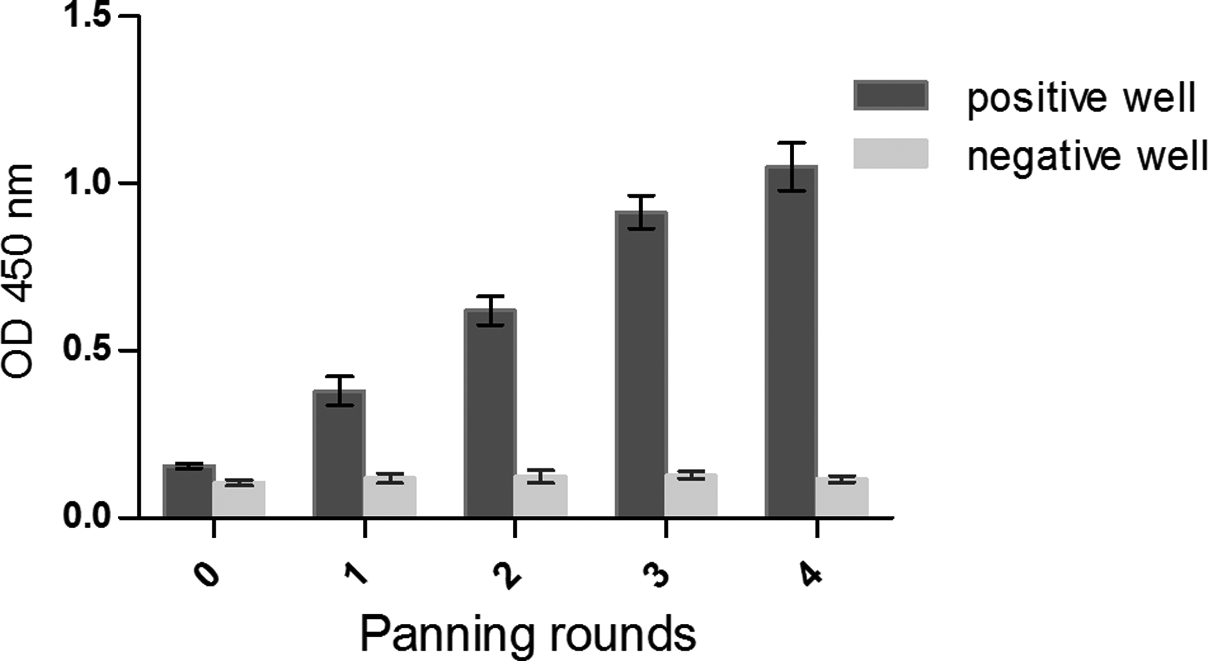
Polyclonal phage ELISA results. The highest signal intensity was observed after third and fourth rounds of panning. Error bar represent mean±SD of triplicate assay.
Enrichment ratio was determined by input and output phages titration. cfu, colony-forming unit.
Screening of human IgG-specific VHH by PE-ELISA
Periplasmic extract results showed that 8 of 45 screened clones were specific to immobilized human IgG and showed high signal in ELISA experiment. All positive clones were sequenced and their sequences were aligned using MEGA5 multiple sequence alignment program.(10) Sequence analysis evaluated three unique sequences that had been selected (data not shown). One clone, named R1Nb, was chosen for soluble expression and further characterization analysis.
Expression and purification
C-terminal His-tagged nanobody was expressed in periplasmic space of WK6 E. coli with 1 mM IPTG and purified using immobilized metal affinity chromatography by Ni+-NTA resin. The purified protein dialyzed against PBS to remove the rest of imidazole. The purity of R1Nb nanobody was evaluated with Coomassie Brilliant Blue stained 15% SDS-PAGE under reducing conditions (Fig. 2A). Western blot analysis was performed with anti-His HRP. Western blot detected a 15 kDa band of nanobody, which confirmed successful expression of R1Nb nanobody (Fig. 2B).
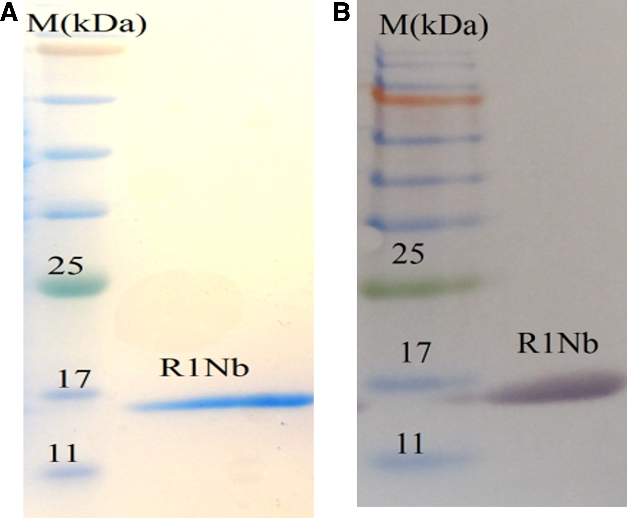
Purification results of R1Nb. (
Specificity and affinity
Specificity and cross-reactivity of R1Nb with a variety of proteins were determined by ELISA. As can be seen in Table 2, R1Nb was highly specific for human IgG antigen and did not cross-react with other antigens. Affinity of R1Nb was determined by enzyme-linked immunosorbent assay as described by Beatty and colleagues.(9) Analysis revealed that the constant affinity for R1Nb was 7.5 nM.
The assay was performed in triplicate; data represent mean of OD value±standard deviation. Selected nanobodies specifically react with human IgG and do not react with other antigens (hVEGF, mVEGF, EGF, skim milk, casein, and BSA). hVEGF, human vascular endothelial growth factor; mVEGF, mouse vascular endothelial growth factor; EGF, epidermal growth factor; BSA, bovine serum albumin.
Discussion
In this study, we employed dromedary immune VHH library and generated and characterized specific nanobody against human IgG using phage display technique. We performed four biopanning selection procedures on immobilized human IgG. To obtain anti-human IgG nanobody with high affinity and specificity, we increased the stringency of the biopanning procedure. The progress of panning was monitored by polyclonal phage ELISA and results of polyclonal phage ELISA revealed the success of the panning. Enrichment of nanobody library was estimated through input and output phage comparison. Periplasmic extract ELISA (PE-ELISA) was performed on immobilized human IgG to select the positive binders from the non-specific binders. In the screening process, eight clones that showed high signal intensity in ELISA were selected and sequenced. Sequencing results revealed that three unique nanobodies were selected according to their CDR3 site. A clone, called R1Nb, was selected and sub-cloned into pHEN6c expression vector to produce soluble nanobody. His-tagged fusion nanobody was purified using nickel affinity chromatography. Specificity results showed that R1Nb was highly specific for its antigen (human IgG). The calculated affinity for R1Nb was about 7.5 nM.
The single domain nature of nanobodies makes them good therapeutics to recognize the hinder region of tumor-specific solid tumors, which are not recognized by conventional monoclonal antibodies (MAbs).(2) Moreover nanobodies can be expressed in bacteria with high yield; therefore their production is cheaper than conventional MAbs, which are normally produced in mammalian cells. Many studies have developed different nanobodies against various antigens with various diagnostic or therapeutic applications.(11–17) In our laboratory the nanobodies have been developed as a research tool and for use in diagnostic or therapeutic application.(12,17) Despite the benefit and advantages of nanobodies over conventional MAbs, they have limited in vivo half-life because of their small size.(5) The small size and single domain of the nanobody allow them to combine two or more different antigen-binding nanobodies in one molecule.(7) In some studies, it has been evaluated that the combination of nanobody with albumin-binding nanobody improved in vivo half-life extension.(6) Combination of antigen-specific nanobody with IgG-binding nanobody is another approach for increasing the in vivo half-life of the nanobody. For this reason, the main aim of this study was isolation and identification of human IgG nanobody for the first time. It is suggested that in the future the combination of the anti-human IgG nanobody with various nanobodies will improve their half-life.
Conclusion
In conclusion, the anti-human IgG nanobody named R1Nb is easily isolated from immune VHH library by phage display technique. To this end, R1Nb showed very high specificity and affinity to its target (human IgG). Therefore R1Nb has potential as a research tool and can be used for diagnostic or drug development aims.
Footnotes
Acknowledgments
We acknowledge with deep respect the Pasteur Institute of Iran for funding this research.
Author Disclosure Statement
The authors have no financial interests to disclose.