




Abstract
When abrin-a was combined with several polyclonal antibodies (PAb), the detection limit could be increased. In this way, a monoclonal antibody (capture) and polyclonal antibody (detection) sandwich enzyme-linked immunosorbent assay (ELISA) and a colloidal gold-PAb conjugate-based immunochromatographic assay for detection of abrin-a were developed. The ELISA had a detection limit of 3.9 ng/mL for abrin-a in standard solution and 7.8 ng/mL in soybean milk, and was more sensitive than polyclonal antibody (capture) and monoclonal antibody (detection) ELISA, which had a detection limit of 15.6 ng/mL. The test strip had a detection range of 50 to 500 ng/mL for abrin-a and a detection limit in standard solution or soybean milk samples of 50 ng/mL. However, the test strip had a reduced detection capability compared with a colloidal gold-monoclonal antibody conjugate-based immunochromatographic assay test strip, which had a lower detection limit of 10 ng/mL. The developed ELISAs and test strip show the specificity towards abrin-a and have no cross-reactivity towards abrin-b, -c, -d, ricin, or the agglutinins from either castor beans or rosary peas.
Introduction
A
Despite the toxicity of abrin-a, few in vitro methods for its detection have been published. Immunological detection methods for abrin-a include radioimmunoassay (RIA) and enzyme-linked immunosorbent assay (ELISA).(6–9) Colloidal gold-based immunochromatographic assays provide a rapid, simple, and inexpensive means for detection and have been employed in the detection of proteins, nucleic acids, hormones, pesticides, drugs, and toxic compounds.(10,11) In this format, abrin-a can only combine with one MAb; however, if abrin-a could be combined with several polyclonal antibodies (PAb),(12) the detection limit could be increased. In this study, a monoclonal antibody (capture) and polyclonal antibody (detection) sandwich ELISA and a colloidal gold-PAb conjugate-based immunochromatographic assay for detection of abrin-a were developed, and the characteristics of the ELISA and test strip were analyzed.
Materials and Methods
Materials and reagents
Abrin-a, -b, -c, and -d were purified from the seeds of Abrus precatorius L.(4) Abrus agglutinin, ricin, and ricin agglutinin were obtained from Sigma (St. Louis, MO). The mouse anti-abrin-a MAb and rabbit anti-abrin-a PAb were produced in our laboratory.(9) The goat anti-rabbit IgG, horseradish peroxidase (HRP)-labeled goat anti-rabbit IgG, and HRP-labeled goat anti-mouse IgG was purchased from Beijing Dingguo Biotechnology Development Center (Beijing, China).
Nitrocellulose (NC) membranes were purchased from Sartorius (Gottingen, Germany). Glass paper was obtained from Ahlstrom (Helsinki, Finland). PVC sheets, absorbent paper, and adhesive tape were purchased from Shanghai Jiening Bio (Shanghai, China). HAuCl4·3H2O, sodium citrate, and bovine serum albumin (BSA) were from Sigma. The 96-well ELISA plates were from Corning Costar (Corning, NY). Other reagents were of analytical purity, and double distilled water was used for all experiments.
ELISA analysis for abrin-a
As abrin-a is a 64 kDa glycoprotein consisting of two polypeptide chains, sandwich ELISAs consisting of monoclonal capture and rabbit polyclonal detector antibodies (mono-poly) and rabbit polyclonal capture and monoclonal detector antibodies (poly-mono) were developed. For the mono-poly ELISA, 100 μL of 2 μg/mL mouse anti-abrin-a MAb in 50 mM carbonate buffer (pH 9.6) was incubated in ELISA plates at 4°C overnight. After washing with PBS, the wells were blocked with 3% BSA (w/v) in PBS (pH 7.4) for 1 h at room temperature. After removing the blocking reagent, 100 μL abrin-a standard solutions were prepared by serial dilution of 1 μg/mL abrin-a solution from 1× to 2048×, respectively, with PBST (containing 1% BSA (w/v), 2 μg/mL rabbit anti-abrin-a PAb) and incubated for 1 h at 37°C under shaking, and were added and incubated for 1 h at 37°C. After washing, 100 μL HRP-labeled goat anti-rabbit IgG was added and incubated for 1 h at 37°C. After washing, the enzymatic reaction was developed with TMB substrate (Sigma) and stopped by adding 1 M H2SO4. The optical density was measured at 450 nm (OD450) using a microtiter plate reader. The detection limits of the sandwich ELISA was determined as the ratio of OD450 values of samples and negative control toward 2.1. (9) For the poly-mono ELISA, after incubating 100 μL 2 μg/mL rabbit anti-abrin-a PAb in ELISA plates at 4°C overnight, the wells were blocked with 3% BSA (w/v) in PBS (pH 7.4) for 1 h at room temperature. After washing, abrin-a standard solutions were prepared by serial dilution of 1 μg/mL abrin-a solution from 1× to 2048×, respectively, with 1% BSA (w/v) in PBST, and were added to the wells (100 μL per well) and incubated for 1 h at 37°C. After washing, 100 μL 2 μg/mL mouse anti-abrin-a MAb (diluted in PBST with 1% BSA (w/v)) was added to each well and incubated for 1 h at 37°C. Plates were washed and incubated with 100 μL HRP-labeled goat anti-mouse IgG for 1 h at 37°C. After washing, the enzymatic reaction was developed and read at 450 nm.
Preparation of colloidal gold and colloidal gold probe
The procedure used to prepare the 40 nm colloidal gold suspension has been described previously.(13) Briefly, 1 mL of a 1% (w/v) sodium citrate solution was added to 100 mL boiling deionized water. When the mixture was heated to boiling again, 1 mL of a 1% (w/v) HAuCl4·3H2O solution was added under constant stirring. After the color of the solution changed to wine-red (in about 2 min), the solution was boiled for another 10 min. The heating source was then removed, and the solution was restored to the original volume after cooling. The obtained gold colloidal was supplemented with 0.02% (w/v) of sodium azide and stored at 4°C. The particle diameter was checked by transmission electron microscopy (TEM, H-7650).
The colloid gold solution was adjusted to pH 8.8 using 0.02 M K2CO3. With gentle stirring, a total of 0.06 mL of purified 4 mg/mL rabbit anti-abrin-a PAb was added dropwise to 10 mL of colloidal gold solution. The mixture was gently mixed for 10 min, then blocked with 1.1 mL of 10% (w/v) PEG 20000. The mixture was gently mixed for 15 min and centrifuged at 10,000 g for 30 min. The gold pellets were suspended in 1 mL dilution buffer (20 mM Tris/HCI buffer (pH 8.8) containing 1% (w/v) BSA, 3% (w/v) sucrose, and 0.02% sodium azide (w/v)), and stored at 4°C until use.
Immunochromatographic test strips for abrin-a
As abrin-a is a 64 kDa glycoprotein consisting of two polypeptide chains, a sandwich immunoassay format was used. The preparation and assembly of immunochromatographic test strips are described as follows (Fig. 1A). BIODOT (XYZ-3) was used to dispense two lines on the NC membrane (25 × 300 mm). Briefly, mouse anti-abrin-a MAb (1.2 mg/mL) was dispensed around the bottom as the test line (1 μL/1 cm line) while goat anti- rabbit IgG (1 mg/mL) was dispensed at the upper position as the control line (1 μL/1 cm line). The distance between the two lines was 5 mm. The conjugate pad was made of glass fiber and was treated with 0.1% Tween-20 for 10 min and dried at 60°C, saturated with the colloidal gold probe, and dried for 1 h at 37°C. The sample pad was saturated with 50 mmol/L phosphate-buffered saline (PBS) solution (pH 7.2), containing 0.3% Tween-20 and 0.5% (w/v) PVP, and dried at 37°C. The NC membrane, gold conjugate pad, sample pad, and absorbent pad were laminated and pasted onto the PVC plate, which was then put into the strip card. All strips were sealed in a plastic bag and stored at 4°C until use.
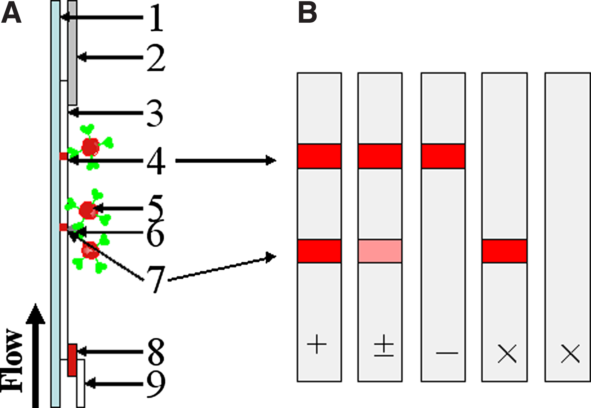
Schematic of immunochromatographic assay for abrin-a. (
Each 100 μL portion of standard solution with different concentrations was pipetted into the well of the sample pad of the test strip. When abrin-a is present in the sample solution, abrin-a and the colloidal gold probe combine to form a conjugate that would be captured by the mouse anti-abrin-a MAb (test line). Excess colloidal gold probe would react with the goat anti-rabbit antibody (control region), and two red bands on the test line and control line would appear. However, if there were less abrin-a in the sample solution, fewer colloidal gold probe conjugates would form and be captured on the test line. Thus, the degree of density of gold color on the test line would be weaker. Therefore, if the sample solution contains no abrin-a or very low levels of abrin-a, only the control line will be visible. If no line is present in the test strip or only one line is observed in the test region, the test is considered invalid (Fig. 1B).
Cross-reactivity of ELISA and test strip
Samples containing 1 μg/mL of abrin-b, abrin-c, abrin-d, abrus agglutinin, ricin, and ricin agglutinin were also assayed by mono-poly ELISA and test strip for purposes of evaluating cross-reactivity.
Analysis of abrin-a in simulated samples
Soybean milk obtained from a local supermarket was spiked with different concentrations of abrin-a for mono-poly ELISA and test strip analysis, with three repetitions for each sample.
Results and Discussion
ELISA analysis for abrin-a
A dynamic range was determined by the mono-poly ELISA for abrin-a standard concentration range of 3.9 to 62.5 ng/mL (Fig. 2). The detection limit of the poly-mono ELISA was 15.6 ng/mL, which was lower than mono-poly ELISA, but had a wider dynamic range friom 16.5 to 500 ng/mL. Compared with monoclonal detector antibodies, abrin-a loaded higher amounts of polyclonal detector antibodies. Thus the signal amplification of the colored product related to the concentration of the target was enhanced significantly. The sensitivity of the mono-poly ELISA was four times higher than that of poly-mono ELISA. The ELISA had no cross-reactivity with abrin-b, abrin-c, abrin-d, abrus agglutinin, ricin, and ricin agglutinin when 1 μg/mL of each toxin was tested, whereas the detection limit for abrin-a in soybean milk was 7.8 ng/mL. Although the soybean milk reduced the sensitivity of the ELISA, the increases in detection limit did not affect the utility of the assays to detect the toxin at levels less than those that would pose a health threat.
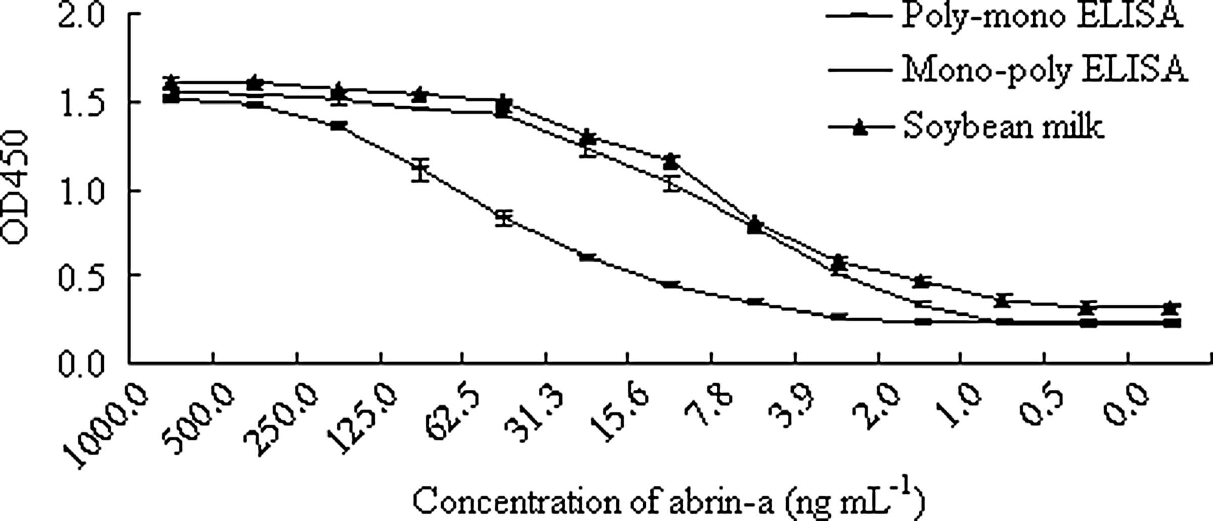
ELISA for abrin-a. Results represent the mean ± standard error of three replicates.
Characterization of colloidal gold particles and colloidal gold probe
The TEM images of the prepared colloidal gold probe revealed that the diameter distribution range of colloidal gold particles was approximately 35–40 nm (Fig. 3A). The optimum pH of the colloidal gold probe was determined by 10% NaCl (w/v).(14) Fifty μg rabbit anti-abrin-a polyclonal antibody was added to 1.0 mL colloidal gold solution with the pH adjusted using 0.02 M K2CO3. The volumes of these samples were adjusted to 1.040 mL with double distilled water and 100 μL of 10% NaCl (w/v) solution was added to each, agitated after 60 min. The minimum pH effective to prevent aggregation was pH 8.8. The pH for preparation of colloidal gold-PAb conjugates was higher than that for the monoclonal antibody conjugates, which has an optimum pH of 8.2.(13) This may be due to the polyclonal antibody conjugate containing a large number of different antibodies against different antigenic determinants of abrin-a, while as a uniform antigen captor, the monoclonal antibody should have a single optimum pH for conjugation to colloidal gold particles. At a pH of 8.8, the optimum amount of rabbit anti-abrin-a polyclonal antibody coated on the colloidal gold was 24 μg/mL selected by 10% NaCl (w/v). The TEM images of the prepared colloidal gold probe are shown in Figure 3B.
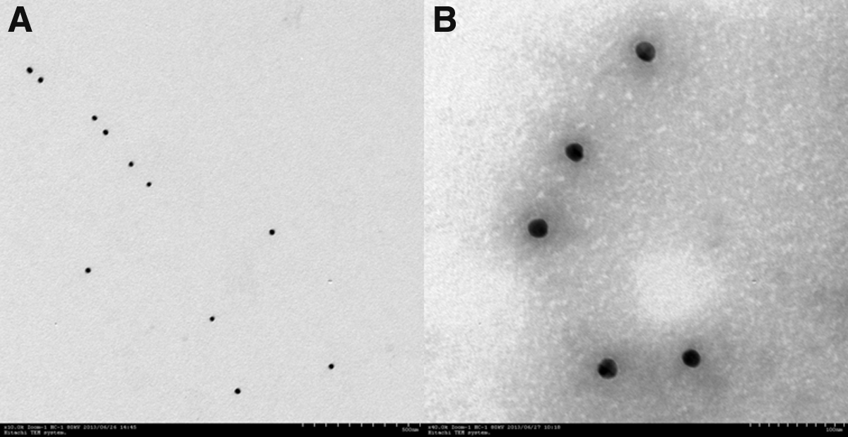
Transmission electron microscopy (TEM) image of colloidal gold and colloidal gold probe. (
Immunochromatographic test strips for abrin-a
Abrin-a standard solutions at concentrations of 0, 10, 50, 100, and 1000 ng/mL were assayed by the test strip. The colloidal gold-PAb conjugate-based test strip demonstrated that the red color intensity was proportional to abrin-a in the range of 50–500 ng/mL (Fig. 4A), and the detection limit was 50 ng/mL. However, the test strip using the colloidal gold-MAb conjugate demonstrated a wider range of 10 to 1000 ng/mL, with a more sensitive detection limit of 10 ng/mL (Fig. 4B). Mono-poly ELISA had a better detection limit for abrin-a, because abrin-a was effectively captured by the amount of rabbit anti-abrin-a PAb after incubation for 1 h at 37°C under shaking. Thus, there should be no steric hindrance for MAb capturing of the conjugate formed by abrin-a and PAb. Considering that the PAbs were prepared for colloidal gold probes, they may contain high levels of interfering IgG for abrin-a, that could make abrin-a have a lower association rate with PAb colloidal gold probes and a low level of detection sensitivity may result. (15) It suggests that an earlier treatment by affinity chromatography to purify abrin-a specificity PAb was necessary for the test strip. Additionally, in the visual assay in nitrocellulose, signal is measured from only approximately the top 10 μm of the NC membrane, and the remaining signal in the system is lost. While the abrin-a colloidal gold-PAb conjugate, as a giant molecule conjugate, would tend to bind with the monoclonal antibody in the bottom layer of the NC membrane, it also could cause the lower detection limit.(16) The cross-reactivity of the test strip was analyzed using several toxins such as abrin-b, abrin-c, abrin-d, abrus agglutinin, ricin, and ricin agglutinin. When 1 μg/mL of each toxin was tested, no band was revealed in the test region. The test strip was used to detect abrin-a in soybean milk. False negatives were detected by the test strip (detection limit of 50 ng/mL) (Table 1), even though the simulated samples should have a weak influence on the test strip. To get an exact result, analysis using the test strip should be performed more than two times.
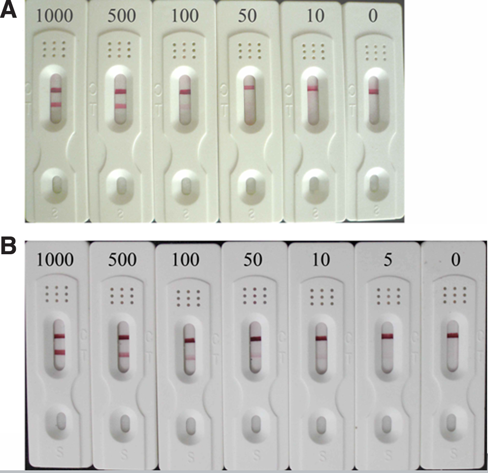
Detection limit of the test strip. Abrin-a standard solutions at concentrations of 0, 10, 50, 100, 500, and 1000 ng/mL were assayed. (
+, positive, a bright visible red bands on test line; ±, positive, a weak visible red bands on test line; –, negative, no visible red band on test line.
Conclusion
In this study, a monoclonal antibody (capture) and polyclonal antibody (detection) sandwich ELISA and a colloidal gold-PAb conjugate-based immunochromatographic assay for detection of abrin-a were developed. A dynamic range was determined by the mono-poly ELISA for abrin-a standard concentration range of 3.9 to 62.5 ng/mL. The detection limit of the mono-poly ELISA was 7.8 ng/mL for abrin-a in soybean milk. With the immunochromatographic assay, the red color intensity was proportional to abrin-a in the range of 50 to 500 ng/mL, with a detection limit of 50 ng/mL.
Footnotes
Acknowledgments
This work was supported by the National “863” Project (2007AA10Z440 and 2007AA10Z426) of China. The authors acknowledge the assistance of International Science Editing.
Author Disclosure Statement
The authors have no financial interests to disclose.