
Abstract
Kallikrein-related peptidase 6 (KLK6) is a serine protease, and one of fifteen kallikrein members located on chromosome 19. KLK6 is associated with the regulation of axonal growth following spinal injury, tumor cell metastasis, and alpha synuclein aggregate pathologies like Parkinson's, making KLK6 a potentially important biomarker. We generated a KLK6 expression vector for large-scale expression in Escherichia coli. After affinity chromatography purification and SDS-PAGE confirmation, mice were immunized with the purified protein for development of monoclonal B cell populations. Our immunizations generated five hybridomas (1D8, 2E3, 3B7, 5G8, and 5H12) against KLK6. Isotyping analysis revealed that clone 2E3 was IgG2b, while the other four clones were IgG1. Further studies found that clone 5G8 could be used to visualize specific KLK6 bands by Western blot analysis from ovarian cancer patient serum and plasma, and also in mouse liver lysates. Finally, we established a sandwich ELISA pair and determined their sensitivity for KLK6 to be 10 ng/mL. These findings establish an essential tool for the detection and analysis of KLK6.
Introduction
I
At present, the neurological studies of KLK6 have been focused primarily in disease and pathological states given its general association with neurodegenerative diseases. Decreased cerebrospinal fluid levels of KLK mark a possible new risk factor for Alzheimer's disease, not to mention a potential diagnostic tool.(6–8) KLK6's role in tumorigenesis has also been evaluated in diseases like ovarian and breast cancer. Studies by Ghosh and colleagues have found that recombinant KLK6 could lead to the degradation of fibrinogen, gelatin, fibronectin, and laminin, thereby promoting tumor cell invasion and metastasis.(9) These findings were further supported by recent data in studies by Klucky and colleagues.(10) Additionally, KLK6's expression enhanced colon cancer cell migration through laminin and Matrigel, implying a potential role in tumor metastasis and extravasation from the original tumor environment.(11)
KLKs may also prove to be promising markers for ovarian cancer, as KLK6 is seen at increased levels in cancerous ovarian tissues and its expression is negatively correlated with ovarian cancer prognosis.(12) Serum KLK6 levels are elevated in ovarian cancer patients; this level is independent of CA125 expression and could therefore serve as an auxiliary diagnostic marker.(13,14) KLK6 levels are increased in primary breast cancer cell lines, contrasting with metastatic breast cancer cell lines, which show lower KLK6 expression.(15) KLK6 expression in tumors is significantly reduced due to the hypermethylation of specific CpG sequences located in proximal promoter.(15) Additionally, several studies have recently identified abnormal expression of KLK6 in pancreatic ductal adenocarcinoma, gastric cancers, and colon cancers, suggesting its role in tumorigenesis may not be restricted to ovarian and breast cancers.(16–18)
In order to best study KLK6 in samples, however, it is necessary to develop specific KLK6 monoclonal antibodies (MAb) and diagnostic platforms to detect its distribution and levels in various cell lines, tissues, and in vivo samples. In this report, we developed MAbs against a recombinant KLK6 protein. These MAbs were shown to be specific for KLK6 via Western blotting, and were also used to detect the KLK6 antigen in a sandwich antibody based enzyme-linked immunosorbent assay (ELISA). The development of an ELISA specific platform allows for the quantitative measurement of the KLK6 protein levels in human samples and should help lead to future clinical testing methods for neurologic disease patients, as well as cancer patients.
Materials and Methods
Materials
The expression vector pET28a (5369bp) was acquired from Novagen (Darmstadt, Germany), and Escherichia coli BL21 was acquired from Takara (Dalian, China). Primers were synthesized Genewiz (Suzhou, China), and DMEM medium and fetal bovine serum were purchased from Gibco (Guangzhou, China). KLK3, KLK6, KLK10, KLK11 proteins, and the Rapid Mouse Isotyping Kit-Gold Series, LFM-ISO, were purchased from RayBiotech (Guangzhou, China). BCA Protein Assay Kit was purchased from Thermo Scientific (Waltham, MA). HiTrap Protein A HP was purchased from GE Healthcare (Atlanta, GA). All other chemicals and reagents used throughout the experiments were from Sigma-Aldrich (St. Louis, MO).
Animal experiments
All animal work was performed according to relevant national and international guidelines. The experiments were approved by the Institutional Animal Care and Use Committee at South China Agricultural University (certification no. CNAS BL0011).
Cloning of KLK6 gene
The cloning procedure was carried out according to standard techniques.(19) A KLK6 PCR product was generated using forward primer (5′-AAACATATG GAGGAGCAGAATAAGTTG-3′) and reverse primer (5′-AAACTCGAGTCA CTTGGCCTAATGGTTT-3′), which included the desired fragment of KLK6 protein and Nde I and Xho I excision sites to allow insertion in the pET28a expression vector. The construct pET28a/KLK6 was verified by PCR with specific primers (5′-AAA
Protein expression
Two colonies were selected for small-scale expression analysis. After induction with 0.8 mM isopropyl β-D-1-thiogalactopyranoside (IPTG) for 4 h at 37°C, the bacterial lysates were sonicated, separated by high speed centrifugation (20,000 g, 20 min at 4°C), and analyzed by SDS-PAGE (12% polyacrylamide gel).(20) The recombinant construct pET28a/KLK6 was transformed into E. coli (DE3), the transformants were inoculated into 0.5 L Luria-Bertani (LB) broth medium supplemented with 50 μg/mL kanamycin, and then grown at 37°C for large-scale protein expression. The culture was induced by the addition of 0.8 mM IPTG until the A600 reached 0.6. The cells were harvested after incubation at 37°C for 4 h; pellets were washed twice with cold PBS, and then kept frozen at −80°C until use.
Protein purification
Bacterial cell pellets were resuspended (2.5 mL/g wet weight of pellet) in lysis buffer containing lysozyme and DNase. After 20 min on ice, the pellet lysate was subjected to sonication to break cells and centrifuged at 20,000 g for 30 min at 4°C to remove cell debris. The precipitate was successively dissolved in 4 and 8 M urea with stirring at room temperature for 30 min and centrifuged at 20,000 g for 10 min at 4°C. The soluble fraction was loaded to a His-Bind Nickel column (GE) to purify the protein according to the manufacturer's recommendations. The eluted fractions were pooled and desalted immediately through dialysis in desalting buffer (20 mM Tris-HCl [pH 8.0], 0.5 M NaCl). The purity of the KLK6 protein was determined by SDS-PAGE.
Preparation of anti-KLK6 monoclonal antibodies
The immunization protocol consisted of small immunogen doses occurring three times over 6 weeks. Female BALB/c mice (6–8 weeks old; Guangdong Medical Laboratory Animal Center, China) were subcutaneously injected with 100 μg KLK6 protein emulsified with equal volume of Freund's complete adjuvant for initial immunization. The mice received the same injections with 50 μg antigen mixed with incomplete Freund's adjuvant at intervals of 2 weeks following primary immunization. Immunity was assessed by indirect ELISA after the third immunization. Finally, a booster immunization was given intraperitoneally with 50 μg antigen at 3 days before cell fusion, when titers of anti-serum were highest.
Immunized mouse splenocytes and myeloma sp2/0 cells (Center of Laboratorial Animals in South China Agricultural University, Guangzhou, China) were washed twice with serum-free DMEM medium, mixed at a ratio of 5:1 in a 50 mL centrifuge tube, and then had a slow addition of 1 mL 50% polyethylene glycol (PEG). Cells were incubated for 1 min, at which point 30 mL DMEM medium were added gently to quench PEG activity. Subsequently, the cells were centrifuged and resuspended in HAT medium containing 20% serum and then added to 96-well plates. Three days after fusion, half of culture supernatant was replaced with HAT medium; 7 days later, the entire supernatant was switched to HT medium containing 20% serum. After 3 days in HT medium, the culture supernatants were tested for activity in an indirect ELISA. Positive hybridoma cells were subcloned by limiting dilution. The resulting clones were validated for KLK6 specificity, expanded in culture, and stored in liquid nitrogen until needed.
Subtype identification of antibody
Antibody subclasses were screened via the Rapid Mouse Isotyping Kit-Gold Series (LFM-ISO, RayBiotech). Fifty μL hybridoma culture supernatant was added onto the sample pad where indicated or the strip was physically dipped into the culture supernatant. After 10 min, the strip was removed from the fluid and placed on a flat and clean surface to allow development of the red bands to indicate the respective Ig isotypes, as described in the manufacturer's manual.
Preparation of ascites and purification of MAb
Preparation of MAb was done by the ascites method, with the hybridoma injected into the peritoneum of mice. Briefly, female BALB/c mice (8–10 weeks old) were intraperitoneally treated with 0.5 mL saxoline, then 7 days later implanted with 1 × 10⁶ hybridoma cells. Ascites fluid was then collected by syringe and centrifuged to remove any impurities. At 4°C, ammonium sulfate (50% saturation) was added slowly with constant stirring. Solution was incubated at 4°C, stirred slowly for 30 min, and then allowed to stand at 4°C for 1 h. Sample was centrifuged again to remove supernatants and resuspended with 50 mL PBS. Purification was done by HiTrap Protein A HP affinity columns (GE Healthcare), according to the manufacturer's instructions.
Western blot analysis
Western blot analysis was performed as previously described(21) using the Li-Cor Odyssey infrared imaging system (Lincoln, NE). 0.1 μg of KLK6 protein or 40 μg of cell and tissue lysates were mixed with 5x sample loading buffer, heated at 100°C for 10 min, and separated by 12% SDS-PAGE.(20) Separated proteins in the gels were electrophoretically transferred onto nitrocellulose membranes at 200 mA for 2 h and incubated in blocking buffer (Odyssey) for 1 h. The membranes were then incubated with the anti-KLK6 antibodies at 1:1000 overnight at 4°C in PBS-T (PBS containing 0.05% Tween-20 [pH 7.4]). Membranes were then washed in PBS-T washing buffer three times. IRDye 800CW Donkey anti-mouse IgG (Li-Cor) was used as a secondary antibody at 1:15,000 in PBS-T and incubated for 1 h. Li-Cor Odyssey Imaging System was then used for detection of targets according to the manufacturer's instructions.
Sandwich ELISA
Two antibodies with distinct antigen binding sites were then identified. Polystyrene EIA/RIA flat bottom plates were coated overnight at 4°C with 100 ng of anti-KLK6 MAb. Plates were washed with PBS-T washing buffer, then blocked with PBS-T plus 5% skim milk (blocking solution) at 37°C for 2 h. Plates were washed and incubated for 1 h with sample or KLK6 recombinant protein diluted in the blocking solution. Following washing, biotin-MAb was added and incubated for 1 h, washed, then incubated with conjugated HRP-Streptavidin for 1 h. Plates were washed again and TMB substrate solution was added. The reaction was stopped 10 min later with 2 M sulfuric acid (100 μL/well, 0.5 mol/L). Absorbance at 450 nm was measured in a microplate reader (Biotek).
Results
Construction and expression of KLK6 vector
The constructed expression vector pET28a/KLK6 is depicted in Figure 1A, and contains a His-tag and the indicated genomic sequence. The KLK sequence we chose to use was Glu17-Lys244 based on the Uniprot Accession #Q92876. This sequence was confirmed by single colony PCR (Fig. 1B) and DNA sequencing. DNA sequences of KLK6 cloned and PET28a vector were digested and connected with Nde I and Xho I simultaneously. Finally, this gene was cloned into a PET28a vector containing a His-tag and expressed in E. coli.

Construction and confirmation of the recombinant plasmid pET28a/KLK6. (
The validated and E. coli inserted constructs were then subjected to a pilot expression test to determine both the level and purity of the KLK6 protein. As shown in Figure 2A, KLK6 protein was indeed expressed at reasonable levels in E. coli, and was expressed predominantly in the insoluble fraction (pellet, lanes 3 and 4), but not in the soluble fraction (culture supernatant, lanes 1 and 2). Further purification of the insoluble fraction via affinity chromatography resulted in recombinant KLK6 purity reaching at least 95%, as assayed by SDS-PAGE and Image J software analysis (Fig. 2B).
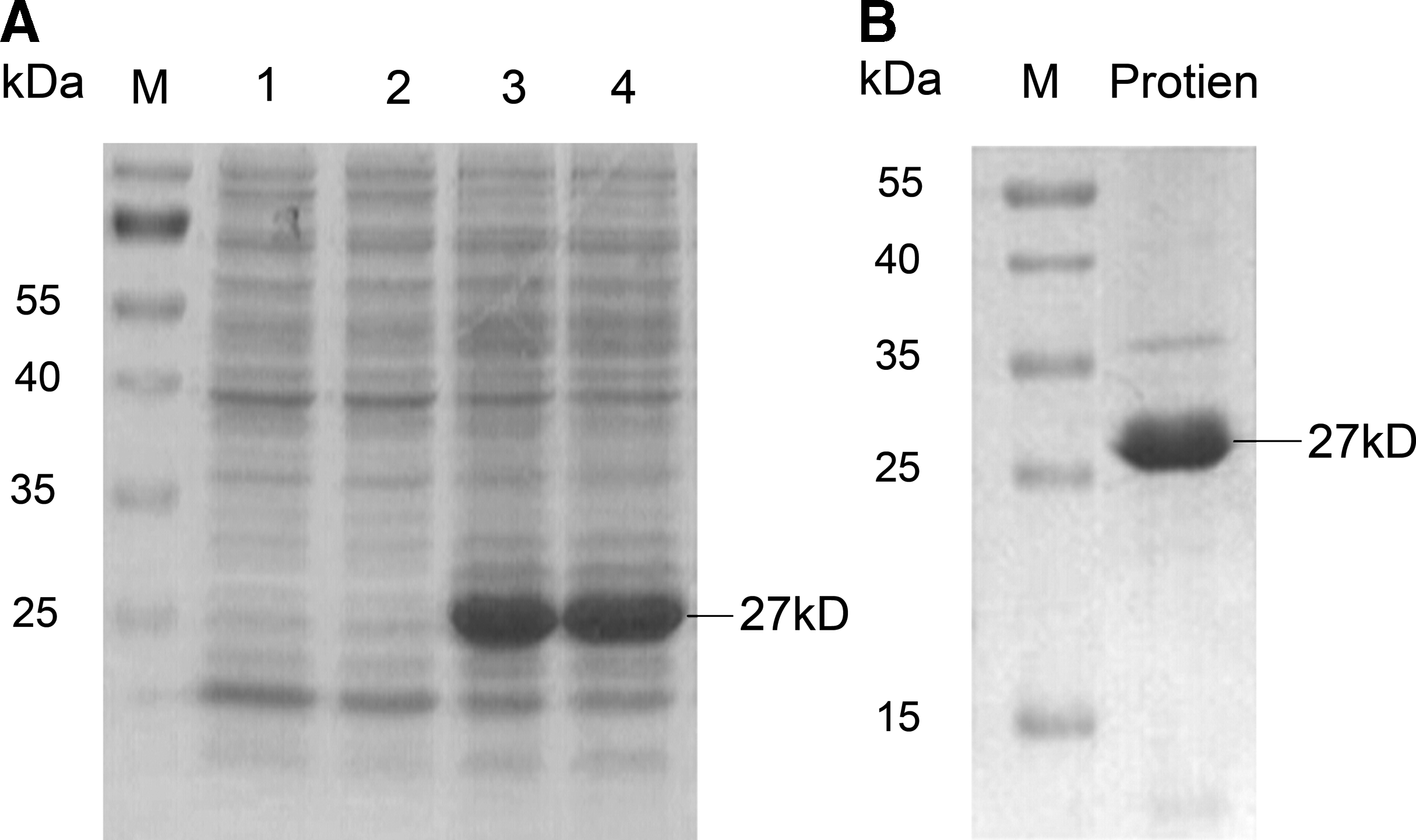
Expression and purification of KLK6 recombinant protein. (
Preparation of antibody
With the purified protein in hand, we set out to immunize mice in order to create monoclonal B cell populations. Mice were immunized, and antigen boosted for four rounds to allow the development and expansion of B cell populations against KLK6. Two mice were selected and tail bled to determine the KLK6 antibody titer by indirect ELISA. ELISA testing determined the titer of the mice to be 40,000 and 80,000, respectively (Fig. 3). The mouse with the highest KLK6 titer was then selected for B cell purification and fusion techniques to generate monoclonal hybridomas. Following B cell hybridoma fusion and three rounds of limiting dilutions to isolate out individual clones, five hybridomas, clones 1D8, 2E3, 3B7, 5G8, and 5H12 respectively, were confirmed to be stable secreters of anti-KLK6 antibodies.
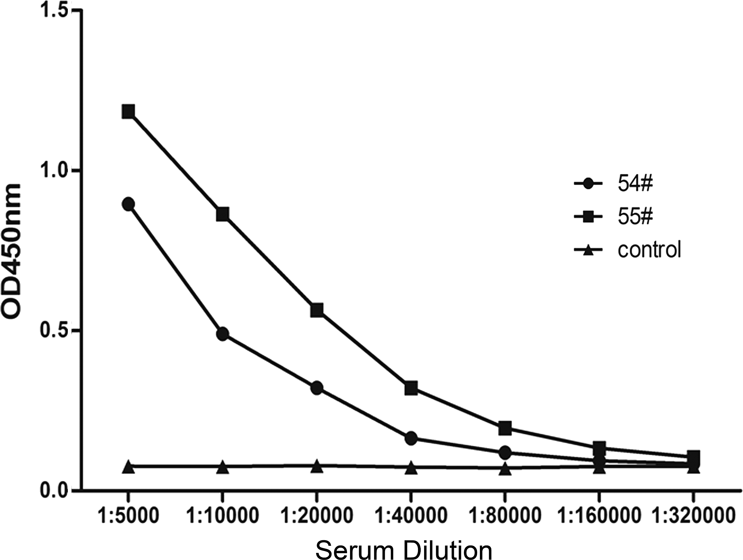
Antisera titers of immunized mice as measured by indirect ELISA. Serum serially diluted began at 1:5000 and added in recombinant KLK6 protein-coated 96-well ELISA plates. Number indicates the titer of immunized mice. Serum from pre-immune mice served as negative control.
Isotype characterization
A commercial rapid mouse isotyping antibody array kit (RayBiotech) was used to determine the antibody isotypes of the generated antibodies. Cell culture supernatant from each hybridoma clone was added onto the sample pad of strip 2 (green, IgG (IgG1 /IgG2a/IgG2b/IgG3) isotyping strip) and allowed to incubate until colored bands appear, which indicate the corresponding isotype. The IgG isotype strips indicated that the isotype of MAb 2E3 was IgG2b, while the other four clones were of the IgG1 isotype (Fig. 4).
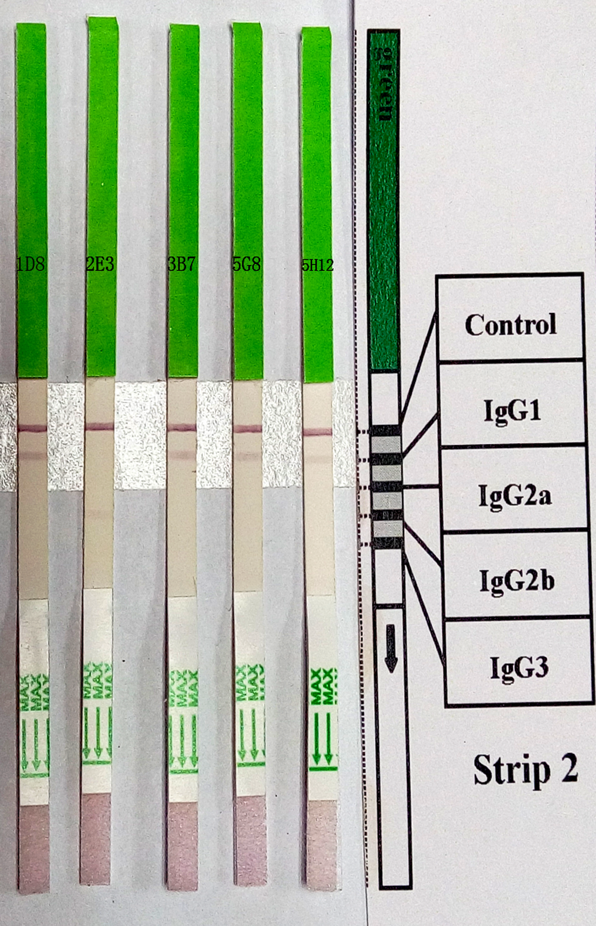
Classes and subclasses of MAbs were determined by Rapid Mouse Isotyping Kit-Gold Series. Each strip was dipped in the indicated hybridoma culture supernate and allowed to develop 10 min. Isotype was determined by comparison to the provided chart, showing positive control “line” and the isotype identified “line.”
Characterization of antibodies
Following validation of our hybridomas, these cells were expanded so that large-scale ascites antibody production could begin. Mice were sensitized with saxoline and implanted with hybridoma cells (clones 1D8 and 5G8) intraperitoneally. Following abdominal swelling, ascites was collected from the peritoneal cavity and purified by saturated ammonium sulfate and Hi-Trap protein affinity columns. The concentration of the collected and purified antibody was 4 mg/mL, as measured by a BCA assay. One clone, 5G8, was then confirmed for specificity by Western blot analysis against KLK6. Recombinant protein, ovarian cancer patient serum or plasma, or mouse liver tissue lysate were all probed with this MAb for antigen binding. As expected, the purified ascites showed significant KLK6 binding at 1:1000 dilution, confirming the presence of anti-KLK6 antibodies produced from the hybridomas (Fig. 5A). To determine the specificity of our generated monoclonal antibodies against other KLK family proteins, we tested them by indirect ELISA for cross-reactivity. Microtiter plates were coated with 1 μg/mL of KLK6, or other KLK6 family members (KLK3, KLK10, and KLK11). These wells were then probed with our MAb. As seen in Figure 5B, the cross-reactivity of our KLK6 monoclonal antibody (5G8) was overall negative for related proteins, implying a strong specificity for KLK6.
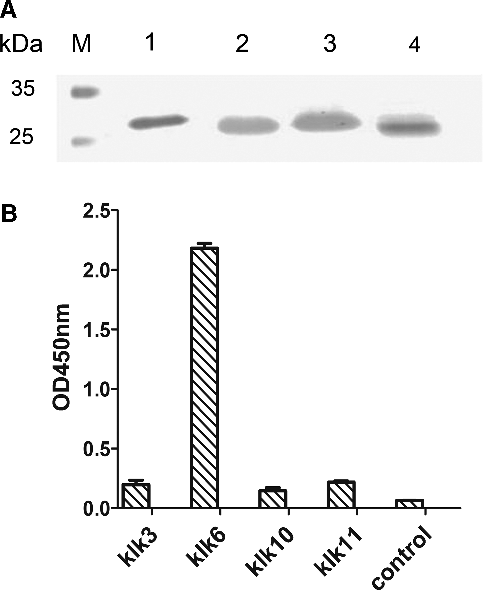
Characterization of monoclonal antibody 5G8. (
Sandwich ELISA development and validation
Finally, with our production of several monoclonal antibodies to KLK6, we set out to develop a sandwich ELISA pair for the quantitative detection of KLK6 in various sample types. In order to identify a potential sandwich antibody pair, unlabeled capture antibody (clone1D8) was coated onto microtiter plates and a secondary antibody was chosen to be the detection antibody (5G8) to attempt to make a valid pair. The result, as shown in Figure 6, identified a good correlation between antigen levels and OD seen in the developed ELISA, with an R2 = 0.999. Sensitivity of the assay was determined by 20 times the average zero concentration OD, plus 2 standard deviations. As seen in Table 1, the minimum limit of KLK6 that could be detected in our custom ELISA was in the 10 ng/mL range.
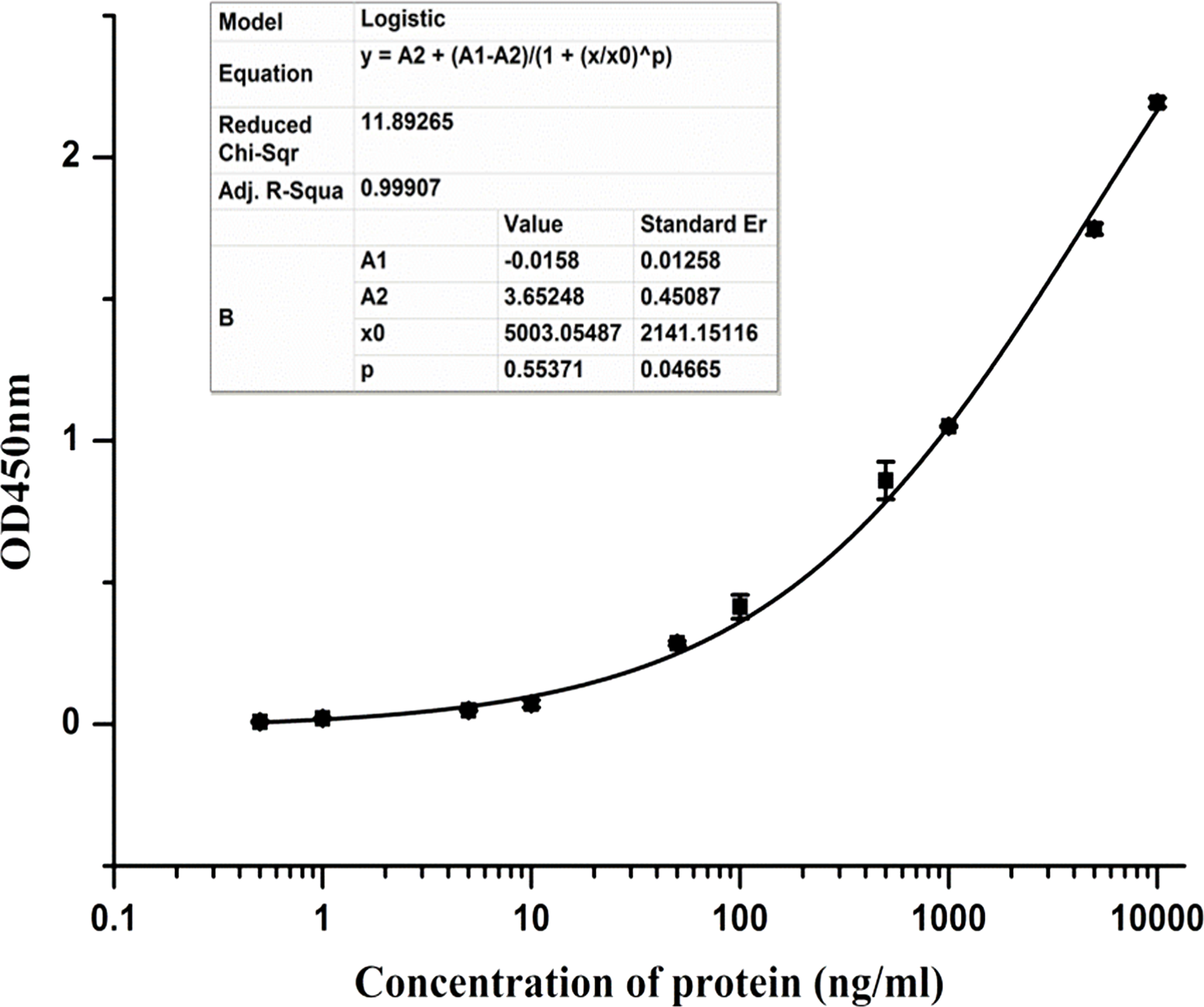
Establishment of sandwich ELISA. 1D8 MAb was used as capture antibody, and detecting one is 5G8. Recombinant KLK6 protein was serial dilution from 10,000 ng/mL to 1 ng/mL. Curve equation was Y = 3.652 – (0.016 + 3.582) / (1 + (X/5003.055) × 0.554); R2 = 0.999.
Capture antibody, 1D8; detection antibody, 5G8. N = 3.
Discussion
Kallikreins are a subgroup of 15 highly conserved serine proteases. These genes are localized to chromosome 19 q13.3-q13.4 and characterized by high sequence similarities including exon and intron organizations, terminal codons, and general exon organization, among others.(22) KLK family members' potential role to serve as biomarkers for various cancers, as well as even protein expression patterns as they relate to an underlying disease, is still under intense investigation.
The interest in this family group began, and continues, following the discovery and identification of prostate specific antigen (PSA, also KLK3), and it is now routinely used for prostate cancer diagnostics purposes.(23) KLK6 in particular, however, has been shown recently to play an important role in the development, invasion, and metastasis of tumors. Numerous studies have indicated that abnormal expression of KLK6 in tumor tissues often negatively correlated with the prognosis of the patient.(24) For example, in ovarian cancer patients, the tumor is difficult to detect until late stage disease when the disease prognosis is extremely poor. Of all the KLKs reported in ovarian cancer cells (KLKs 2-11 and 13-15), or those abnormally expressed in serum (KLKs 5-8, 10, and 13), KLK6 was reported to have the most promising potential as a disease biomarker.(22) Its utility for disease detection may not be limited entirely to cancer, as recent evidence indicates its expression levels also are altered in certain neurological diseases, such as Alzheimer's and Parkinson's. Therefore the clinical detection and quantification of KLK6 could be particularly important and informative for patient care.
Quantitative real-time PCR is another method for detecting KLK6,(25) but can be a time-consuming and relatively inefficient platform. Additionally, how DNA and/or mRNA translate to protein is not always directly correlative from assay to assay; therefore a genetic evaluation of KLK6 expression may give incomplete information about protein expression levels. As such, direct physical detection of KLK6 protein provides a much more accurate measure of physiologic protein levels.
Our development of several KLK6-specific monoclonal antibodies, as well as the final development of a functional and sensitive sandwich ELISA, provides an excellent detection platform for this critical protein in many liquid sample types. These methods for antibody and ELISA development, as well as the developed KLK6 testing platform, lay the foundation for future screening of at-risk and diseased patients, and also to determine the biomarker potential KLK6 proteins may hold.
Footnotes
Acknowledgments
We express our thanks for the support of RayBiotech innovative research fund, Program of hundred leading innovators and entrepreneurs (LCY201111), Guangdong innovative research team program (201001s0104659419), UK-China (Guangzhou) Healthtech Open Innovation (2012Q-P182), Guangzhou Municipal Innovation Fund (2013J4400170), Foundation of Enterprise University Research Institute Cooperation of Guangdong Province and Ministry of Education of China (2012B090600021), Special program for the development of technology business incubators in Guangzhou (2013J4200016), and Foundation of Enterprise University Research Institute Cooperation of Guangdong Province and Ministry of Education of China (2012B091000145).
Author Disclosure Statement
All authors, except Yun Xi, are employees of RayBiotech, Inc.