
Abstract
First identified as a high-affinity kinase-deficient receptor for class-3 semaphorins and vascular endothelial growth factor (VEGF) families, Neuropilin2 (NRP2) is a transmembrane non-tyrosine-kinase glycoprotein that has a vital function in neuronal patterning. Furthermore, NRP2 expression is often upregulated in cancer tissues and correlated with poor prognosis. In the present study, we report the establishment of a monoclonal antibody specific for NRP2b1b2 domain (NRP2 MAb) through hybridoma method. NRP2 MAb is measured to have a titer of 5.12 × 10⁵ against NRP2b1b2 in indirect ELISA. Western blotting, flow cytometry, and immunofluorescence analysis indicate that NRP2 MAb can combine full-length NRP2 in LoVo and SW480 cells. Besides helping further understand NRP2-related pathological mechanisms and cell-signaling pathways, NRP2 MAb may act as a therapeutic agent for cancer in the future.
Introduction
A 110–130 kDa single-pass transmembrane glycoprotein, NRP2 was first isolated by RT-PCR and gene transfer in 1997. NRP2 belongs to the NRP family. In humans, NRP2 is encoded by 112 kb genes located on chromosome 2q34. Structurally, NRP2 is composed of an extracellular domain, transmembrane domain, and a short intracellular domain. The extracellular domain is divided into three parts: a1/a2 domains, b1/b2 domains, and c-domain. a1/a2 domains are responsible for class-3 semaphorin binding, whereas b1/b2 domains are suggested for both VEGF and class-3 semaphorin binding, and c-domain is involved in dimerization of the receptor.(1–3)
NRP2 is well known for its role in facilitating axonal guidance during the development of the neuronal system. Additionally, it is expressed in vascular endothelial cells and lymphatic endothelium where it affects proliferation, migration, angiogenesis, and the formation of small lymphatic vessels and capillaries.(4,5) Knockout of NRP2 results in a reduction of small lymphatic vessels and capillaries. Homozygous NRP2 mutants show absence or severe reduction of small lymphatic vessels and capillaries during development.(6–9) Thus NRP2 MAb may be useful in inhibiting lymphangiogenesis in tumors since lymphatic EC express solely NRP2. Recent studies have identified NRP-2 as a polysialylated protein expressed in human dendritic cells modulating DC-T cell interactions.(10) Moreover, NRP2 was also located in focal adhesions on laminin and necessary for the association of α6β1 integrin with the cytoskeleton.
Nearly all tumor cells express NRP2 such as osteosarcomas, melanomas, lung cancers, brain tumors, colon cancers, pancreatic cancers, breast cancers, and bladder cancers.(11–14) However, NRP2 has low expression in normal tissues. NRP2 expression could be used as a diagnostic and prognostic marker of these tumors.(15–18) All these features point to NRP2 as an attractive target for cancer therapy.
However, the NRP2 signaling pathway has just been studied. The mechanism by which NRP2 regulates the interaction of α6β1 integrin with laminin to form focal adhesions involves PKC activation.(18) NRP2 mediates colorectal carcinoma (CRC) lymphangiogenesis via the VEGF-C/VEGFR3 pathway.(19) NRP2 is upregulated during TGF-β1-driven epithelial-mesenchymal transition (EMT) in lung cancer cells.(20) An antibody against NRP2 that blocks VEGFC binding disrupts VEGFC-induced lymphatic EC migration.(21) Interrupting the expression of NRP2 by siRNA can inhibit the growth of colon cancer cells.(22) In a word, NRP2 may regulate tumor progression by several mechanisms, not only by angiogenesis but also lymphangiogenesis, metastasis, and EMT.
In this study, we used the synthetic peptide sequence NRP2b1b2, which responded to b1b2 domain of the human NRP2 to immunize mice and successfully established a hybridoma cell line stably secreting NRP2 MAb. The monoclonal antibody was produced by ascites and purified by rProteinA affinity column. Enzyme-linked immunosorbent assay (ELISA) was conducted to determine the titer of the MAb, and Western blotting (WB), flow cytometry, and confocal microscope were applied to identify the specificity of the MAb.
As NRP2 plays an important role in tumor growth, angiogenesis, lymphangiogenesis, epithelial-mesenchymal transition, and metastasis, it may represent a promising target for novel anti-tumor therapies.
Materials and Methods
Reagents
Dulbecco's modified eagle medium (DMEM) and fetal bovine serum (FBS) were purchased from Gibco (Grand Island, NY). Freund's adjuvant (complete and incomplete), HT (hypoxanthine, thymidine), HAT (hypoxanthine, aminopterin, thymidine), goat anti-mouse horseradish peroxidase (HRP)-conjugated secondary antibody, goat anti-mouse fluorescein isothiocyanate (FITC)-conjugated secondary antibody, goat anti-mouse tetramethyl rhodamine isothiocyanate (TRITC)-conjugated secondary antibody, and polyethylene glycol 1450 (PEG1450) were from Sigma Chemicals (St. Louis, MO). O-phenylenediamine (OPD) and enhanced chemiluminescence (ECL) were purchased from Sangon (Shanghai, China). rProtein A Sepharose column was purchased from GE Healthcare (Uppsala, Sweden).
Materials
The synthetic peptide sequence (NRP2b1b2) was kindly provided by Professor Craig W. Vander Kooi (Department of Molecular and Cellular Biochemistry and Center for Structural Biology, University of Kentucky). SP2/0 cell line, human colorectal carcinoma SW480 cell line, LoVo cell line, and BALB/c mice (8 weeks old, female) were obtained from Cancer Research Center of Xiamen University (Xiamen, China).
Immunization of mice
Eight BALB/c mice (female, 6–8 weeks old) were chosen and each was subcutaneously injected with 50 μg NRP2 (in 0.5 mL PBS) fully emulsified with 0.5 mL Freund's complete adjuvant. Two weeks later, each mouse was boosted with 50 μg NRP2 in Freund's incomplete adjuvant. The booster injection was repeated every 2 weeks for three injection times. Serum from the tail vena was monitored for their antibody titers against NRP2 by indirect ELISA. Mice with sustained antibody titers above 1 × 10⁴ were selected and intravenously injected with 25 μg NRP2 without Freund's adjuvant 3 days before cell fusion.
Establishment of hybridomas
Mouse myeloma cells SP2/0 in good condition were fused with splenocytes from the selected mice at the cell ratio of 1:5 (SP2/0:splenocyte) in 50% PEG. We performed cell fusion according to the established protocol of Kohler and Milstein. The fusion cells were cultured in HAT medium in 96-well plates in a 37°C, 5% CO2 incubator for 2 weeks. Cell colonies were selected and their supernatants were screened for antibody titers against NRP2 by indirect ELISA. The hybridoma cell colonies were cultured and recloned by limiting dilution method in HT medium for 2 weeks.
Indirect ELISA
To conduct indirect noncompetitive ELISA, 4 μg/mL at 4°C.[AU: supply missing words] The plates were blocked with 5% skim milk at 37°C for 1 h and washed with PBS containing 0.05% Tween-20 (PBST) three times. The serum or culture supernatants of hybridoma cells were incubated in 96-well plates at 37°C for 1 h. After washing, goat anti-mouse horseradish peroxidase (HRP)-conjugated secondary antibody was added and incubated at 37°C for 1 h. OPD with 0.04% hydrogen peroxide (H2O2) was added to develop color for 10 min and the optical density (OD) was measured at 490nm by microplate reader (model 680, Bio-Rad, Tokyo, Japan).
MAb production
The hybridoma cell clones secreting NRP2 MAb were screened and those with the highest titer was massively cultured for injection in high glucose-DMEM containing 10% FBS. Before the hybridoma injection, 20 BALB/c mice were intraperitoneally injected with sterile paraffin oil (0.5 mL per mouse). After 7 days, each mouse was injected with 3 × 10⁵ hybridoma cells suspended by basic H-DMEM. 7 to 10 days later, ascites were collected and centrifuged at 10,000 g for 15 min to acquire the supernatant.
MAb purification
The ascites collected were further purified by rProteinA Sepharose column according to the manufacturer's protocol to obtain purified MAb. To assess the purity, the purified NRP2 MAb was subjected to sodium salt-polyacrylamide gel electrophoresis (SDS-PAGE) and was analyzed by Quantity One software (Bio-Rad).
Isotype analysis
The class and subclass of the NRP2 MAb were identified by a mouse monoclonal antibody isotyping reagent (Sigma) according to the manufacturer's directions.
Titer analysis
For the titer analysis, the ascites, the purified NRP2 MAb, and SP2/0 mouse ascites (negative control) were serially diluted and tested by indirect ELISA described above. Data were analyzed and graphed with OriginPro 8.1 software (OriginLab, Northampton, MA).
Western blot analysis
SW480 and LoVo cells were lysed in 1 mL RIPA solution (containing 1% Triton X-100, 1% deoxycholate, and 0.1% SDS; Beyotime Biotechnology, Haimen, China) supplemented with a protease inhibitor (PMSF, Sangon, Shanghai, China) for 30 min at 4°C, then centrifuged at 12,000 g for 30 min at 4°C to obtain the total cell extracts. The protein concentration in the cell extracts was determined by modified Bradford protein assay.
For the Western blot experiment, 15 μL total cell extracts were directly loaded for the SDS-PAGE analysis. Then proteins were electrophoretically transferred to PVDF membranes by MiniPROTEAN (Bio-Rad, Hercules, CA). The PVDF membranes were blocked with TBST (10 mM Tris-HCl [pH 7.4], 150 mM NaCl, and 0.1% Tween-20) containing 5% skim milk for 2 h at 37°C, then washed with TBST three times and incubated with goat anti-mouse IgG-labeled peroxidase (diluted 1:5000) for 1 h at RT. Proteins of interest were visualized with ECL in Kodak Image Station 4000R (Carestream Health, Rochester, NY).
Flow cytometry
Flow cytometry was used to determine the affinity of NRP2 MAb for NRP2 molecule on SW480 and LoVo cells. The cells in exponential phase were collected and washed with PBS three times. The cells were then incubated with NRP2 MAb and IgG (as isotype control) respectively for 1 h at 37°C. Following three washes, goat anti-mouse fluorescein isothiocyanate (FITC)-conjugated secondary antibody was added and incubated for another 30 min at 37°C and washed again. Results were performed on a FAC Scan flow cytometer (Partec, Munster, Germany). Data were analyzed with FlowJo flow cytometry analysis software (Tree Star, Ashland, OR).
Confocal microscopy analysis
SW480 and LoVo cells were respectively cultured on glass cover slides overnight at 4°C and fixed with 4% paraformaldehyde at room temperature (RT) for 15 min. We covered the slides with blocking solution (containing 1% BSA, 5% goat serum, 0.2% NaN3) at RT for 1 h to block the non-specific binding. Then slides were washed three times with PBS and incubated with the diluted NRP2 MAb (1:100) at 37°C for 2 h. After washing, the cells were mounted with goat anti-mouse FITC-conjugated secondary antibody and restained with Hoechst 33258 (Beyotime Biotechnology). The slides with cells were incubated without the NRP2 MAb as control. The fluorescence images were captured at excitation laser of 360nm and emission laser of 460nm for Hoechst33258, and at excitation laser of 488nm and emission laser of 530nm for FITC by using Olympus FV1000 Inverted Confocal Fluorescence Microscope (Olympus, Columbia, SC).
Results
MAb production
Ten to 12 days after cell fusion, supernatants of growing colonies were detected for their ability to secrete specific antibodies by indirect ELISA. Three colonies with high titers (designated E4, E8, and C3) were chosen to subclone to 96-well plates. To ensure their stability and specificity, the three hybridoma cells were experimented three times with subcloning and detecting. Then the hybridoma cells with the highest titer were chosen to produce NRP2 MAb ascites.
MAb purification
The ascites were purified by rProtein A Sepharose column to acquire purified NRP2 MAb. The concentration of the purified NRP2 MAb is 2 mg/mL, analyzed by the Bradford protein assay. Then the purified NRP2 MAb was loaded for the SDS-PAGE analysis. Figure 1 shows two bands in lane 4 with molecular masses of ∼50 kDa and ∼25 kDa, which fit the molecular weight of the heavy chain and light chain of antibody. The purity of the NRP2 MAb was analyzed by Quantity One software (Bio-Rad, Tokyo, Japan) and the result of the purity is about 95%.

SDS-PAGE analysis of the purified NRP2 MAb. Lane 1, marker; lane 2, bovine serum albumin (BSA); lane 3, ascites; lane 4, purified NRP2 MAb.
Isotype analysis
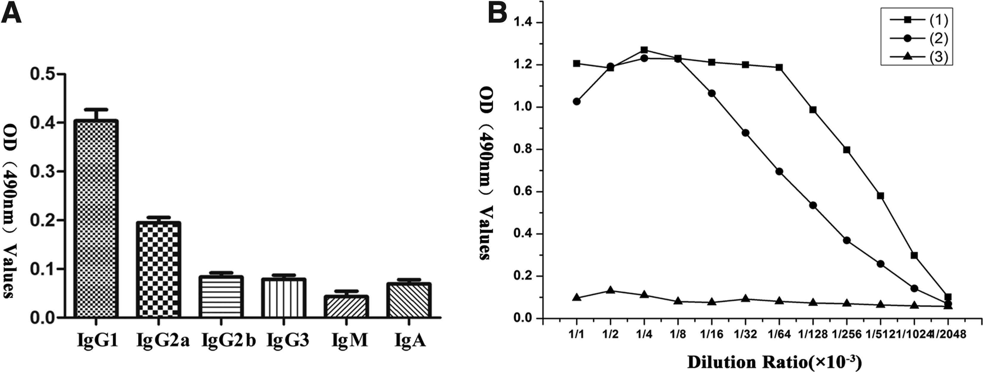
Isotype analysis for the hybridoma culture supernatants by anti-isotype reagent indicated that the isotype of NRP2 MAb is IgG1. As shown in Figure 2A, OD490nm values in IgG1 was much higher than OD490nm values in IgG2a, IgG2b, IgG3, IgM, and IgA.
(
Titer analysis of MAb
The ascites and purified NRP2 MAb were serially diluted to measure the titers against NRP2b1b2 by indirect ELISA. As shown in Figure 2B, with the increased dilution ratio of NRP2 MAb, OD490nm value was not changed in IgG control, but obviously reduced in ascites and purified MAb. The ascites and the purified NRP2 MAb have titers of 1.02 × 10⁶ and 2.56 × 10⁵, respectively.
Western blot analysis
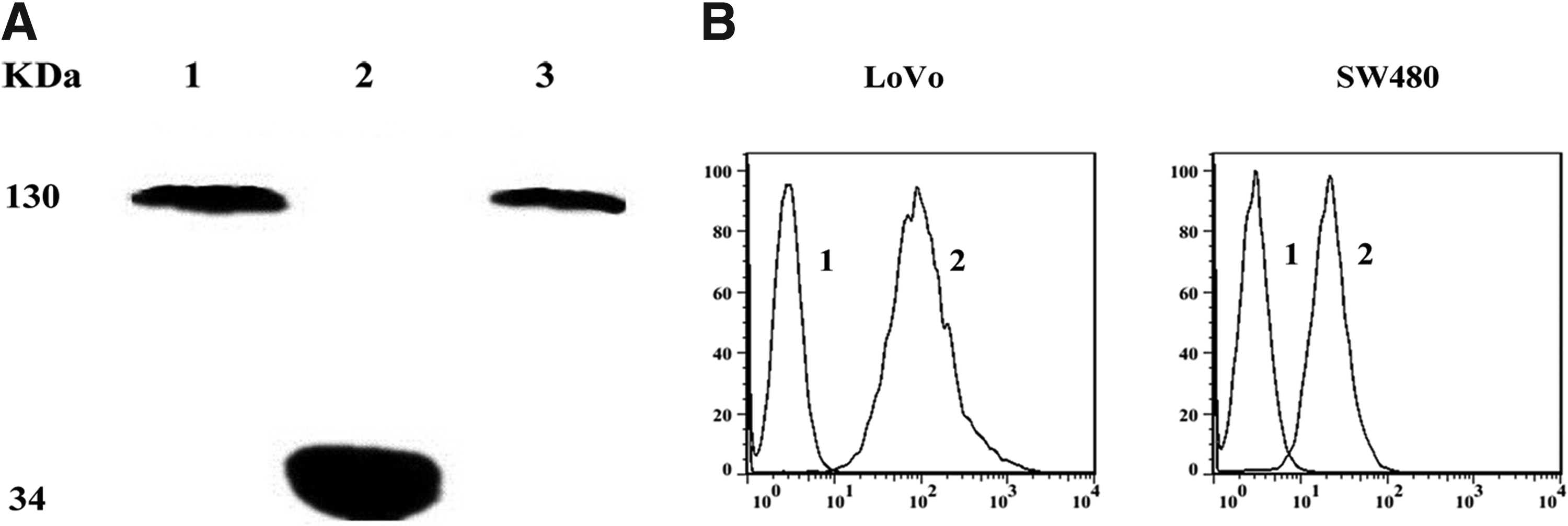
Western blot analysis showed that NRP2 MAb yielded a positive signal with the total cell extracts of LoVo and SW480 cells at a molecular weight of 130 kDa and reacted with NRP2b1b2 at a molecular weight of 34 kDa (Fig. 3A).
(
Flow cytometry
The affinity of NRP2 MAb for NRP2 expressed on LoVo and SW480 cells were analyzed by flow cytometry (Fig. 3B). The results showed that a much higher fluorescence intensity was detected compared with the control. It demonstrated that our antibody has the ability to combine the membrane-associated NRP2.
Confocal microscopy analysis
Binding specificity of the NRP2 MAb was detected in both LoVo and SW480 cells by confocal microscopy. The images showed NRP2 protein mainly localized to the membrane in both LoVo and SW480 cells with the treatment of NRP2 MAb; the control had no signal by comparison (Fig. 4).

Immunofluorescence of LoVo and SW480 cells with or without NRP2 MAb.
Discussion
Firstly identified as a mediator of neuronal guidance, NRP2 has also been described as a mediator of angiogenesis and tumor progression.(1–3) NRP2 may regulate the process of tumor through signal pathways, including PI3K/AKT, integrin α9β1/FAK/Erk, and TGF-β1.(18–20) Surprisingly, targeting NRP2 like siRNA and small molecule polypeptide drugs showed their effectiveness in vivo and in vitro.(21,22) However few MAbs are available to research its signal pathway and related function.
In our lab, we obtained NRP2 MAb targeting b1b2 domain of NRP2 with the synthetic peptide sequence NRP2b1b2 as immunogen. Firstly, we successfully developed a stable hybridoma secreting NRP2 MAb. Secondly, we produced the MAb by ascites and purified ascites by rProteinA sepharose column. Titer analysis of the purified NRP2 MAb showed that this MAb had high affinity for NRP2b1b2. Additionally, Western blotting showed that the NRP2 MAb can combine NRP2b1b2 and the full-length linear structure of NRP2 in colon cancer cells (LoVo and SW480). These two kinds of colon cancer cells both expressed NRP2, the grey level of LoVo cell being much stronger than that of SW480 cell, showing that LoVo cells express higher NRP2 than SW480. Furthermore, flow cytometry analysis and immunofluorescence indicated that the NRP2 MAb had high affinity for the spatial configuration of NRP2 in colon cancer cells. The results were in accordance with previous references.(19–22) Therefore, our produced NRP2 MAb may not only become a molecular probe in detecting these tumors but may also lay the foundation for the development of a new anti-tumor strategy targeting NRP2.
In conclusion, we developed a NRP2 MAb that may be a molecular probe to detect NRP2 in colon cancer samples and applied to further study the functions, mechanisms, and related signal pathways of NRP2 in tumor.
Footnotes
Acknowledgments
This work was supported by the National Natural Science Foundation of China (81472458, 31401180, 81172970); Science and Technology Foundation of Fujian Province (2013Y0080, 2013J01384, 2014R10361); and the Science and Technology Foundation of Xiamen, Fujian Province (3502Z20143016).
Author Disclosure Statement
The authors have no financial interests to disclose.