
Abstract
Death receptor 5 (DR5) can selectively induce cell death in a wide variety of tumor cells. However, at least certain versions of the recombinant soluble TRAIL (sTRAIL) or anti-DR5 monoclonal antibody (mAb) are also shown to cause apoptosis in normal cells (especially in hepatocytes), hampering its clinical use for cancer therapy. Recently, the development of small recombinant antibody fragments as high-affinity therapeutic reagents with reduced immunogenicity has come under the spotlight. A popular format of engineered recombinant antibody fragment is the single-chain fixed-variable (scFv) molecule, in which the VH and VL regions of the parental antibody are joined by a polypeptide linker. The scFv fragment retains the target specificity and antigen binding affinity of the intact antibody, whereas it can be genetically designed and produced in large quantities by ectopically expressing both VH and VL regions from a single cDNA in cells. In this study, an aDR5scFv was constructed and expressed, and it was conformed so that it could recognize and bind eDR5 specifically. The therapeutic effects on human lung adenocarcinoma cells lines 973 in vitro and in vivo were detected by MTT assay, flow cytometry, hematoxylin and eosin staining, and TUNEL assay. aDR5scFv was able to induce 973 cell apoptosis in an in vitro system. The protein expressions of caspase-3, Bax, and cytochrome c were raised, and aDR5scFv also inhibited tumor growth in mice with its effect as well as with radiotherapy. It is concluded that aDR5scFv could possibly be considered as a novel therapeutic candidate for the treatment of tumors.
Introduction
T
However, certain versions of recombinant soluble TRAIL (sTRAIL) or anti-DR5 mAb are also shown to cause cytotoxicity in hepatocytes,(7) and TRAIL resistance is widely found in cancer cells, leading to limitations in utilizing TRAIL or anti-DR5 mAb as a therapeutic agent for cancer.(8–10) A critical drawback is associated with human immunoreactions to murine antibodies. The immunogenicity of rodent mAbs sometimes can be successfully reduced by antibody humanization. First, VH and VL region genes were copied, and then linked with human Fc fragment, or construction of the single-chain fixed-variable (scFv) molecule, in which the VH and VL regions are joined by a polypeptide linker. The scFv fragment retains the target specificity and antigen binding affinity of the intact antibody. The scFv is smaller in size than its intact antibody, so it shows improved pharmacokinetics in tumor penetration and is better tolerated by the host immune system.(11,12) Juan Shi took advantage of the anti-DR5 (AD5-10) mAb and combined the scFv fragment technique with rAAV vector–mediated gene transfer to achieve tumor suppression in mice. However, the apoptotic effect of aDR5scFv antibody fragments on tumor cells in vivo and in vitro was moderate, maybe because of the relatively low affinity of the fragment with DR5.(13) In this study, aDR5scFv was constructed and expressed, and its specific identification and function in lung adenocarcinoma cells lines 973 were conformed in vitro and in vivo to compare its function with ionizing radiation.
Materials and Methods
aDR5scFv and antitumor agents
The plasmid pEt22b+ for the expression of aDR5scFv was constructed previously in the authors' laboratory. Murine immunoglobulin G (IgG) was purchased from Sangon Biotech Co. Ltd. (Shang Hai, China) and used as isotypematched controls. Phosphate-buffered saline (PBS) was used as a diluent for antibodies.
Cell lines and cell culture
Human lung adenocarcinoma cells lines 973 (non-small-cell lung cancer [NSCLC]) were purchased from American Type Culture Collection (ATCC, Manassas, VA). The 973 cells were cultured in Dulbecco's modified Eagle's medium (DMEM; Gibco BRL, Grand Island, NY), supplemented with 10% heat-inactivated fetal bovine serum (FBS) and 1% penicillin-streptomycin. Cell lines were cultured at 37°C under 5% CO2.
Reagents
Rabbit polyclonal antibodies to caspase-3, cytochrome c, Bax, and horseradish peroxidase-conjugated anti-rabbit IgG (Epit Mics Co. Ltd., Burlingame, CA), and mouse polyclone antibody to β-actin (Santa Cruz Biotechnology, Inc., Paso Robles, CA) were used for Western blotting.
Preparation of aDR5scFv
Variable region sequences of heavy chain and light chain to murine anti-human DR5 monoclonal antibody were acquired by reverse transcription polymerase chain reaction, and then linked through a flexible linker peptide and cloned into the expression vector after being expressed by Escherichia coli and purified using affinity chromatography.
Enzyme-linked immunosorbent assay
Purified horseradish peroxidase-conjugated aDR5scFv was diluted in a coating buffer (0.05 M carbonate-bicarbonate buffer, pH 9.6) to a final concentration of 4 μg/mL. Each well of 96-well microtiter plates was coated with 100 μL of the protein overnight at 4°C. Plates were washed to remove unbound peptides, and binding intensity of peptides was determined spectrophotometrically. The cutoff value was defined as the mean value plus three standard deviations of the mean optical density.
In vitro proliferation assay
The 973 cells were seeded in 96-well plates in triplicate (1 × 10⁴ cells/well) and were treated with aDR5scFv or aDR5mAb. After attachment, the cells were exposed to DMEM with 0.5% FBS for 4 h, and then the effects of aDR5scFv on 973 proliferation were evaluated using the 3-(4,5-dimethylthiazol-2-yl)-2,5-diphenyltetrazolium bromide (MTT; Bio Basic, Inc., Konrad Cres, Markham, Canada) colorimetric assay. Briefly, the medium was removed and replaced with medium containing 5 mg/mL of MTT and incubated for 4 h. The medium was then aspirated, and the product was solubilized with dimethyl sulfoxide. Absorbance was measured at 570 nm for each well using a microplate reader (Thermo Labsystems, Waltham, MA) according to the manufacturer's instructions.
Clonogenic survival
The effect of vinorelbine on the radiosensitivity of the 973 cells was assessed using standard clonogenic survival assays. Before irradiation, cells were plated in 25-cm2 flasks in growth media. Cells were irradiated (0, 1, 2, 4, 6, 8, and 10 Gy), and colonies were allowed to develop for 10–14 days. The colonies were stained with 4% formaldehyde in PBS containing 0.05% crystal violet. Colonies containing >50 cells were counted. The surviving fraction was calculated. Cell survival curves were fitted using a linear quadratic equation.
Morphological studies with Hoechst 33342
The grown 973 cells were treated with aDR5scFv for 4 h. Cells were washed twice, stained with 10 μg/mL Hoechst 33342, and incubated for 15 min at room temperature. Images were recorded using an inverted fluorescence microscope (Olympus, Tokyo, Japan) with an excitation wavelength of 350 nm (blue fluorescence).
Detection of apoptotic cells
The cell lines (1 × 10⁵) were treated with aDR5scFv for 4 h. Apoptosis was determined by flow cytometric analysis using an annexin V fluorescein isothiocyanate (FITC) apoptosis detection kit I. Finally, 400 μL of binding buffer was added to each tube, and the stained cells were analyzed by flow cytometry (BD Diagnostics, Franklin Lakes, NJ) with Winmdi software within 1 h. Ten thousand cells were recorded per assay.
In vivo xenograft models
Tumors were generated in female nude mice (Animal Center for WTLH, Beijing, China) by subcutaneous (s.c.) injection of 973 cells (5 × 10⁵ cells in 50 μL PBS) into the right hind leg of each mouse. Tumor measurements were converted to tumor volume (V) using the formula (l × w2 × 0.52), where l and w are the length and width, respectively. Measurements were made with a Vernier caliper. All procedures were carried out following approval of the Institutional Animal Care Committee. All tumor-bearing mice were divided randomly into groups of six, and treatment was initiated on day 10 when tumor volume reached about 40–50 mm3 (designated as day 0). Control animals were treated with injections of PBS, or with three times irradiation of 5 Gy each time.
Results
Yield and solubility of purified aDR5scFv protein
The results demonstrated that IPTG-induced BL21 (DE3) pLysS (0.6 mmo1/L) at 30°C for 12 h appeared to be an optimal condition for the expression of aDR5scFv. Purification products of aDR5scFv were examined using sodium dodecyl sulfate polyacrylamide gel electrophoresis (SDS–PAGE), as well as Western blotting analysis. A predominant protein of 30 kDa, corresponding to the expected molecular weight of aDR5scFv, was detected as a single band by SDS-PAGE (Fig. 1A), suggesting that the purification protocol achieved a high degree of purity for the target protein.
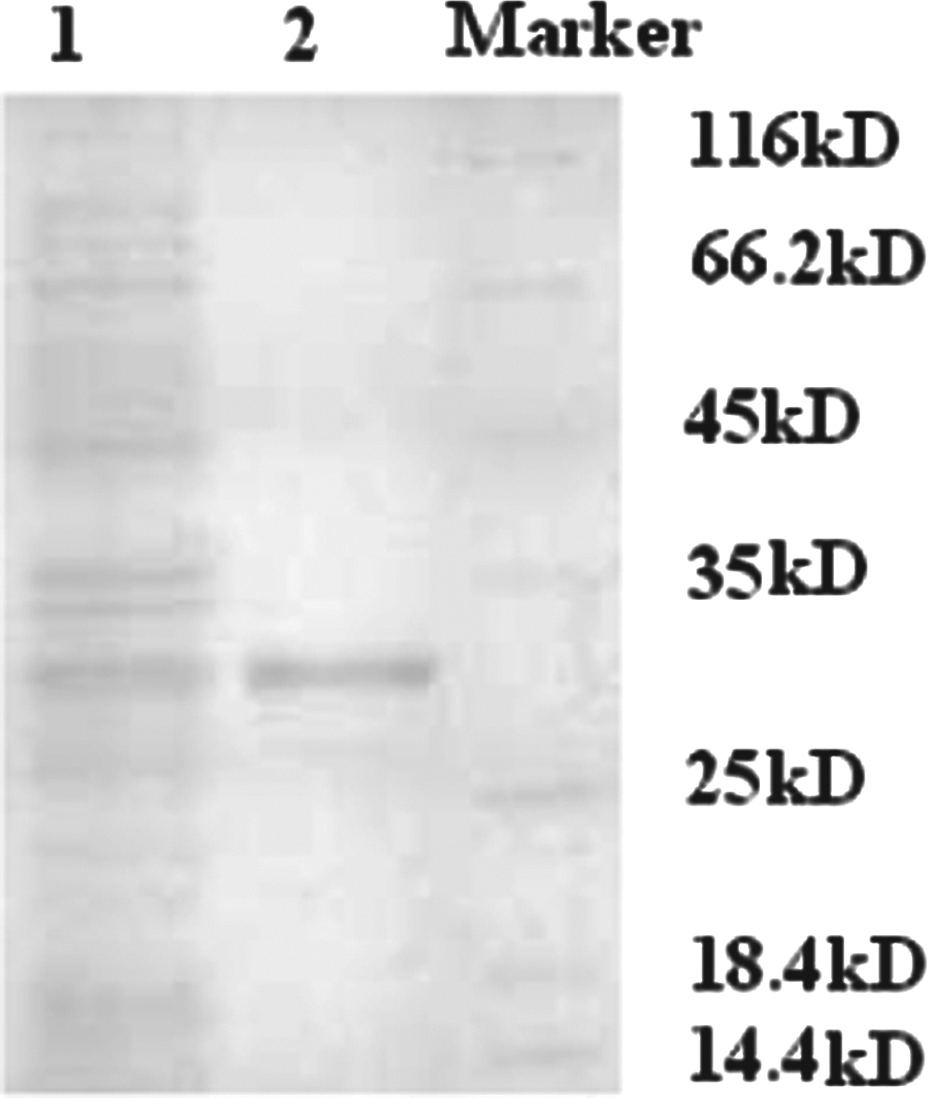
Identification of the specification of anti-human death receptor 5 (DR5) single-chain fixed-variable (scFv) molecules by enzyme-linked immunosorbent assay. Protein expression graph of gene engineering Escherichia coli with aDR5-scFv. (1) Expressional protein induced 12 h by IPTG; (2) purified scFv protein.
Identification of the specification of aDR5scFv binding to DR5 by enzyme-linked immunosorbent assay
In order to validate the specification of aDR5scFv, an indirect enzyme-linked immunosorbent assay was performed to detect if aDR5scfv could bind with DR5. The extracellular domain of human DR5 (eDR5) is coated as antigen, aDR5scFv were two rounds of limiting dilution. As shown in Jingjing Yang's research 14 , the titer of aDR5scFv is 1.2 × 10⁴. From the analytical results of the antibody-adding method, there is the same antigen site on the surface of the aDR5scFv as aDR5mAb, which can form antigen-antibody complexes and induce cell apoptosis by death receptor pathway.
Cytotoxicity of the aDR5scFv and radiotherapy among tumor cell line 973
The cytotoxic activities of aDR5scFv were tested in human lung adenocarcinoma cells lines 973. The 973 cells were treated with different concentrations of aDR5scFv (0, 0.225, 0.45, 0.9, and 1.2 mg/mL) for 4 h. At each dose point, the viability of the cells was determined by MTT assay. When the cells were treated with aDR5scFv at 0.225 mg/mL for 4 h, the cell death rate was 28.8 ± 1.11%, and when the concentration of aDR5scFv increased to 0.9 mg/mL, the cell death rate was 65.31 ± 1.15%. The inhibition has presented dose dependence, suggesting that aDR5scFv could inhibit 973 cell proliferation and that aDR5scFv has the same inhibitory activity as anti-DR5 mAb (Fig. 2A).

Cell viability of human tumor cell lines 973. (
The effect of vinorelbine on the radiosensitivity of 973 cells was assessed using standard clonogenic survival assays. Before irradiation, cells were plated in 25-cm2 flasks in growth media. Cells were irradiated (0, 1, 2, 4, 6, 8, and 10 Gy), and colonies were allowed to develop for 10–14 days. The colonies were stained with 4% formaldehyde in PBS containing 0.05% crystal violet. Colonies containing >50 cells were counted. Surviving fraction was calculated. The SF2 of radiotherapy was 0.559. Cell survival curves were fitted using a linear quadratic equation. The concentration of aDR5scFv increased to 0.9 mg/mL, and the cell death rate was 65.31 ± 1.15%—higher than the cell death of cells treated with 2 Gy.
aDR5scFv induces 973 cells apoptosis by death receptor pathway
First, the morphology change of 973 cells was observed. The 973 cells were treated with 0.45 mg/mL aDR5scFv for 4 h. Next, cells were washed twice and stained with 10 μg/mL Hoechst 33342 before being incubated for 15 min at room temperature, after which cells were observed under a fluorescence microscope. The cells shrank, developed bubble-like blebs on their surface, as well as chromatin compaction and segregation, condensation of cytoplasm, and nuclear fragmentation. Apoptotic cells broke into smaller pieces called apoptotic bodies. However, the same morphology changes in the control group were not observed (Fig. 3A).
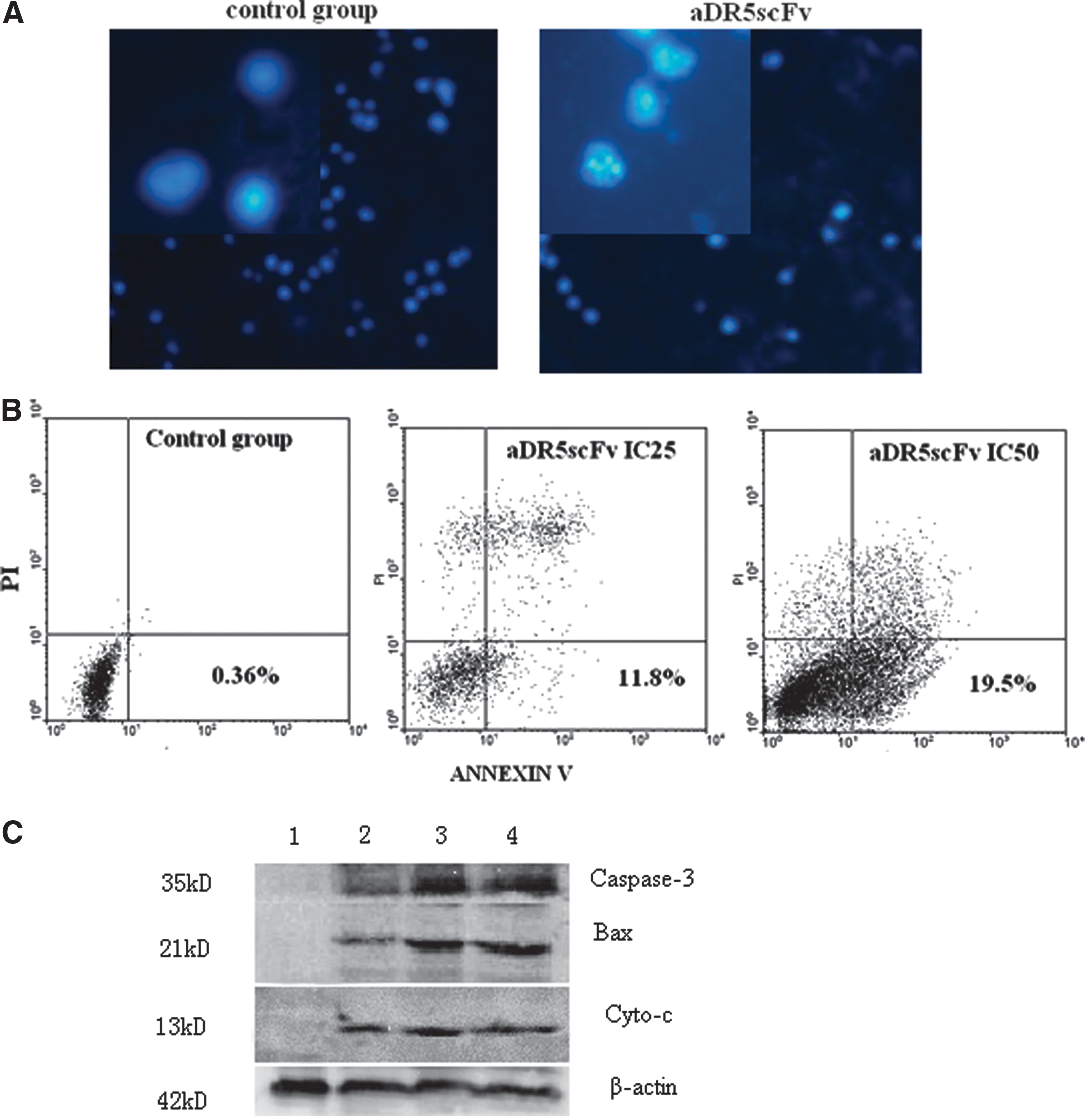
aDR5scFv induced apoptosis in 973 cells. (
Annexin-V/PI staining assays were combined to investigate whether aDR5scFv can induce human cells apoptosis. Furthermore, the apoptotic rate of the cell lines was assessed by different concentrations of aDR5scFv(0, 0.225, and 0.45 mg/mL) for 4 h using Annexin V/PI staining assay by fluorescence-activated cell sorting. As shown in Figure 4B, aDR5scFv induced apoptosis in 973 cells in a dose- and time-dependent manner. The result was correlated with the MTT assay.

Tumor growth suppression in nude mice treated with aDR5scFv. The 973 cells (5 × 10⁵ cells in 50 μL of phosphate-buffered saline [PBS]) were injected subcutaneously into the right hind leg of each nude mouse. When the tumor volume reached about 50 mm3, the mice were injected intraperitoneally daily with 0.45 mg/kg aDR5scFv every other day three times, or 24 h before radiation (on days 11, 13, and 15) in the combined treatment group. The tumor volume of the mice was measured every other day. Control animals were treated with injections of PBS, or three times by irradiation of 5 Gy each time. (
To clarify further the apoptotic molecular mechanisms of human lung adenocarcinoma 973 cells induced by aDR5scFv treatment, the total proteins of the 973 cells that were treated by 0.45 mg/mL aDR5scFv were analyzed for different time periods (0, 4, 7, and 12 h) using Western blot assay. Analysis of the expression of the proteins involved in the apoptosis pathway revealed that the level of caspase-3 and Bax was different at each time point. Compared with the normal group, the expression of caspase-3 and Bax increased gradually according to the treatment time. The expression of cytochrome c did not change obviously (Fig. 3C).
aDR5scFv and radiotherapy suppressed the growth of tumor xenografts by inducing tumor cell apoptosis
To analyze the therapeutic potential of the aDR5scFv in vivo, a tumor model was established in BALB/c nude mice by s.c. inoculation of human lung cancer 973 cells (1 × 10⁶ per injection) in the right dorsal flank of the mice. When the tumors reached 50 mm3, the animals were divided into six groups and received a PIPT injection of aDR5scFv or PBS as control. The tumor volume was measured twice a week over a period of 21 days. The result showed that the mean tumor volume, which was 0.126 cm3 in the control group, reduced to 0.101 mm3 in mice injected with aDR5scFv, demonstrating a substantial 20% decrease in tumor growth (p < 0.01; Fig. 4A and B).
As it was found that aDR5scFv can prolong mice survival and decrease tumor growth, the next step was to research the mechanism related to it. Hematoxylin and eosin stain showed that the treatment groups had increased necrosis or apoptosis cells compared with the control group. The amount of necrotic cells in the group receiving the combination treatment increased. The apoptotic and necrotic cell population of the aDR5scFv and radiotherapy groups was less than that of the aDR5scFv + radiotherapy group (Fig. 4C).
The histopathologic changes in the lung, liver, spleen, and kidney of the animals used in this study were also examined to investigate the cytotoxicity of aDR5scFvs toward normal tissues. No obvious lesions were found in any of the organs (data not shown).
Moreover, the aDR5scFv-injected mice did not show any sign of systemic toxicity as estimated by body weight, gross appearance, and behavior.
To determine whether treatment with aDR5scFv led to apoptosis in tumor cells, TUNEL assays were carried out in lung tumor sections acquired from aDR5scFv or injected mouse xenografts. Tumor tissues undergoing apoptosis were subsequently examined using a fluorescence microscope after TUNEL staining. Green indicates dUTP end-labeled by FITC. Results showed that more TUNEL-positive sites were detected among the tissues treated with aDR5scFv + radiotherapy than among tissue treated with the single agent alone. The concentration of aDR5scFv was associated with an accumulation of apoptotic cells within the tumors (Fig. 4D).
Discussion
Death receptor 5 (DR5) were expressed in many tumor cells. DR5 could selectively induce cell death by trigging TRAIL or agonistic mAbs specifically targeting DR5.(15,16) Five kinds of anti-DR5 mAbs were obtained, which could induce tumor cells apoptosis, such as MGC803, HL60, and BEL7402.(6) In order to reduce immunogenicity of rodent mAbs and improve pharmacokinetics in tumor penetration, VH and VL regions of mAb targeting DR5 were used. The present study developed a novel therapeutic strategy by expressing and investigating an agonistic anti-DR5 scFv antibody protein. It showed that this anti-DR5scFv can specifically and stably bind to eDR5. The titer of aDR5scFv is 1.2 × 10⁻⁴. From the analytical results of the antibody-adding method, aDR5scFv had the same antigen site as anti-DR5 mAb from which VH and VL chain genes were obtained.
This aDR5scFv showed higher cytotoxic activity in 973 cell lines. It was speculated that an underlying mechanism might be involved in antibody-induced specific conformational changes that were sufficient to stimulate receptor aggregation. However, further studies are required to confirm this hypothesis.
Next, it was investigated whether the apoptotic pathway involved in the cell death was caused by aDR5scFv. Several lines of evidence were presented in the study that indicated that apoptosis was the mode of cell death caused by aDR5scFv. Morphological changes were clearly observed after 4 h. Consistent with this observation, nuclear fragments were also evident. The result of cell apoptosis analysis assessed by flow cytometry indicated that the apoptosis was the main mode of death of 973 cells treated with aDR5scFv. The expression level of various intracellular apoptotic factors was analyzed, including caspase-3, Bax, and cytochrome c, and it was shown that the expression of intracellular Bax, cytochrome c and caspase-3 protein was increased in aDR5scFv-treated 973 cells. This result was consistent with the mechanism of DR5-mediated apoptosis induction by anti-DR5 mAb.(6,7)
Ionizing radiation (IR) could enhance TRAIL-induced apoptosis in human osteosarcoma cells, and was associated with upregulations of DR5.(17) IR was identified as having synergy with TRAIL in either prostate or pancreatic cancer cells, or both.(18) It was shown that aDR5scFv alone or in combination with radiotherapy could inhibit the tumor growth in vivo. Combining aDR5scFv and radiotherapy displayed an obvious suppressive effect on 973 tumor growth compared with a single treatment (p < 0.05). Cell apoptosis induced dramatically after exposure to aDR5scFv + IR compared with either treatment alone. The effect of aDR5scFv on tumor growth and survival in murine H22 hepatocellular carcinoma tumor model was studied using aDR5scFv, EPI, or aDR5scFv + EPI. It was shown that aDR5scFv induces good inhibition on the proliferation of H22 cells, especially in aDR5scFv + EPI group. It was speculated that an antitumor effect of aDR5scFv was also very promising in vivo.
This study took advantage of specific aDR5scFv in vitro and combined it with IR in vivo to achieve tumor suppression in 973 cells, providing a novel strategy with significant clinical potential for cancer therapy.
Footnotes
Acknowledgments
This work was partially supported by the Natural Science Foundation of China (Grant No. 81072472) and the 985 Funds of Medical College of Xiamen University.
Author Disclosure Statement
The authors have no financial interests to disclose.