
Abstract
The existence of a developed network of suppressory factors and cells against an immune response in different cancers has been proven; regulatory T cells are a typical issue. Therefore their depletion, elimination, or suppression has been assessed in different research studies that were not entirely successful. By applying an improved vaccine against regulatory T cells, we have evaluated the B cell response elicited by the vaccine in an experimental design. A previously described DNA vaccine and recombinant protein of Foxp3-Fc fusion were produced and used in the vaccination regimen. DNA construct and respective protein were injected into C57BL/6 mice. After 2 weeks, serum levels of IgG antibody and its subtypes against Foxp3 were investigated by ELISA. To produce recombinant Foxp3 for ELISA antigen coating, pET24a-Foxp3 vector was transformed into Escherichia coli strain BL21 as host cells. Afterward, protein was expressed and then purified using Ni-NTA agarose. SDS-PAGE and Western blot analysis were carried out to confirm protein expression. The expression analysis of Foxp3 was confirmed by SDS-PAGE followed by Western blot analysis. FOXP3-Fc DNA vaccine/fusion protein vaccination regimen could induce T helper–dependent humoral responses. Due to the effectiveness of Foxp3-Fc(IgG) in inducing humoral responses, it would be expected to be useful in developing vaccines in tumor therapies for the removal of regulatory T cells as a strategy for increasing the efficiency of other means of immunotherapy.
Introduction
O
Regulatory T cells are characterized by concurrent expression of CD4, CD25, and Foxp3.(1) Unlike in humans, Foxp3 expression may be a sufficient identifier of this population in mice,(3,4) which could be a proper candidate in designing vaccines against Tregs.
Increased numbers and/or enhanced functionality of Tregs have been demonstrated in different types of solid tumors.(5–8) It has also been presented that Treg depletion can efficiently increase vaccine-mediated antitumor response and survival in cancer patients.(9)
Dendritic cells, which are the most powerful antigen-presenting cells (APCs), employ a various array of receptors to sense danger signals and maturate.(10) Targeting Fcγ receptors is proposed as an efficacious strategy to deliver antigen to DCs that leads to DC activation and antigen processing towards MHCI, MHCII pathways, and B cell activation.(10,11)
The main purpose of this study is vaccination with plasmid coding for Foxp3 sequence fused to Fc fragment of mice IgG and its respective recombinant fusion protein, Foxp3-Fc, to observe whether B cells are induced through present vaccination methods besides targeting DCs in vivo.
Materials and Methods
DNA constructs and recombinant proteins as immunogens
DNA construct containing fragment C (Fc) portion of IgG fused to Foxp3 was designed. DNA construct pIRES2-EGFP-Foxp3-Fc (IgG) was transfected into HEK cells to investigate its expression through fluorescent microscopy and flow cytometry. Its specific expression was also assessed by Western blot.(12) Expression of Foxp3-Fc by pET21a vector harboring the corresponding gene (pET21a-Foxp3-IgG2Fc) in BL21-AI strain of E. coli under the control of IPTG-inducible (and detailed steps for confirmation of recombinant fusion protein by SDS-PAGE followed by Western blot analysis and purification through SDS-PAGE reverse staining) have been described previously.(12) The protein concentration was determined with BCA assay and Nanodrop spectrophotometry analyzer (Thermo Scientific, Fremont, CA).
Recombinant Foxp3 production for antigen coating
For expression of recombinant protein Foxp3, pET24a-Foxp3 plasmid containing truncated Foxp3 sequence (which lacks a polypeptide segment of nuclear localization signal that leads to inefficient functional properties) was ligated in pET24a expression vector; it was transformed into competent E. coli BL21 (DE3). Transformed cells were grown at 37°C in LB medium containing kanamycin (50 μg/mL) until the exponential phase (OD600 nm = 0.6), followed by induction with 1 mM IPTG (Fermentas, Vilnius, Lithuania). Samples were collected after 3 h and analyzed by SDS-PAGE to follow the protein expression. To purify the recombinant protein, E. coli BL21 (DE3) containing pET24a-Foxp3 plasmid was grown in large scale, and the pellets of bacterial cells expressing protein were harvested and resuspended in lysis buffer (8 M urea, 0.1 M NaH2PO4, and 0.01 M Tris [pH 8.0]) containing protease inhibitors. Cell suspension was sonicated and centrifuged for 20 min at 10,000 rpm. After centrifugation, the recombinant protein (∼42 kDa) was purified from supernatant under denaturing conditions via its His-tag using Ni-nitrilotriacetic acid (Ni-NTA) affinity chromatography (Ni-NTA Agarose, Hilden, Qiagen, Germany) according to the manufacturer's instructions.(13,14) The output fractions were analyzed by SDS-PAGE and the quantity of protein was determined with BCA protein assay and Nanodrop analyzer. The Foxp3 protein solution was dialyzed against 0.1 M phosphate-buffered saline (PBS, pH 7.4) for 72 h to remove urea, filtered (0.22 μm, Sartorius, Goettingen, Germany), and stored at −70°C until use.
Western blot analysis
For Western blot analysis, the separated proteins by SDS-PAGE gel were transferred to a nitrocellulose membrane (Schleicher & Schuell, Keene, NH). The membrane was then blocked in Tris-buffered saline (TBS) containing 5% bovine serum albumin (BSA) overnight at 4°C and washed three times with TBS containing 0.05% Tween-20 (TBST). Afterward, the nitrocellulose membrane was incubated for 2 h at room temperature with mouse anti-his tag antibody (Qiagen) diluted 1:10,000 in TBS-T. Then the membrane was washed with TBS-T and incubated with goat anti-mouse immunoglobulin G (heavy and light chain) horseradish peroxidase (HRP) conjugate antibody (diluted 1:5000 in TBS-T) for 2 h at room temperature. After washing three times, the membrane was treated using DAB solution (Sigma, St. Louis, MO) and placed in darkness until the appearance of the protein band.(15,16)
Experimental groups, immunization procedures, and bleeding
Six- to eight-week-old female C57/BL6 mice were purchased from Pasteur Institute of Iran and kept in standard conditions. The mice were assigned into three groups (n = 10) and immunized with different immunogens, as summarized in Table 1. The mice were immunized intramuscularly (into the quadriceps muscles of leg) with 100 μg of the plasmids (pIRES2-EGFP-Foxp3 or pIRES2-EGFP-Foxp3-Fc(IgG2)) in the final volume of 100 μL (1μg/mL) and subcutaneously (s.c.) in the back of the neck with 30 μg of purified protein immunogen containing Foxp3 or Foxp3-Fc formulated in incomplete Freund's adjuvant (Sigma). Vaccination was performed as heterologous prime/boost immunization using purified plasmid DNA prepared by EndoFree Plasmid Giga Kit (Qiagen, Victoria, Australia) in the first immunization followed by two protein administrations on the second and third immunization dates (Table 1). Control group was injected by pIRES2-EGFP and BSA (Table 1). Two weeks after the last immunization, the mice were bled and serum samples were collected and stored at −70°C until use.
Values in parentheses indicate number of days of animal immunization.
Performing ELISA for specific antibody and subclasses
The enzyme-linked immunosorbent assay (ELISA) was used to determine the presence of anti-Foxp3 antibodies in the sera of immunized mice. Ninety-six well micro-titer plates (Extragene, Taiwan) were coated with 100 μL of 10 μ/mL of recombinant Foxp3 protein (1 μg/well) diluted in PBS, and incubated overnight at 4°C. Each plate was washed five times with PBS–T and blocked with PBS containing 5% BSA (blocking buffer) for 2 h at 37°C. Following blocking and washing, mouse sera were added (diluted in blocking buffer 1:500) and plates were incubated for 1 h at 37°C. The plates were then washed five times and incubated with HRP-conjugated anti-mouse IgG (Sigma) diluted 1:10,000 (as secondary antibody) at 37°C for 1 h. To develop the reaction, plates were washed and incubated with tetramethylbenzidine (TMB) as the substrate for 30 min at room temperature in dark conditions. The reaction was stopped with 100 μL of 2N HCL.
To determine the antibody subtypes, biotinylated antibodies against mouse IgG1, IgG2a, IgG2b, or IgG3 (Sigma, 1:1000 dilution), streptavidin-HRP conjugate (Sigma, 1:3000 dilution), and consequently TMB substrate were used for detection. In both experiments, absorbance was read at an optical density of 450 nm by ELISA reader.
Statistical analysis
Data were summarized using descriptive statistical methods. One-way analysis of variance (ANOVA) was used to compare the mean values. A p value <0.05 or 0.001 represented a significant difference. All data were analyzed and depicted using SPSS Software (v. 20.0) and Graph pad Prism 5.
Results
SDS page analysis of Foxp3
Cells harboring pET24a-Foxp3 plasmid were cultured at 37°C in the presence of IPTG. The whole cell lysates were analyzed by 12% SDS-PAGE. One major band appeared at the ∼42 kDa position in the case of IPTG induction, which was the expected position of Foxp3 (Fig. 1A). Induction of the cells at 37°C for 3 h and IPTG with dosage of 1.0 mmol/L was optimal to achieve the highest level of Foxp3 expression. Afterward, recombinant protein was carefully purified with Ni-NTA affinity chromatography under denaturing conditions (Fig. 1B).

(
Western blot analysis of Foxp3
Western blot analysis was performed to detect the expression of the desired protein. The major band observed in SDS-PAGE (42 kDa) (Fig. 2) was confirmed as Foxp3 protein by Western blot analysis with mouse serum anti-His-tag, which indicates the apparent molecular mass of 42 kDa.

Western blot analysis of recombinant Foxp3 protein probed by anti-His (1:10,000). Lane 1, negative control (BL21 transformed with pET24a+); lane 2, pellet of IPTG-induced bacteria; lane M, protein MW marker in kDa.
Humoral immune responses
Total IgG
To determine whether Foxp3-Fc immunization can induce antibody responses, specific antibody production was measured by an optimized ELISA method with sera obtained from three vaccinated groups 2 weeks after the last vaccination (Table 1). All animals immunized with recombinant vaccines (Groups II and III) were able to break humoral tolerance to produce the specific antibody anti-Foxp3. As depicted in Figure 3, the highest antibody titers were observed in the third group of immunization—Foxp3-IgG2Fc vaccinated group (p value = 0.004)—although total IgG titers were also significant in the second group of vaccination compared to control (p value = 0.03). Taken together, this result indicates the superiority of Foxp3-Fc in inducing B cell responses.
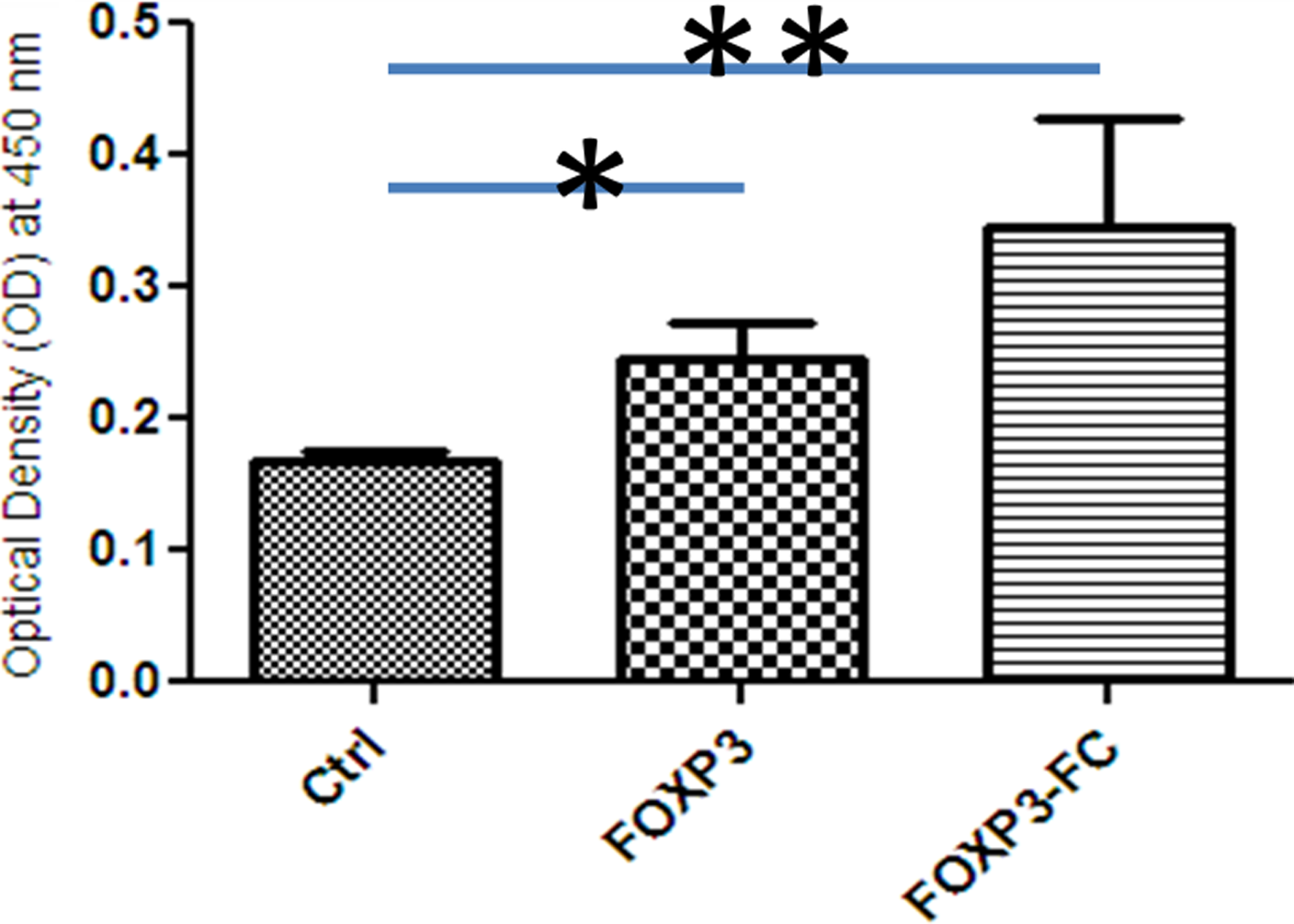
Comparison of specific total IgG antibody (anti-Foxp3) levels in sera of control and immunized mice indicates significant increase in Foxp3 and Foxp3-Fc groups. mean ± SD,*p ≤ 0.05, **p ≤ 0.001, n = 10.
IgG subclasses
Assessment of subclass antibodies of IgG may elucidate the direction of immunological responses; therefore subclass antibodies were measured following total IgG antibody measurement. As shown in Figure 4, IgG2b subclass, which is one of the T helper 1–dependent subclasses, has the highest serum level in the second and third groups of vaccination compared to the other subclasses in their respective groups. The increase of IgG2b is significant compared to other subclasses in both vaccinated groups II and III (p value <0.0001). Despite the fact that the serum level of IgG3 was also augmented in group III compared to IgG2a subclass, its increase was not significant compared to its level in the control group.

Comparison of Foxp3 specific IgG subclass (IgG1, IgG2a, IgG2b, IgG3) levels in sera of vaccinated and control groups. See text for detailed procedure and results. mean±S.D,*p ≤ 0.05, ***p ≤ 0.0001.
Discussion
APCs are the pioneering cells in inducing and launching B and T cell responses activated by DNA and recombinant protein vaccination in pathologic conditions such as cancers.(11) However the results of immunotherapeutic treatments in most tumors such as melanoma are not acceptable. This weak antitumor reactivity is due to chronic inflammation in the tumor microenvironment characterized by soluble mediators and infiltration of suppressory cells such as regulatory T cells, which are known as CD4+ CD25+ Foxp3+ T cells.(17) Despite other methods of active vaccination against Foxp3,(4) vaccination protocol of DNA vaccine/recombinant protein in the present study could break tolerance in T helper–dependent humoral response; DNA vaccine as used in the vaccination regimen in this research can launch both routes of antigen presentation through presenting its containing gene by either MHC I to cytotoxic T cells or MHC II to helper T cells.(18) In this study, we used a receptor-mediated internalization process to augment DC antigen presentation(11) to evaluate induction of B cell response. The results of this study demonstrated a more efficient B cell response by Foxp3-Fc DNA/protein vaccination than by Foxp3 DNA/protein vaccination.
Via interaction with activatory or inhibitory Fcγ receptors (FcγRs), antibodies have crucial roles in managing cellular immunity. Fcγ R involvement can facilitate receptor cross-linking on antigens or induce retrogate FcγR signals to activate or inhibit antibody-dependent internalization of antigens.(19)
Antigen-presenting cells express both activatory and inhibitory FcγRs. Affinity of IgG subtypes to FcγRs is different from each other. Mouse IgG2a and IgG2b have higher affinity for activating than inhibiting FcγRs.(19) As we mentioned in our results, IgG2b subclass had the highest level in immunized mice that improves the FcγR-mediated internalization of recombinant Foxp3-Fc. On the other hand, the recombinant fusion protein of Foxp3-Fc contains an Fc portion in itself that aids its uptake by APCs intrinsically.(11,20) Consequently, we had an improved elevated humoral response in the Foxp3-Fc vaccinated group, especially in IgG2b subclass, which indicates a T helper 1–mediated immune response alteration. Thus, we should proceed with further evaluation or other consequences of immunization by our constructs in vivo and to see whether they can induce a functional immune response against Tregs.
Conclusion
In summary, Foxp3-IgG2Fc DNA vaccine and recombinant protein were used for vaccination in an experimental study to assess the humoral response. The results showed an increase in T helper 1–dependent antibody response (cellular immunity alteration). In this regard, recombinant Foxp3 protein were produced and implicated for antigen coating in an ELISA test. Future studies may include investigating other aspects of Foxp3-IgG2FC as a potential vaccine against regulatory T cells, including regulatory T cells contained in spleen, CTL assay, and tumoral challenge.
Footnotes
Acknowledgments
This work was supported by the Faculty of Advanced Medical Science, Tabriz University of Medical Science, Department of Hepatitis and AIDS, the Pasteur Institute of Iran, Department of Immunology, Faculty of Medicine, Tehran University of Medical Science and Iran National Science Foundation (INSF).
Author Disclosure Statement
The authors have no financial interests to disclose.