




Abstract
The truncated spike protein (S1, 390-789aa) of porcine epidemic diarrhea virus was expressed as a recombinant protein in Baculorvirus expression system. Female BALB/c mice were immunized with the purified recombinant S1 (rS1) protein and two monoclonal antibodies, designated 8A3A10 and 12C4G12, were generated by hybridoma technique. The two monoclonal antibodies specifically reacted with PEDV rS1 protein, as proven by Western blot and indirect immunofluorescence assay. 8A3A10 monoclonal antibody showed a neutralizing activity to PEDV, as demonstrated in the results of fluorescent focus neutralization assay. These two newly established monoclonal antibodies may work as a useful tool to study the pathogenic mechanisms of PED virus and to develop diagnostic reagents.
Introduction
P
PEDV is an enveloped, single-stranded, positive-sense RNA virus, belonging to the family Coronaviridae and order Nidovirales, which contains four major structural proteins: spike protein, nucleocapsid protein, membrane protein, and small envelope protein.(3) The spike protein is a type I glycoprotein peplomer on the viral surface mediating viral entry in the natural host, which can be divided into S1 (1-789aa) and S2 (790-1383aa) domains. The S1 protein contains several neutralizing epitopes and has been the primary target for the development of effective vaccines against PEDV.(4) Therefore, to generate monoclonal antibodies (MAb) with neutralizing activity, rS1 protein was expressed in Baculorvirus expression system and the anti-rS1 protein MAbs were obtained by hybridoma technique. The neutralizing activity of MAbs was next evaluated by fluorescent focus neutralization assay.
Materials and Methods
Vector and cell line
The truncated PEDV S1 gene (390aa-789aa) was amplified by PCR and inserted into pFastBac I plasmid (Invitrogen, Carlsbad, CA). After transformation into DH10Bac cells, the white recombinant bacmids were selected and subjected to bacmid DNA isolation. The recombinant bacmid DNA was then used to transfect Sf9 insect cells for subsequent rS1 protein expression and purification. The rS1 protein was purified from the culture supernatant by immunoaffinity chromatography using Ni-NTA His Bind Resin (Novagen, Madison, WI) according to the manufacturer's instructions. Vero cells were grown in Dulbecco Modified Eagle Medium (DMEM, Gibco, Grand Island, NY) supplemented with 5% fetal bovine serum (Hyclone, Logan, UT) and antibiotics (100 U/mL penicillin and 100 μg/mL streptomycin).
Immunization of mice
Six-week-old female BALB/c mice were immunized with purified rS1 protein (100 μg/mouse) emulsified in equal volume of Freund's complete adjuvant via hypodermic injection at four different sites on the back. After 2 weeks, another hypodermic immunization was administrated by using 100 μg purified rS1 protein emulsified in Freund's incomplete adjuvant in 1:1 proportion. The equal amount of antigen without adjuvant was injected into mice on day 42 for the boost.
Generation of anti-rS1 protein-specific MAb
After multiple immunizations, the anti-rS1 protein serum titer of mice was determined by using indirect ELISA, as described previously.(5) Spleen cells from the immunized mice with the highest anti-rS1 serum antibody titer were fused with SP2/0 myeloma cells by using 50% PEG 1500 as a fusion agent. The hybridoma cells were cultured in 96-well plates at 37°C in a humidified 5% CO2 incubator in HAT screening culture medium. The supernatant of the fusion cells was then subjected to indirect ELISA to detect anti-rS1 antibody production. Positive hybridomas were selected and cloned for at least three rounds by limiting dilution and injected into paraffin-treated BALB/c mice to gain abundant ascetic fluid. Two monoclonal antibodies (designated 8A3A10 and 12C4G12) were identified, and their immunoglobulin subclasses were determined by the mouse MAb isotyping kit (Sigma, St. Louis, MO).
Purification of MAbs
The ascetic fluid was purified by affinity binding with protein G resin. Briefly, 10 mL binding/wash buffer (20 mM Na2HPO4 and 0.15 M NaCl, pH 8.0) was used to equilibrate 5 mL column of protein G. The ascetic fluid sample was then applied to the column for binding. After washing the column with 30 mL binding/wash buffer, the purified MAb was eluted by appropriate volume of elute buffer (0.1 M glycine, pH 2.5) and neutralized to pH 7.4 with neutralization buffer (1 M Tris-HCl, pH 8.5). The final concentration of MAbs was adjusted to 4 mg/mL for further use.
Western blot analysis
The specificity of anti-rS1 MAbs was analyzed by Western blot. The purified rS1 protein was separated on 10% SDS-polyacrylamide gel electrophoresis (SDS-PAGE) and transferred to nitrocellulose (NC) membrane. The membrane was blocked overnight with 5% non-fat milk in TBST buffer (TBS with 0.05% Tween-20), then incubated with corresponding MAb (1:100 diluted in TBST) at 37°C for 1 h. After washing three times with TBST, the HRP-conjugated goat anti-mouse IgG as second antibody (1:5000 diluted in TBST) was added onto the NC membrane at room temperature for 1 h. After washing, the substrate 3, 3-diami-nobenzidine tetrahydrochloride hydrate (DAB) was used for color development.
Indirect fluorescence assay
Vero cells were inoculated with PEDV HN1303 strain at a multiplicity of infection (MOI) of 0.05 and were cultured in DMEM containing trypsin (20 μg/mL) at 37°C for 24 h until CPE appeared. Cells were washed once with PBS and were fixed in cold acetone for 30 min at 4°C. The fixed cells then reacted with MAb for 1 h. After washing three times with PBS, FITC-conjugated goat anti-mouse IgG (1:1000 dilution in PBS) was added into the wells at 37°C for 1 h. The cells were observed under fluorescence microscopy.
Fluorescent focus neutralization assay
Serial two-fold dilutions of supernatants of MAb in duplicate were prepared in DMEM plus 20 μg/mL trypsin and then mixed with an equal volume of 500 TCID50 of PEDV HN1303 strain. After incubation for 1 h at 37°C in a humidified CO2 atmosphere, the mixture was inoculated onto Vero cells and incubated for 2 h at 37°C in 96-well plates. Plates were washed with DMEM/trypsin medium and incubated for 24 h to allow for replication of non-neutralized virus. Cells were fixed with 80% acetone and incubated with 8A3A10 MAb for 1 h. After washing three times with PBS, FITC-conjugated goat anti-mouse IgG (1:1000 dilution in PBS) was added into the wells at 37°C for 1 h. Endpoint neutralization titers were determined as the highest MAb dilution, resulting in a 90% or greater reduction in fluorescent foci relative to controls.
Results and Discussion
Expression and purification of rS1 protein
The S1 gene fragment (1170bp-2367bp) was amplified and inserted into pFastBac I vector. The sequencing results confirmed the expression plasmid containing S1 gene fragment in the correct direction and reading code frame (data not shown). The plasmid was transformed into DH10Bac cells and the positive recombinant bacmid was selected by white/blue colony screening. The recombinant bacmid was then transfected into sf9 insect cells. After 3 days, the supernatant containing the recombinant PEDV rS1 protein was collected when apparent CPE was observed. SDS-PAGE analysis showed that the recombinant rS1 protein was successfully expressed in the culture supernatant (Fig. 1). The recombinant protein was next purified by immunoaffinity chromatography and was used for mice immunization.
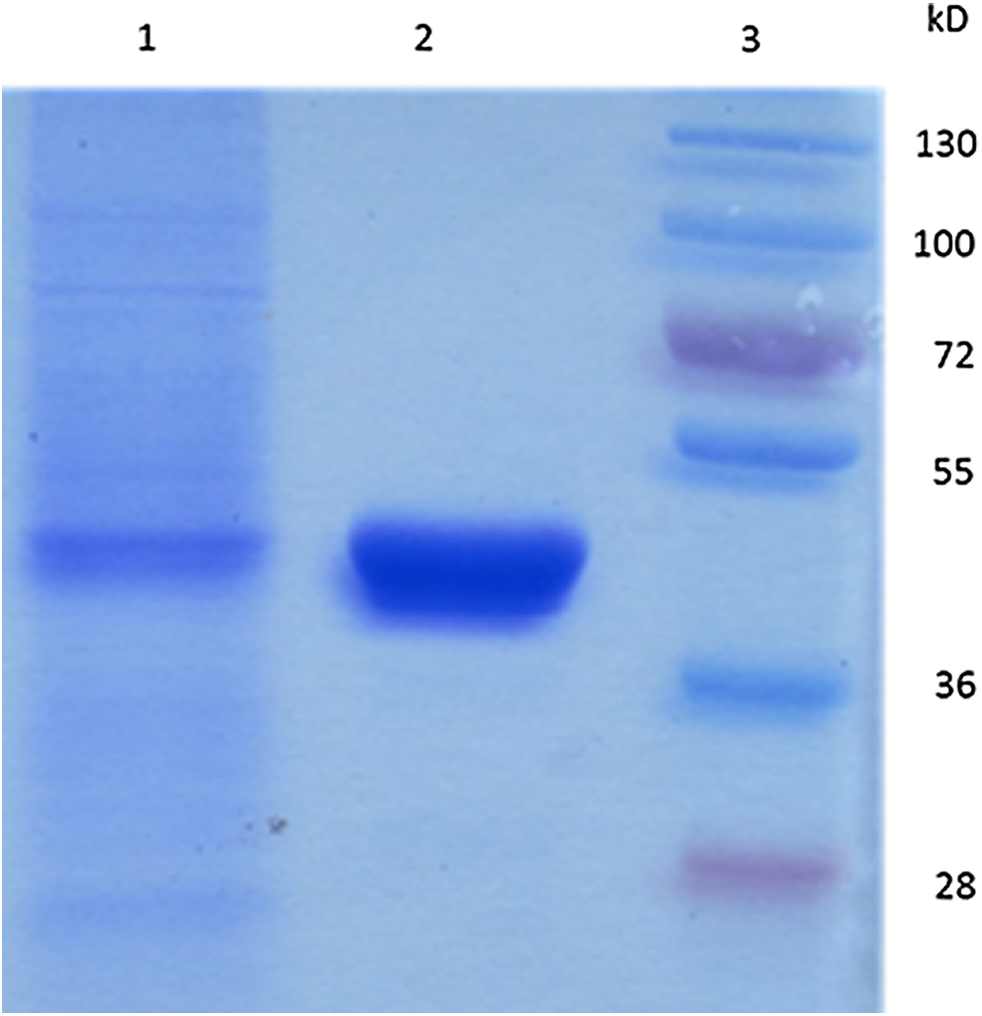
Expression and purification of rS1 protein analysis by SDS-PAGE. Lane 1, supernatant of sf9 insect cell culture; lane 2, purified S1 protein using immunoaffinity chromatography; lane 3, protein marker.
Generation of MAbs against PEDV rS1 protein
Indirect ELISA results showed that the antibody titers of immunized mice ranged from 1:6400 to 1:12,800 as compared to the negative control. The mouse that had the highest antibody titer was selected for splenic cell isolation. After fusion with SP2/0 myeloma cells, two positive hybridoma clones (designated 8A3A10 and 12C4G12) were identified. The isotypes of 8A3A10 and 12C4G12 were identified as IgG2a/κ and IgG1/κ, respectively.
Determination of MAb reactivity
Reactivity of the antibodies was analyzed by Western blot and IFA. As shown in Figure 2, both MAbs recognized PEDV rS1 protein with one major band at estimated protein molecular weight (Fig. 2, lanes 2, 3). However, the supernatant of SP2/0 myeloma cells had no such reactivity (Fig. 2, lane 1). As expected, the two MAbs also had no reaction with supernatant of sf9 cells (Fig. 2, lanes 4, 5). IFA was further used to test the reactivity of the MAbs, and the results showed that both MAbs reacted with PEDV-infected Vero cells exclusively while SP2/0 cell culture supernatant had no reactivity (Fig. 3). Therefore, the above results confirmed the reactivity of these two MAbs to PEDV rS1 protein.
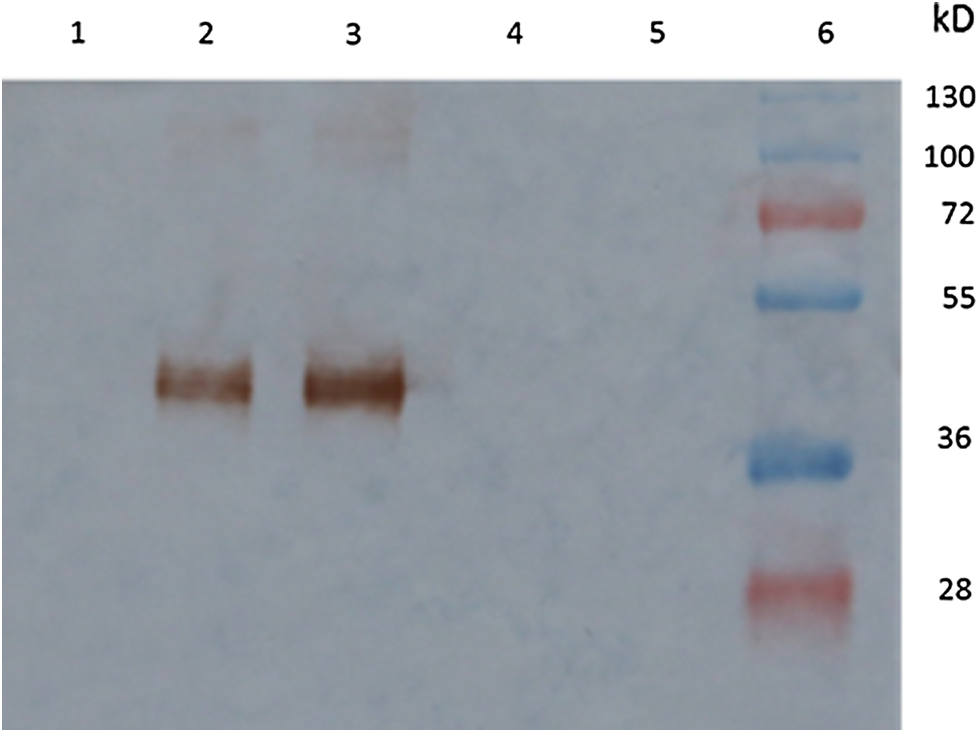
Specificity of monoclonal antibodies against rS1 protein analyzed by Western blot. Lane 1, supernatant of SP2/0 cells does not react with rS1 protein; lane 2, 8A3A10 MAb reacts with rS1 protein; lane 3, 12C4G12 MAb reacts with rS1 protein; lane 4, 8A3A10 MAb does not react with supernatant of sf9 cells; lane 5, 12C4G12 MAb does not react with supernatant of sf9 cells; lane 6, protein marker.

Specificity of monoclonal antibodies against S1 protein analyzed by IFA. (
Neutralizing ability of two MAbs
PEDV S1 protein contains three neutralizing epitopes (499-638aa, 748-755aa, and 764-771aa).(4) To test if the two generated MAbs have a neutralizing activity, fluorescent focus neutralization (FFN) assay was performed. It showed that MAb 8A3A10 (1:256 diluted) could completely neutralize 500 TCID50 PEDV virus after 1 h incubation at 37°C, as no fluorescence was detected 24 h post-infection (Table 1). By contrast, MAb 12C4G12 (1:4 diluted) did not show any neutralizing activity since apparent fluorescence appeared 24 h post-infection.
Ten microscopic fields of each dilution of samples were observed under fluorescence microscope. Positive control, Vero cells were infected with PEDV; negative control, healthy Vero cells. +, positive fluorescence signal; –, negative signal (90% or greater reduction of fluorescent foci relative to controls).
The PEDV S1 protein was previously expressed in Escherichia coli and was used to immunize mice for hybridoma production.(5,6) However, the recombinant protein expressed by prokaryocytes lacks post-translational modifications and molecular folding, which may hinder the recognition of neutralizing epitopes by antibodies. Therefore, in this study, the truncated recombinant S1 protein was expressed in Baculorvirus expression system for generation of the monoclonal antibodies. Indirect ELISA indicated that the purified rS1 protein could induce a high level of antibody titer in immunized mice. Two MAbs were generated by hybridoma technique and showed decent reactivity to the PEDV rS1 protein, as shown by the results of the Western blot and IFA. One of the MAbs (8A3A10) showed good neutralizing activity since it completely neutralized 500 TCID50 PEDV virus at 1:256 dilution.
In conclusion, the generation of neutralizing monoclonal antibodies against PEDV might be useful in the understanding of basic aspects of PEDV infection biology and immunity.
Footnotes
Acknowledgments
This work was supported by grants from the Major Science and Technology Program in Henan Province (no. 131100110200), Innovation Scientists and Technicians Troop Construction Projects of Henan Province (no. 142101510001), Talents Plan for Scientific and Technological Innovation in Henan Province (no. 144200510002), and Science and Technology Innovation Team in Henan Province (no. C20130005).
Author Disclosure Statement
The authors have no financial interests to disclose.