
Abstract
The maltose binding protein (MBP) is a commonly used protein tag. Two monoclonal antibodies (mAbs) were generated against the MBP by immunizing mice with purified 6xHis-tagged MBP (6xHis-MBP). A nontoxic adjuvant cocktail of poly(I:C) and anti-CD40 mAb was used. The two mAbs, 3D7 and 2A1, are demonstrated to be effective in immunoprecipitation, immunoblotting, western blot hybridization, and the ELISA assay. These two mAbs are available individually or in combination at cost through the Developmental Studies Hybridoma Bank, a nonprofit National Resource created by the National Institutes of Health.
Introduction
T
We demonstrate here that the two anti-MBP mAbs work effectively in immunoblot assays of native MBP, western blot hybridization of denatured recombinant MBP and wild type MBP, IP, and the ELISA assay. Both mAbs, 2A1 and 3D7, as well as the recombinant MBP (6xhistidine [6xHis]-MBP) and two negative controls 6xHis-green fluorescent protein (GFP) and 6xHis-glyceraldehyde 3-phosphate dehydrogenase (GAPDH), are now available to basic researchers at cost through the Developmental Studies Hybridoma Bank, a nonprofit National Resource created by the National Institutes of Health.
Materials and Methods
Producing the recombinant protein immunogens
The plasmid vector pDB.His.MBP for bacterial expression of a 6xHis-tagged MBP (6xHis-MBP) was obtained from the DNASU plasmid repository at the Biodesign Institute of Arizona State University. The recombinant 6xHis-MBP was expressed in E.coli strain BL21 (Life Technologies, Carlsbad, CA) and purified from the cell lysates using nickel magnetic beads (
Mice and immunization
Two 8-week-old female BALB/c mice were immunized by intraperitoneal injection of 50 μg of purified recombinant 6xHis-MBP, combined with 50 μg of poly(I:C) HMW VacciGrade (InvivoGen, San Diego, CA) and 50 μg of anti-CD40 mAb (BioXCell, West Lebanon, NH) as the adjuvant,(13,14) in 200 μL of phosphate buffer solution (PBS; Gibco, Grand Island, NY). Mice were boosted 2 weeks after initial immunization by injection of 50 μg of recombinant 6xHis-MBP and 50 μg of poly(I:C) in 200 μL of PBS. Mice were bled 2 weeks after the boost, and sera tested at ≥1:500 dilutions for anti-MBP reactivity by western blot analysis. Three days before fusion, the mouse with the highest anti-MBP antibody titer was injected intravascularly with 50 μg of 6xHis-MBP and 50 μg of poly(I:C) in 100 μL of PBS.
Hybridoma fusion
The hybridoma fusion was performed as described previously(14) with minor modification. In brief, spleen cells of the immunized mouse and Sp2/0-Ag14 myeloma cells (CRL-1581; ATCC, Manassas, VA)(15) were washed, passed through a 70 mm filter, and resuspended in serum-free RPMI 1640 medium (HyClone, Logan, Utah). Cells were mixed at a ratio of five nucleated splenocytes to one myeloma cell (5:1) and pelleted. The cell pellet was dispersed with gentle mixing and warming to 37°C for 1 minute. Then, 1 mL of prewarmed 50% (v/v) PEG-RPMI 1640 medium was gently added. Cells were gently mixed for two additional minutes at 37°C and 15 mL of warm serum-free RPMI 1640 medium added dropwise over the next 90 seconds. Cells were incubated without agitation for 8 minutes at room temperature, placed in a 37°C water bath for 2 minutes, and pelleted by centrifugation at 200 g for 5 minutes. The supernatant was removed and the pellet dispersed with gentle mixing. Cells were gently dispersed by pipetting into 30 mL of the hybridoma culture medium IMDM (HyClone), containing 15% fetal bovine serum (FBS; Hyclone), 20% RPMI 1640 medium conditioned by giant cell tumor (TIB-223; ATCC),(16) and 1× HT-Hybri-Max. The suspension of cells was transferred to T75 flasks. Cells were incubated overnight at 37°C and 100 μL of the cell suspension added to each well in 96-well tissue culture plates. Hybridomas were selected by adding 100 μL of 2× hypoxanthine-aminopterin-thymidine (HAT; Sigma-Aldrich, St. Louis, MO) medium to each well and exchanging 100 μL of medium with 100 μL of fresh 1× HAT medium twice every 3 days. When cells grew to 25%–50% confluency, supernatants were tested for mouse IgG production by dot blot analysis (Bio-Rad, Hercules, CA). Positive hybridomas were expanded and supernatants retested for MBP reactivity by western blot analysis. Hybridomas producing anti-MBP-specific antibodies were further expanded and then subcloned by fluorescence-activated cell sorting using propidium iodide (Life Technologies) staining for viability. Antibody isotypes were determined from culture supernatants using the Mouse mAb Isotyping Test Kit (AbD Serotec, Raleigh, NC), according to the manufacturer's protocols.
Dot blot
One microgram of each protein preparation was spotted onto dry nitrocellulose membranes and dried. Membranes were blocked with 5% w/v nonfat dry milk in 1× Tris-buffered saline with Tween-20 (TBS-T) (50 mM Tris-HCI, pH 7.6, 150 mM NaCl, 0.005% Tween-20) for 1 hour at room temperature. Membranes were then incubated in the blocking buffer containing 1 μg/mL of anti-MBP mAbs, obtained directly from hybridoma culture supernatants or rabbit polyclonal anti-MBP antibody AB-559988 (Thermo Fisher Scientific, Rockford, IL), on a rocker for 1 hour at room temperature. Membranes were then washed thrice in 1× TBS-T, incubated with either IR Dye 800-conjugated goat anti-mouse or goat anti-rabbit antibody (Li-COR Biosciences, Lincoln, NE) at a 1:10,000 dilution in the dark on a rocker for 1 hour at room temperature. The membranes were then washed thrice with 1× TBS-T and scanned in an Odyssey scanner (Li-COR Biosciences).
Western blot
Denatured samples of 1–2 μg of purified proteins or 2–5 μg of cell lysates were separated in a Tris/Glycine/sodium dodecyl sulfate–polyacrylamide gel electrophoresis (SDS-PAGE) gel (Bio-Rad) with dual-color molecular weight markers (Bio-Rad). The gel was then blotted onto a nitrocellulose membrane. After blocking, the membranes were incubated with 0.5–1 μg/mL of anti-MBP antibodies in blocking buffer for 1 hour at room temperature. Membranes were washed thrice with 1× TBS-T, incubated with either IR Dye 800-conjugated goat anti-mouse or goat anti-rabbit antibody (Li-COR Biosciences) at a 1:10,000 dilution in the dark on a rocker for 1 hour at room temperature. The membranes were finally washed three additional times with 1× TBS-T and scanned in an Odyssey scanner (Li-COR Biosciences).
Immunoprecipitation
Recombinant MBP protein fused to a fragment of the mouse Ror2 receptor (MBP-Ror2)(12) was expressed from the bacterial vector pMal-Ror2 in E. coli BL21 strain. This served as the test protein for anti-MBP mAbs. Overnight, E. coli suspension cultures were inoculated in 200 mL of LB medium (1% Bacto-tryptone, 0.5% yeast extract, and 1% NaCl) supplemented with 100 μg/mL ampicillin to OD600 = 0.1. This culture was grown in a water bath shaker at 37°C at 250 rpm until the OD600 reached 0.3–0.4. The expression of MBP-Ror2 protein was induced by further incubation with 1 mM IPTG for 3 hours. Cells were pelleted in 10 tubes and one of the pellets resuspended in 800 μL of lysis buffer (50 mM NaH2PO4 [pH 7.4], 150 mM NaCl, 1 mM EDTA, 5% glycerol, 0.2% NP-40,10 mM MgCl2,1 unit DNase I, 2 mg/mL RNase, 0.1 mg/mL lysozyme, and protease inhibitor cocktail [Roche]). The suspension was incubated at 37°C for 30 minutes and sonicated thrice for 20 seconds at 1 minute intervals on ice. Cell debris was removed by centrifugation at 10,000 rpm for 5 minutes. Cell lysates were diluted 1:10 with lysis buffer. MBP-Ror2 protein was immunoprecipitated from the diluted cell lysate using Dynabeads Protein G (Life Technologies) bound to the generated anti-MBP mAbs. An aliquot of the supernatants, containing 10 μg of anti-MBP mAbs, was used for each IP reaction. The eluted immunoprecipitates were separated by SDS-PAGE, stained with Coomassie blue, and analyzed also by western blotting. MBP precipitates on the western blot were detected with rabbit polyclonal anti-MBP antibody (Thermo Fisher Scientific). The Odyssey dye IR Dye 800-conjugated goat anti-rabbit antibody (Li-COR Biosciences) was used as the secondary antibody. Protein bands on the western blots were detected with an Odyssey scanner. This analysis was performed thrice with highly similar results.
ELISA
In a 96-well microtiter plate, 2 μg of the purified 6xHis-MBP antigen in 100 μL of 0.05 M sodium carbonate coating buffer (pH 9.6) was added to the top row of wells, and a 1:1 serial dilution was performed through the wells below with sodium carbonate buffer, to a final concentration of 0.016 μg of antigen per well. To coat the wells with the antigen protein, the plates were incubated for 2 hours at room temperature. The supernatant was then removed and the wells washed thrice with 200 μL of 1× TBS-T (50 mM Tris-HCI, pH 7.6, 150 mM NaCl, 0.05% Tween-20), to remove free antigen. The wells were then blocked overnight with 200 μL of blocking buffer (1% BSA in 1× TBS-T) at 4°C. Wells were washed thrice with 200 μL of 1× TBS-T and the wells of two sets of serial dilutions incubated with 0.1, 1, and 10 μg/mL of each of the two anti-MBP mAb in 100 μL of blocking buffer for 1 hour at room temperature. Wells were then washed thrice and incubated with horseradish peroxidase (HRP)-conjugated goat anti-mouse IgG1 (Jackson ImmunoResearch Laboratories, Inc.) for 1 hour at room temperature. After three additional washes, 100 μL of TMB substrate solution was added. After 20 minutes at room temperature, 100 μL of stop solution (0.18 M H2SO4) was added for color development. The absorbance of each well was read at 450 nm in SpectraMax Plus 384, a microplate reader (Molecular Devices, Sunnyvale, CA).
Results
Immunization and generation of hybridomas
Mice were immunized against 6xHis-MBP recombinant protein, using the novel water-soluble vaccine adjuvant cocktail of poly(I:C) and anti-CD40 antibody.(14) The mice were boosted 2 weeks later by the injection of 6xHis-MBP with poly(I:C). Two weeks after the boost, immunization against MBP was confirmed by immunoblot analysis with at least 1:500 diluted sera from immunized mice. Three days before myeloma fusion, one mouse was injected with 6xHis-MBP for a final boost. The immunization process was completed in 5 weeks. B cells from the spleen of the immunized mouse were then fused to Sp2/0-Ag14 myeloma cells.(15,17) After HAT selection, hybridomas were cloned and screened for anti-MBP antibody production. Two hybridoma clones, 2A1 and 3D7, producing anti-MBP antibodies, were expanded and their antibodies tested for performance. The immunoglobulin isotypes of both mAbs were IgG1.
Immunoblot and western blot analyses
The capacity of the two generated anti-MBP mAbs, 2A1 and 3D7, to identify native MBP was tested by an immunoblot assay using the native recombinant MBP proteins 6xHis-MBP and MBP-Ror2 (MBP fused to a portion of the mouse Ror2 receptor).(12) The results were compared to those obtained with a commercially available rabbit polyclonal antibody, RbAb anti-MBP (Thermo Fisher Scientific). The purified recombinant 6xHis-MBP protein, which was used for mouse immunization, and an E. coli lysate containing native MBP-Ror2 protein were dotted on nitrocellulose membrane. Two purified 6xHis-tagged recombinant proteins, 6xHis-GFP and 6xHis-GAPDH, were dotted as negative controls. These controls were also used to test specificity and rule out the possibility that the mAbs recognized the 6xHis-tag. The MBP-Ror2 and 6xHis-MBP dot blots exhibited strong signals with the two mAbs, as well as the commercial polyclonal antibody (Fig. 1A). The dot blots of the two control 6xHis-tagged recombinant proteins, 6xHis-GFP and 6xHis-GAPDH, were negative for all three antibodies (Fig. 1A).
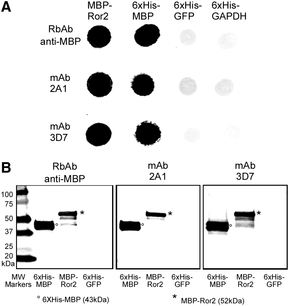
Immunoblot analysis of the two anti-MBP mAbs, 2A1 and 3D7, using native and denatured MBP recombinant proteins.
Next, we tested the ability of the anti-MBP mAbs to bind denatured MBP proteins by western blot analysis. The anti-MBP mAbs recognized denatured 6xHis-MBP (predicted molecular weight, 43 kDa) and MBP-Ror2 (predicted molecular weight, 52 kDa) recombinant proteins, but not the denatured 6xHis-tag of the recombinant 6xHis-GFP protein (predicted molecular weight, 28 kDa) (Fig. 1B). The mAbs also recognized the endogenous untagged MBP of the E. coli strain used to express MBP-Ror2, as a band at 42.5 kDa (Fig. 1B). Results with the commercial anti-MBP polyclonal antibody, RbAb anti-MBP, were similar to those with mAbs 2A1 and 3D7 (Fig. 1B). Both the dot blot and western blot analyses indicated that the two anti-MBP mAbs recognize the MBP, not the 6xHis-tag.
Immunoprecipitation
The two anti-MBP mAbs were tested for IP of the recombinant MBP protein fused to a fragment of mouse Ror2 receptor (MBP-Ror2).(12) IP of MBP was performed with the cell lysate using Dynabeads linked to protein G. As a control, the same IP process was performed with Dynabeads lacking mAbs. Immunoprecipitates were first analyzed by SDS-PAGE gels stained with Coomassie blue (Fig. 2A) and second by western blot hybridization (Fig. 2B). Both of the anti-MBP mAbs precipitated the 52 kDa MBP-Ror2 recombinant protein. Proteins, which were ∼27, 29, and 60 kDa, also stained in SDS-PAGE gels, probably represent proteins that were bound to MBP (Fig. 2A). Western blot hybridization also revealed that the two anti-MBP mAbs precipitated MBP-Ror2, as well as the endogenous 42.5 kDa untagged MBP (Fig. 2B). Control precipitate samples did not show any significant bands either in SDS-PAGE gel stained with Coomassie blue (Fig. 2A) or western blots (Fig. 2B).
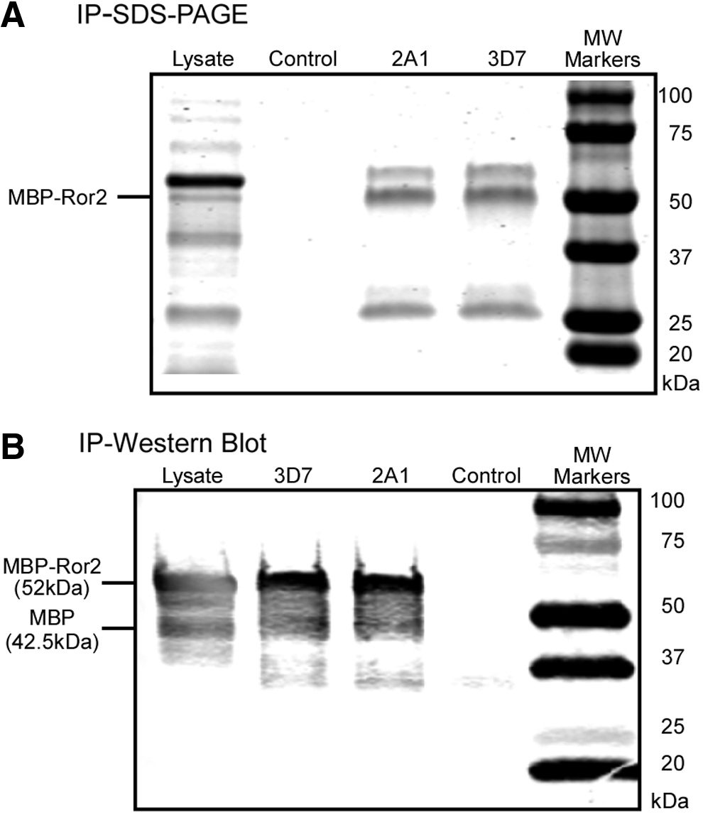
Immunoprecipitation (IP) of MBP-Ror2 recombinant protein by the anti-MBP mAbs, 2A1 and 3D7. IP was tested for MBP-Ror2 in a lysate of the E. coli strain expressing the recombinant protein.
ELISA
We next tested the anti-MBP mAbs for enzyme-linked immunosorbence in the ELISA. Purified 6xHis-MBP protein served as the antigen. Serial dilutions, spanning 2 μg–16 ng of 6xHis-MBP, were used to coat the wells of microtiter plates. Anti-MBP mAbs at concentrations of 0.1, 1.0, and 10.0 μg/mL were used in the assay. Binding was quantified at OD450 using HRP conjugated to goat anti-mouse IgG1. Background values, with 1× TBS alone, were subtracted from test values. Background values were lower than 0.001. Both anti-MBP mAbs, 2A1 and 3D7, bound to the substrate with high affinity (Fig. 3). Binding of 3D7 anti-MBP mAb, however, was stronger compared with 2A1 anti-MBP mAb (Fig. 3). The three concentrations of each mAb did not show significant differences. Therefore, 0.1 μg/mL of mAbs is suitable for a reliable ELISA for both mAbs (Fig. 3). A concentration of 0.1 μg/mL of 3D7 anti-MBP mAb was able to detect 30 ng of MBP antigen (Fig. 3).
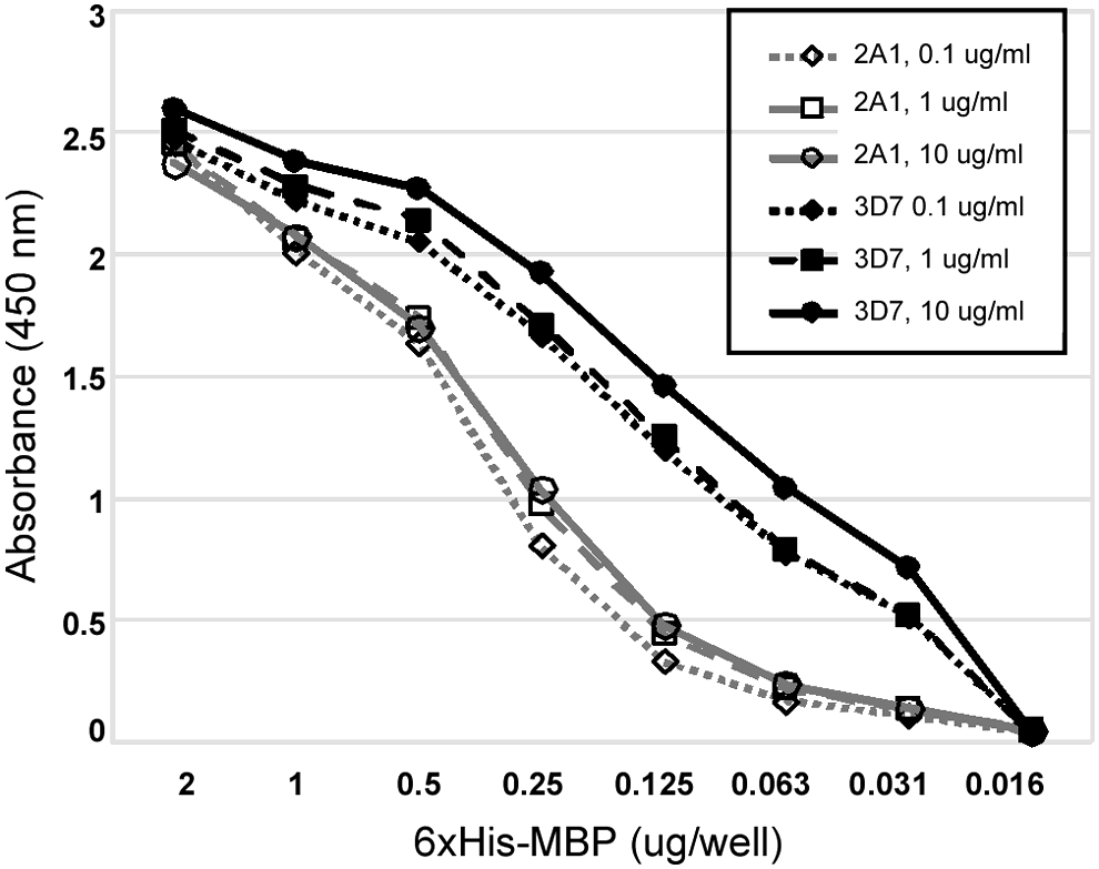
ELISA with anti-MBP mAbs, 2A1 and 3D7. Varying concentrations of purified 6xHis-MBP recombinant protein served as substrate for the ELISA, with anti-MBP mAbs 2A1 and 3D7 in microtiter plates. The mAb concentrations tested were 0.1, 1.0, and 10.0 μg/mL.
Discussion
The detailed characterization of mAbs in basic research has become a major issue given that a significant minority of commercial reagents have proven ineffective, at least in the hands of users. The investment in time and money in the application of an unverified or dysfunctional reagent usually outweighs the initial cost of purchasing it. We have, therefore, begun to generate mAbs that are verified in house. In this study, we have generated two anti-MBP mAbs that effectively identify MBP and MBP recombinant proteins in immunoblots, western blot analysis, and ELISA, and which are effective in IP. Since the mAbs identify the native state of MBP proteins, as well as denatured protein, they should be effective for DNA or chromatin IP (DIP or ChIP) and electrophoretic mobility shift assay, if MBP is tagged to a transcription factor or other DNA-binding protein. The two mAbs, 2A1 and 3D7, can be obtained at cost from the Developmental Studies Hybridoma Bank.
Footnotes
Acknowledgment
This research was funded by the Developmental Studies Hybridoma Bank (DSHB), a nonprofit National Resource created by the NIH. All antibodies can be obtained at cost from the DSHB for use in basic research (
Author Disclosure Statement
No competing financial interests exist.