
Abstract
Podoplanin (PDPN) is a type I transmembrane sialoglycoprotein, which is expressed in several normal cells, including lymphatic endothelial cells throughout the body, podocytes of the kidney, and lung type I alveolar cells of the lung. We have established many monoclonal antibodies (mAbs) against human PDPN, mouse PDPN, and rat PDPN. In addition, we recently produced an anti-rabbit PDPN mAb, PMab-32, which was established by immunizing mice with recombinant proteins of rabbit PDPN. Herein, we compared the reactivity of PMab-32 with that of newly established anti-rabbit PDPN mAbs, PMab-33 and PMab-21, against normal tissues in immunohistochemistry. PMab-32 reacted with podocytes, type I alveolar cells, and lymphatic endothelial cells, whereas PMab-33 detected only podocytes and type I alveolar cells but not lymphatic endothelial cells. PMab-21 was not useful in immunohistochemistry. We identified the epitope of PMab-32 and PMab-33 as Ser61-Ala68 of rabbit PDPN using western blot and flow cytometric analyses. In contrast, the epitope of PMab-21 was identified as Leu44-Glu48, which is corresponding to platelet aggregation-stimulating (PLAG) domain, indicating that Ser61-Ala68 of rabbit PDPN is a more appropriate epitope for immunohistochemistry compared with PLAG domain. PMab-32 could be useful for uncovering the function of rabbit PDPN.
Introduction
P
PDPN, also known as Aggrus,(16) activates platelet aggregation by binding to the C-type lectin-like receptor-2 (CLEC-2) on platelet.(14,17,18) The interaction with CLEC-2 was mainly observed at Glu47 and Asp48 in the platelet aggregation-stimulating (PLAG) domain and the α2,6-linked sialic acid at Thr52 of human PDPN.(19) PDPN possesses many other important functions. The interaction between PDPN and CLEC-2 facilitates blood–lymphatic vessel separation.(20) The actin cytoskeleton is rearranged in CLEC-2-expressing dendritic cells to promote efficient motility along PDPN-expressing stromal surfaces.(21) The signal from CLEC-2 to PDPN controls the contractility of fibroblastic reticular cells and lymph node microarchitecture.(22) The physical elasticity of lymph nodes is maintained by PDPN of stromal fibroblastic reticular cells.(23) PDPN is expressed in human fetal rib and chondrocytes of the proliferative and hypertrophic regions of the growth plate.(24) Binding of the cytoplasmic tail of PDPN to the ezrin, radixin, and moesin proteins may change cytoskeletal organization, which alters the phenotype of PDPN-expressing cells. This may contribute to morphological changes in the rudiment cartilages, which lead to the establishment of the primary ossification centers during the endochondral ossification.
The function of rabbit PDPN has not been clarified because no anti-rabbit PDPN monoclonal antibody (mAb) has been established. In this study, we investigated three anti-rabbit mAbs and determined the critical epitope for immunohistochemistry.
Materials and Methods
Cell lines, rabbit tissues, and animals
Chinese hamster ovary (CHO)-K1 and P3U1 were purchased from the American Type Culture Collection (ATCC, Manassas, VA). CHO-K1 cells were transfected with the rabbit PDPN (rabPDPN)-PA tag plasmid using a Gene Pulser Xcell Electroporation System (Bio-Rad Laboratories, Inc., Philadelphia, PA). CHO-K1, CHO/rabPDPN, and P3U1 were cultured in the RPMI 1640 medium (Nacalai Tesque, Inc., Kyoto, Japan) supplemented with 10% heat-inactivated fetal bovine serum (Thermo Fisher Scientific, Inc., Waltham, MA), 100 units/mL of penicillin, 100 μg/mL of streptomycin, and 25 μg/mL of amphotericin B (Nacalai Tesque, Inc.) at 37°C in a humidified atmosphere of 5% CO2 and 95% air. Female BALB/c mice (4 weeks old) were purchased from CLEA Japan (Tokyo, Japan). Animals for immunization were housed under pathogen-free conditions. The Animal Care and Use Committee of the Tohoku University approved the animal experiments described herein.
Hybridoma production
BALB/c mice were immunized by intraperitoneal (i.p.) injection of 100 μg of recombinant rabbit PDPN(25) or synthesized rabbit PDPN peptides (39–52 amino acids) together with Imject Alum (Thermo Fisher Scientific, Inc.). After several additional immunizations of 50 μg, a booster injection of 50 μg was given i.p. 2 days before spleen cells were harvested. The spleen cells were fused with P3U1 cells using GenomONE-CF (Ishihara Sangyo Kaisha, Ltd., Osaka, Japan) against immunization of recombinant rabbit PDPN or PEG1500 (Roche Diagnostics, Indianapolis, IN) against immunization of synthesized rabbit PDPN peptides. The hybridomas were grown in the RPMI medium with hypoxanthine, aminopterin, and thymidine selection medium supplement (Thermo Fisher Scientific, Inc.).
Enzyme-linked immunosorbent assay
Recombinant proteins of rabbit PDPN or synthesized rabbit PDPN peptides were immobilized on Nunc Maxisorp 96-well immunoplates (Thermo Fisher Scientific, Inc.) at 1 μg/mL for 30 minutes. After blocking with 1% bovine serum albumin (BSA)/phosphate-buffered saline (PBS), the plates were incubated with culture supernatant, followed by 1:3000 diluted peroxidase-conjugated anti-mouse IgG (Dako; Agilent Technologies, Inc., Santa Clara, CA). The enzymatic reaction was conducted with a 1-Step Ultra TMB-ELISA (Thermo Fisher Scientific, Inc.). The optical density was measured at 655 nm using an iMark Microplate Reader (Bio-Rad Laboratories, Inc.).
Immunohistochemical analyses
Rabbit normal tissues were obtained from the Yamagata University,(25) and one rabbit fibrosarcoma tissue was obtained from the North Lab. Four-micrometer-thick histological sections were deparaffinized in xylene and rehydrated. Then, they were autoclaved in citrate buffer (pH 6.0, Dako; Agilent Technologies, Inc.) for 20 minutes. Sections were incubated with PMab-32, PMab-33, and PMab-21 for 1 hour at room temperature, followed by treatment with EnVision+ Kit for 30 minutes (Dako; Agilent Technologies, Inc.). Color was developed using 3,3′-diaminobenzidine tetrahydrochloride (DAB, Dako; Agilent Technologies, Inc.) for 1 minute, and then, the sections were counterstained with hematoxylin (Wako Pure Chemical Industries Ltd., Osaka, Japan).
Production of rabbit PDPN mutants
The amplified rabbit PDPN cDNA was subcloned into a pCAG vector (Wako Pure Chemical Industries Ltd.), and a PA tag(26) was added at the N-terminus. Substitutions of amino acids to alanine in rabbit PDPN were performed using a HotStar HiFidelity polymerase chain reaction (PCR; QIAGEN, Inc., Hilden, Germany) with oligonucleotides containing the desired mutations. PCR fragments bearing the desired mutations were inserted into a pCAG vector using In-Fusion PCR Cloning Kit (Clontech, Palo Alto, CA). CHO-K1 cells were transfected with the plasmids using a Gene Pulser Xcell Electroporation System (Bio-Rad Laboratories, Inc.).
Western blot analyses
Cell lysates (10 μg) were boiled in sodium dodecyl sulfate sample buffer (Nacalai Tesque, Inc.). The proteins were electrophoresed on 5%–20% polyacrylamide gels (Wako Pure Chemical Industries Ltd.) and were transferred onto a polyvinylidene difluoride (PVDF) membrane (EMD Millipore Corp., Billerica, MA). After blocking with 4% skim milk (Nacalai Tesque, Inc.), the membrane was incubated with PMab-32, PMab-33, PMab-21, anti-PA tag (clone: NZ-1), and anti-β-actin (clone AC-15; Sigma-Aldrich Corp., St. Louis, MO) and then with peroxidase-conjugated anti-mouse or anti-rat antibodies (1:1000 diluted, Dako; Agilent Technologies, Inc.) and developed with the Pierce Western Blotting Substrate Plus (Thermo Fisher Scientific, Inc.) or the ImmunoStar LD Chemiluminescence Reagent (Wako Pure Chemical Industries Ltd.) using a Sayaca-Imager (DRC Co. Ltd., Tokyo, Japan). NZ-1 was previously developed against human PDPN(27) and has been used as anti-PA tag mAb.(26) NZ-1 does not cross-react with the other PDPN proteins, such as mouse PDPN, rat PDPN, and rabbit PDPN.
Flow cytometry
Cells were harvested by a brief exposure to 0.25% trypsin/1 mM EDTA (Nacalai Tesque, Inc.). After washing with 0.1% BSA/PBS, the cells were treated with PMab-32 (10 μg/mL), PMab-33 (10 μg/mL), PMab-21 (10 μg/mL), and anti-PA tag (clone: NZ-1, 1 μg/mL) for 30 minutes at 4°C, followed by treatment with Oregon Green-conjugated anti-mouse IgG or anti-rat IgG (1:1000 diluted; Thermo Fisher Scientific, Inc.). Fluorescence data were collected using a Cell Analyzer EC800 (Sony Corp., Tokyo, Japan).
Determination of the binding affinity using flow cytometry
CHO/rabPDPN (2 × 10⁵ cells) were resuspended in 100 μL of serially diluted antibodies (0.0061–100 μg/mL), followed by secondary anti-mouse IgG (Thermo Fisher Scientific, Inc.). Fluorescence data were collected using a Cell Analyzer (EC800; Sony Corp.). The dissociation constants (KD) were obtained by fitting the binding isotherms using the built-in one-site binding models in GraphPad Prism 6 (GraphPad Software, Inc., La Jolla, CA).
Results
Production of mAbs against rabbit PDPN
In this study, we used two different antigens for producing mAbs against rabbit PDPN: one is recombinant rabbit PDPN of full length; the other is the synthetic peptide (39–52 amino acids) of PLAG domains of rabbit PDPN. After immunizing mice with the recombinant protein, which was purified from CHO/rabPDPN cells, the enzyme-linked immunosorbent assay (ELISA) screening was performed. Twenty-four wells reacted with CHO/rabPDPN in flow cytometry. Seventeen of the 24 wells of IgG class were cloned using limiting dilution, and two clones, such as PMab-32 (mouse IgG1, kappa) and PMab-33 (mouse IgG1, kappa), were established. After immunizing mice with the synthetic peptide of PLAG domains of rabbit PDPN, we established 24 clones (PMab-8–PMab-31), which react with CHO/rabPDPN cells in flow cytometry.
Immunohistochemical analysis against normal tissues
First, we investigated whether PMab-32 and PMab-33 detect rabbit PDPN of normal tissues, such as the kidney, lung, and small intestine. As shown in Figure 1, PMab-32 reacted with podocytes of the kidney, type I alveolar cells of the lung, and lymphatic endothelial cells of the small intestine. In contrast, PMab-33 detected podocytes and type I alveolar cells but not lymphatic endothelial cells. Second, we used 24 clones (PMab-8–PMab-31) for immunohistochemistry. However, no mAb stained PDPN-expressing cells. Typical results are shown in Figure 1. One of those clones, PMab-21 did not stain any PDPN-expressing cells, such as podocytes, type I alveolar cells, and lymphatic endothelial cells.
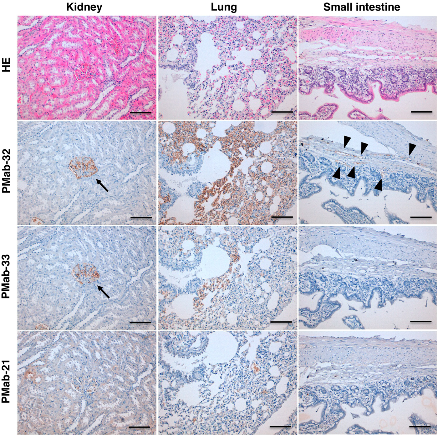
Immunohistochemical analysis against normal tissues by PMab-32, PMab-33, and PMab-21. Sections of the rabbit kidney, lung, and small intestine were incubated with 5 μg/mL of PMab-32, PMab-33, or PMab-21, followed by EnVision+ Kit, and color was developed using DAB and counterstained with hematoxylin. Arrows: glomerulus; arrowheads: lymphatic vessels. Scale bar: 100 μm. DAB, 3,3′-diaminobenzidine tetrahydrochloride.
Epitope mapping of anti-rabbit PDPN mAbs
Because PMab-32 and PMab-33 are useful for immunohistochemical analysis, we performed epitope mapping using western blot analysis. Figure 2A showed that several point mutants, such as S61A, G65A, T67A, and A68G, completely lost the reaction by PMab-32. In contrast, PMab-33 did not recognize the point mutants from Ser61 to Ala68. Flow cytometric analysis revealed that PMab-32 did not react with G65A (Fig. 2B). PMab-33 did not react with S61A and weakly reacted with G65A, T67A, and A68G. Taken together, the epitope of PMab-32 is Ser61-Ala68, and Gly65 is the most important amino acid. In contrast, the epitope of PMab-33 is Ser61-Ala68, and Ser61, Gly65, Thr67, and Ala68 are the most important amino acids. Flow cytometric analysis showed that the KD of PMab-32 and PMab-33 were determined to be 3.1 × 10⁻⁸ M and 9.8 × 10⁻⁸ M, respectively, demonstrating that the apparent binding affinity of PMab-32 is higher than that of PMab-33 (Fig. 2C).

Characterization of PMab-32 and PMab-33.
To determine the epitope of PMab-21, we performed western blot analysis. PMab-21 reaction was lost in point mutations of 44–48 amino acids (Fig. 3A). Flow cytometric analysis revealed that PMab-21 did not react with L44A, G46A, and V47A and weakly reacted with T45A and E48G (Fig. 3B). Taken together, the epitope of PMab-21 is Leu44-Glu48, and Leu44, Gly46, and Val47 are the most important amino acids. Flow cytometric analysis showed that the KD of PMab-21 was determined to be 7.8 × 10⁻⁹ M (Fig. 3C).
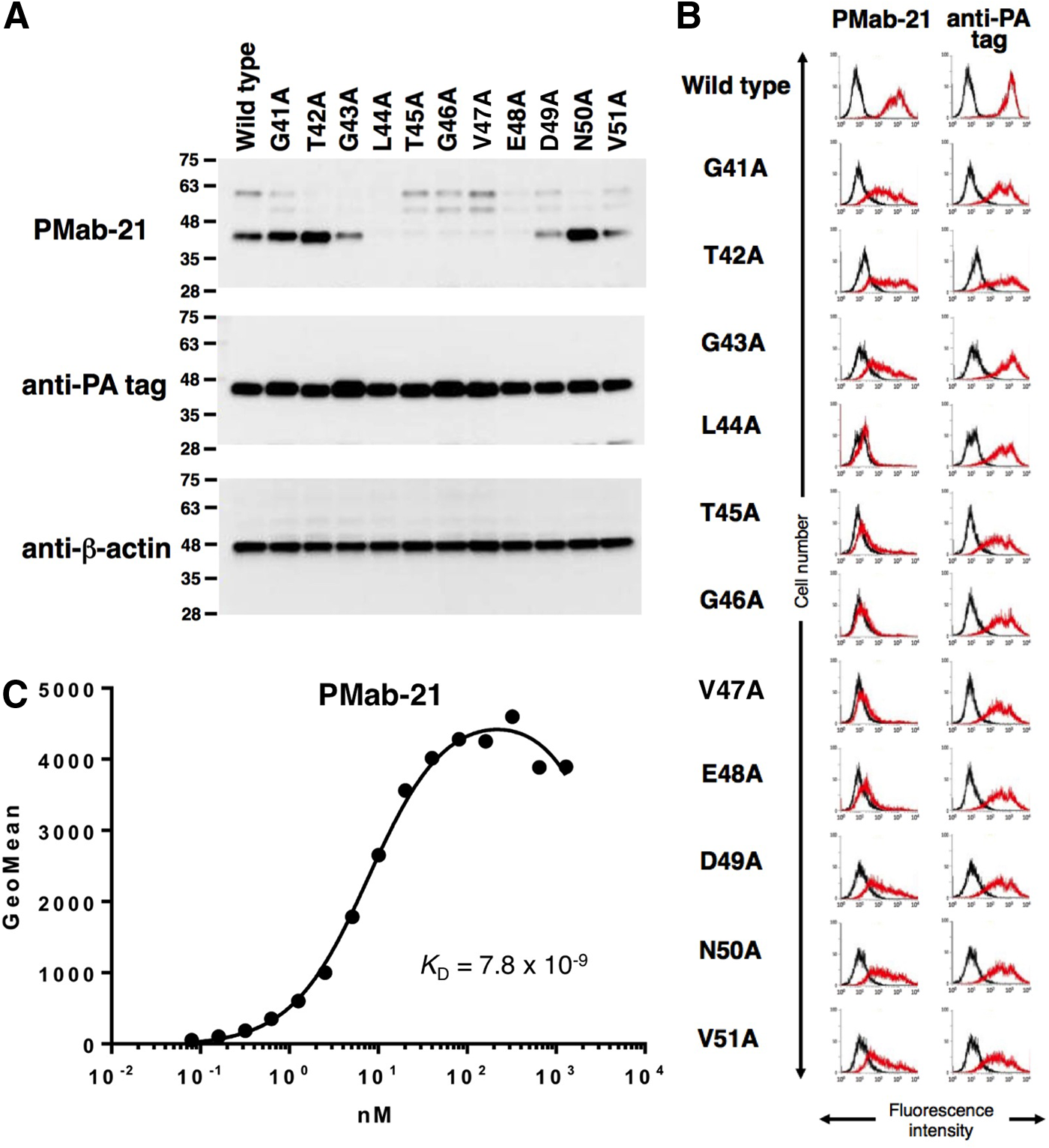
Characterization of PMab-21.
Immunohistochemical analysis against tumors
We finally investigated whether PMab-32 is applicable for immunohistochemical staining against rabbit tumors. PMab-32 stained lymphatic endothelial cells of fibrosarcoma tissues, whereas it did not recognize blood vessels (Fig. 4A). Of interest, PMab-32 also stained tumor cells of rabbit fibrosarcoma in a membranous staining pattern (Fig. 4B, C).
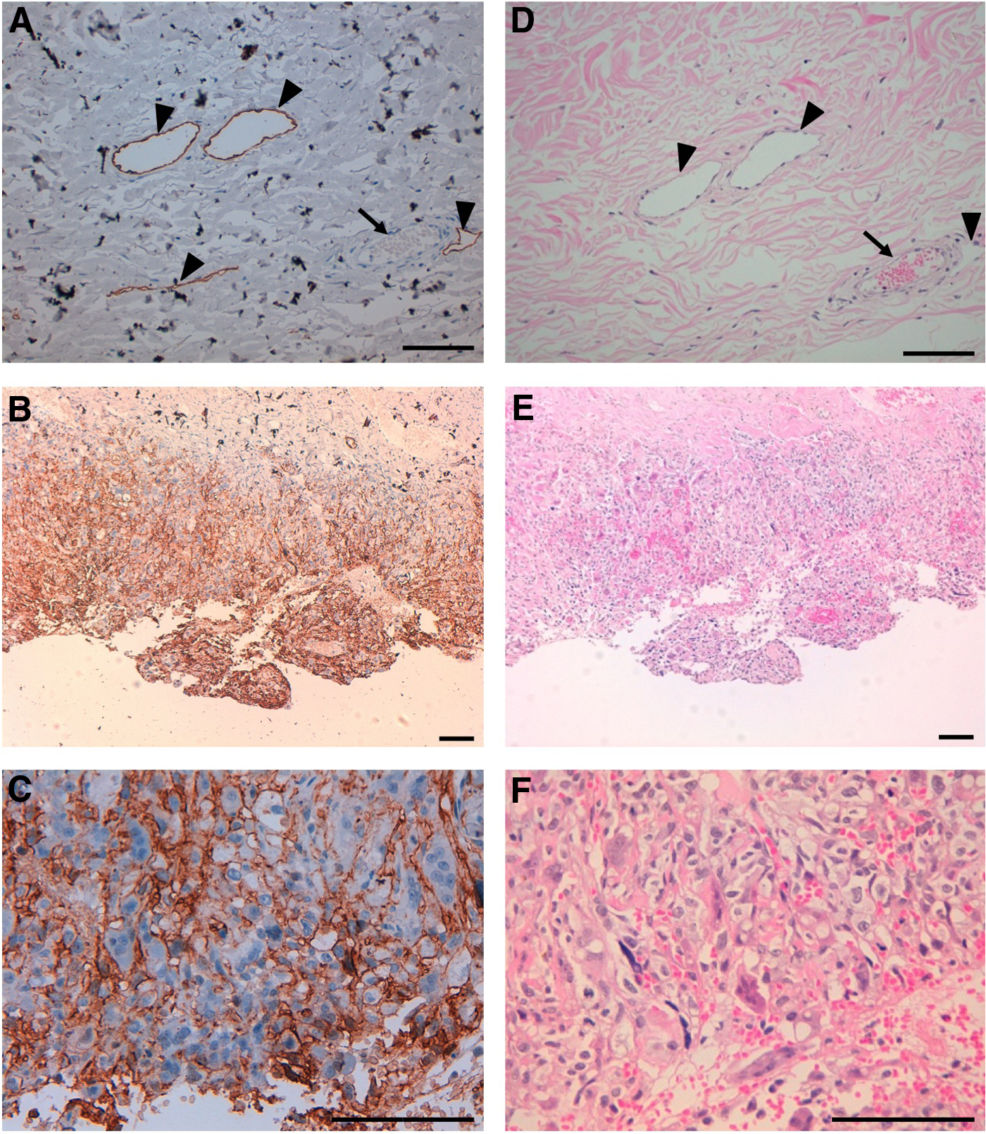
Immunohistochemical analysis against rabbit tumors using PMab-32.
Discussion
We have established mAbs against human PDPN,(6,27–31) mouse PDPN,(32) and rat PDPN.(33) In addition, we previously reported a novel anti-rabbit PDPN mAb, PMab-32, which was established by immunizing mice with recombinant proteins of rabbit PDPN.(25) Furthermore, anti-rabbit PDPN mAbs were established in this study and were well characterized. Many anti-human PDPN mAbs, such as LpMab-9, LpMab-10, NZ-1, and D2-40, react with PLAG domain of human PDPN.(30,31,34) In contrast, anti-human PDPN mAbs against non-PLAG domains, such as LpMab-2, LpMab-3, LpMab-7, and LpMab-17, are also useful for western blot, flow cytometry, and immunohistochemistry.(6,28,29,35) Especially, LpMab-7 possesses a much higher sensitivity in immunohistochemistry compared with NZ-1 and D2-40.(12)
Because anti-rabbit PDPN mAbs have not been established and characterized, we herein investigated three anti-rabbit PDPN mAbs and determined the critical epitope for immunohistochemistry. PMab-32 and PMab-33 are useful to detect rabbit PDPN of normal tissues in immunohistochemical analysis, although PMab-33 did not detect lymphatic endothelial cells. In contrast, 24 clones (PMab-8–PMab-31), which were established by immunizing PLAG domain, did not stain any PDPN-expressing cells (data not shown). The PLAG domains of PDPN have been known to be suitable for producing mAbs, which are useful for immunohistochemical analysis: NZ-1 mAb against human PLAG domain,(27) PMab-1 mAb against mouse PLAG domain,(32) and PMab-2 mAb against rat PLAG domain.(33) Therefore, PLAG domain of rabbit PDPN might be different from those of other species.
Epitope mapping of anti-rabbit PDPN mAbs demonstrated that the critical epitope of anti-rabbit PDPN mAb for immunohistochemistry is Ser61-Ala68, and Gly65 is the most important amino acid. The binding affinity of PMab-21 is much higher than those of PMab-32 and PMab-33, although PMab-21 is not applicable to immunohistochemistry. These results demonstrate that the high binding affinity is not sufficient for the usefulness in immunohistochemistry against rabbit PDPN. PMab-32 also stained lymphatic endothelial cells and tumor cells of fibrosarcoma,(36) demonstrating that PMab-32 could be useful for diagnostic tool for rabbit tumors.
In conclusion, PMab-32 is very useful for not only western blot analysis and flow cytometry but also immunohistochemical analysis. The critical epitope of anti-rabbit PDPN mAb for immunohistochemistry is Ser61-Ala68, and Gly65 is the most important amino acid. PMab-32 could be useful for uncovering the function of rabbit PDPN.
Footnotes
Acknowledgments
We thank Takuro Nakamura, Noriko Saidoh, Hazuki Kanno, Kanae Yoshida, and Eiko Saito for their excellent technical assistance. This work was supported in part by the Platform for Drug Discovery, Informatics, and Structural Life Science (PDIS) from the Japan Agency for Medical Research and Development, AMED (Y.K.); by the Basic Science and Platform Technology Program for Innovative Biological Medicine from AMED (Y.K.); by the Regional Innovation Strategy Support Program from the Ministry of Education, Culture, Sports, Science and Technology (MEXT) of Japan (Y.K.); by JSPS KAKENHI grant numbers 26440019 (M.K.K.) and 25462242 (Y.K.); and by the Takeda Science Foundation (S.O.).
Author Disclosure Statement
No competing financial interests exist.