
Abstract
Camel single domain antibody known as Nanobody™ refers to a novel class of monoclonal antibodies with appropriate pharmacological properties. Nanobody is an antigen-binding site of camel heavy chain antibody also known as VHH. Expression in a microbial system, stability in difficult conditions and extremes of PH, and nanomolar affinity to target an appropriate drug format makes Nanobody a potential for drug discovery. Needs for Nanobody function evaluation in animal models turned our interest to develop anti-mouse vascular endothelial growth factor (mVEGF) Nanobodies using phage display as a potent technique in the isolation of antibodies. Isolation of anti-mVEGF Nanobodies was performed on Camelus dromedarius immune library through four consecutive rounds of biopanning on immobilized mVEGF. Enrichment of the Nanobody library was monitored by polyclonal phage-ELISA, and specific Nanobodies were selected using periplasmic extract-ELISA. Selected Nanobodies were expressed in WK6 Escherichia coli cells and purified using immobilized metal affinity chromatography. Specificity and affinity of selected Nanobodies were evaluated on immobilized mVEGF. Results demonstrated the successful enrichment of the Nanobody library. Two clones named Nb5 and Nb10 were selected through screening procedures according to their signal value in periplasmic extract-ELISA. Selected Nanobodies specifically reacted to mVEGF, but cross-reactivity with other antigens was not observed. Evaluated affinity for the Nanobodies was in nanomolar range. Taken together, according to the results, the selected Nanobodies promise to be a novel tool in research and for further development of diagnostic or therapeutic purposes in pharmaceutical science.
Introduction
V
Materials and Methods
Materials
mVEGF was expressed and purified in our laboratory (Unpublished data). Briefly, the gene of mVEGF codon optimized for expression in Escherichia coli BL-21(DE3) and synthesized by Biomatik Co. Recombinant expression was induced by 1 mM of IPTG (Isopropyl β-D-1-thiogalactopyranoside). Purification of mVEGF was performed by Ni+-NTA resin (QIAGEN) according to the manufacturer's protocol. Anti-M13 HRP conjugated monoclonal antibody and anti-mouse HRP conjugated monoclonal antibody were purchased from Amersham-Pharmacia-Biotech. Anti-His HRP conjugated and Anti- hemagglutinin A (anti-HA) were from Roche. 4-chloro-1-naphthol (4-CN) substrate was purchased from Sigma. The nickel nitrilotriacetic acid (Ni+-NTA) resin was from QIAGEN. pHEN4 and pHEN6 vectors were kindly gifted by Serge Muyldermans (Laboratory of Cellular and Molecular Immunology, Vrije University Brussels, Brussels, Belgium). M13K07 helper phage was from Stratagene.
Phage displayed library amplification
Phagemid (pHEN4) library containing a Nanobody gene from a camel (Camelus dromedarius) immunized by mVEGF was transformed into electro-competent E. coli TG1.(18) Bacterial library with a complexity of 108 was converted to phage library after M13K07 (107 pfu/mL) infection. Nanobody library was displayed on phage as fusion to PIII of phage. Coating of all antigens was done at 4°C.
Enrichment of library
Selection of specific Nanobody against mVEGF was performed using four consecutive rounds of biopanning procedure. A 96-well plate (Nunc; MaxiSorp) was coated overnight with 10 μg/mL of mVEGF in sodium bicarbonate buffer (PH = 9.6), and a well coated only with sodium bicarbonate buffer (PH = 9.6) was used as negative control. On the next day, after blocking (skim milk 4% for 1 hour at RT) and washing with PBST (0.5% [V/V] Tween 20 in phosphate-buffered saline [PBS]), wells were incubated with 1012cfu of phage library at 37°C for 1 hour. Unbounded phage was removed by washing of wells with PBST (0.5% [V/V] Tween 20 in PBS). Bounded phages were eluted by 100 mM triethylamine (TEA, pH 10.0) and incubation was performed for 10 minutes at RT. Triethylamine was neutralized by 1 M Tris- HCl (pH 8.0). Eluted phages (output phages) were amplified into TG1 host (log phase) and rescued by M13K07 helper phage infection. Recombinant phages were purified by PEG/NaCl (20% polyethylene glycol 6000 and 2.5 M NaCl) precipitation. Amplified phages (input phages) were used for further rounds of biopanning. Biopanning was performed for four consecutive rounds according to the abovementioned procedure. Stringency of biopanning conditions increased through increasing of Tween 20 concentration (0.5%, 1%, 2%, and 4%) in washing buffer. After each round of biopanning, titration of eluted phages was performed on TYE medium containing ampicillin for enrichment estimation.
Enrichment estimation
Enrichment of the Nanobody library was monitored by titration and polyclonal phage ELISA. In the polyclonal phage ELISA, a 96-well plate was coated overnight with 1 μg/mL of mVEGF in sodium bicarbonate buffer and also wells coated with sodium bicarbonate buffer as negative control. The next day, wells were blocked with skim milk 4% and incubated for 1 hour at RT. After washing with PBST, wells were incubated at 37°C in RT with 1012 cfu of input phages from each round of biopanning. Anti M13-HRP antibody (1:2500, resuspended in PBS) was added into washed wells and incubated for 1 hour at RT. Peroxide activity of HRP was detected by adding substrate of TMB (3, 3′, 5, 5′-tetramethylbenzidine) and then the reaction was stopped by H2SO4. Finally, absorbance was measured at 450 nm using an ELISA plate reader (Epoch BioTek).
Screening of mVEGF-specific Nanobody
Specific clones against mVEGF were isolated through periplasmic extract ELISA. pHEN4 phagemid contains PelB signal (for secretion of Nanobody into periplasmic space) and HA tag (hemagglutinin, for detection in ELISA). Forty-seven colonies were randomly selected from output phages (fourth round) and cultured in 1 mL of LB broth until reaching log phase (OD600 nm = 0.7–0.9). Expression of Nanobody-PIII fusion was induced by 1 mM of IPTG (Isopropyl β-D-1-thiogalactopyranoside). Periplasmic proteins were extracted by osmotic shock and added to wells containing immobilized mVEGF (1 μg/mL) and negative wells (wells coated with sodium bicarbonate buffer). Bound Nanobodies were detected by anti-HA (1:2000 in PBS) antibody followed by anti-mouse HRP conjugated antibody (1:5000 in PBS). The clones that showed higher signal (three times or more) were considered positive and were sequenced to identify the integrity of the Nanobody gene.
Expression and purification of mVEGF-specific Nanobodies
The sequence of Nanobodies was aligned using MEGA-5 software. Frame work and CDR regions were determined according to IMGT numbering. Nanobody genes were subcloned in pHEN6c expression vector and transformed into E. coli WK6.(18,19) Expression of c-terminal His-tag fusion Nanobodies was induced by 1 mM of IPTG and incubated at 28°C for 16 hour with shaking at 200 rpm. Periplasm extracted Nanobody was purified by Ni+-NTA resin. Yield of protein concentration was determined by absorbance of UV 280 nm. The purity of expressed Nanobodies was determined by Coomassie brilliant blue stained 15% SDS-PAGE and Western blot. Anti-His HRP conjugated antibody (1:500 in PBS) was used for Nanobody detection and 4-CN substrate was used as developer.
Specificity
For analyzing the specificity of Nanobodies to mVEGF, different antigens such as bovine serum albumin (BSA), casein, mVEGF (R&D), hVEGF (R&D), expressed mVEGF, epidermal growth factor (EGF), and basic fibroblast growth factor (R&D) were coated overnight (10 μg/mL). Next day, wells were blocked with skim milk 4% for 1 hour at RT. Then, wells were washed with PBST. Nanobody binding was detected by anti-His HRP conjugated antibody. Absorbance of wells was measured at 450 nm.
Affinity determination
Affinity of Nanobodies was determined according to the procedure of Beatty et al.(20) In a checker board assay, two different concentrations of mVEGF and BSA (negative control) (1 and 10 μg/mL) were selected and coated overnight. Various concentrations of Nanobodies (0.1–100 nM) were added to each well and incubation was performed for 1 hour at 37°C. Nanobody binding was detected by anti-His HRP conjugated (1:500 in PBS) antibody. Affinity was measured according to Beatty's equation:
K aff = n − 1/2(n[Nb1] − [Nb2]) and n = [Ag1]/[Ag2], where [Ag1]: concentration of 10 μg/mL mVEGF, [Ag2]: concentration of 1 μg/mL mVEGF, [Nb1]: concentrations of Nanobody at 50% binding in the Ag1 curves, and [Nb2]: refer to the concentrations of Nanobody at 50% binding in the Ag2 curves.
Results
Enrichment results of Nanobody library
Results of output phage titration are shown in Table 1. These results indicated the success of biopanning and enrichment of the library against immobilized mVEGF. As shown in Table 1, phage particles increased after each round of biopanning, indicating successful enrichment of the library against immobilized mVEGF. Indeed, polyclonal phage ELISA results confirmed the progress of biopanning. With increasing biopanning rounds, a significant increase in signal intensity was observed in the fourth round (Fig. 1). Therefore, to achieve Nanobody with high affinity and specificity to target, colonies in round four were randomly selected and screened.
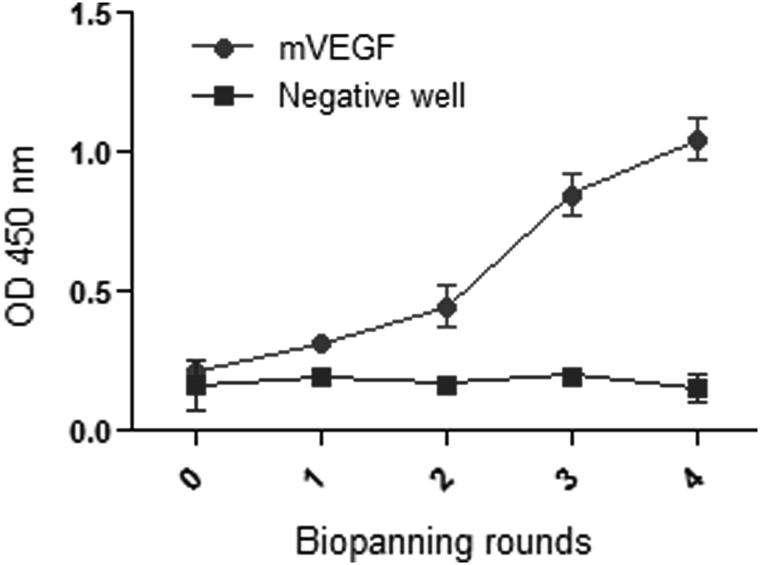
Polyclonal phage ELISA results. The input phages after each round of biopanning were monitored against immobilized mouse vascular endothelial growth factor (mVEGF) and negative well (well containing sodium bicarbonate as blank well). The assay was performed in triplicate ± SD. mVEGF, SD, standard deviation.
Isolation of mVEGF-specific Nanobodies
Screening of mVEGF-specific Nanobody by periplasmic extract ELISA showed that 5 of 47 colonies specifically reacted to immobilized mVEGF according to their high signal intensity (Fig. 2). Two clones named Nb5 (NCBI accession number = KR633139) and Nb10 (NCBI accession number = KR633140) were randomly selected from output phages of the fourth round and then sequenced. Sequencing results revealed that these Nanobodies have a unique sequence with hallmark amino acid of VHH in their frame work 2.
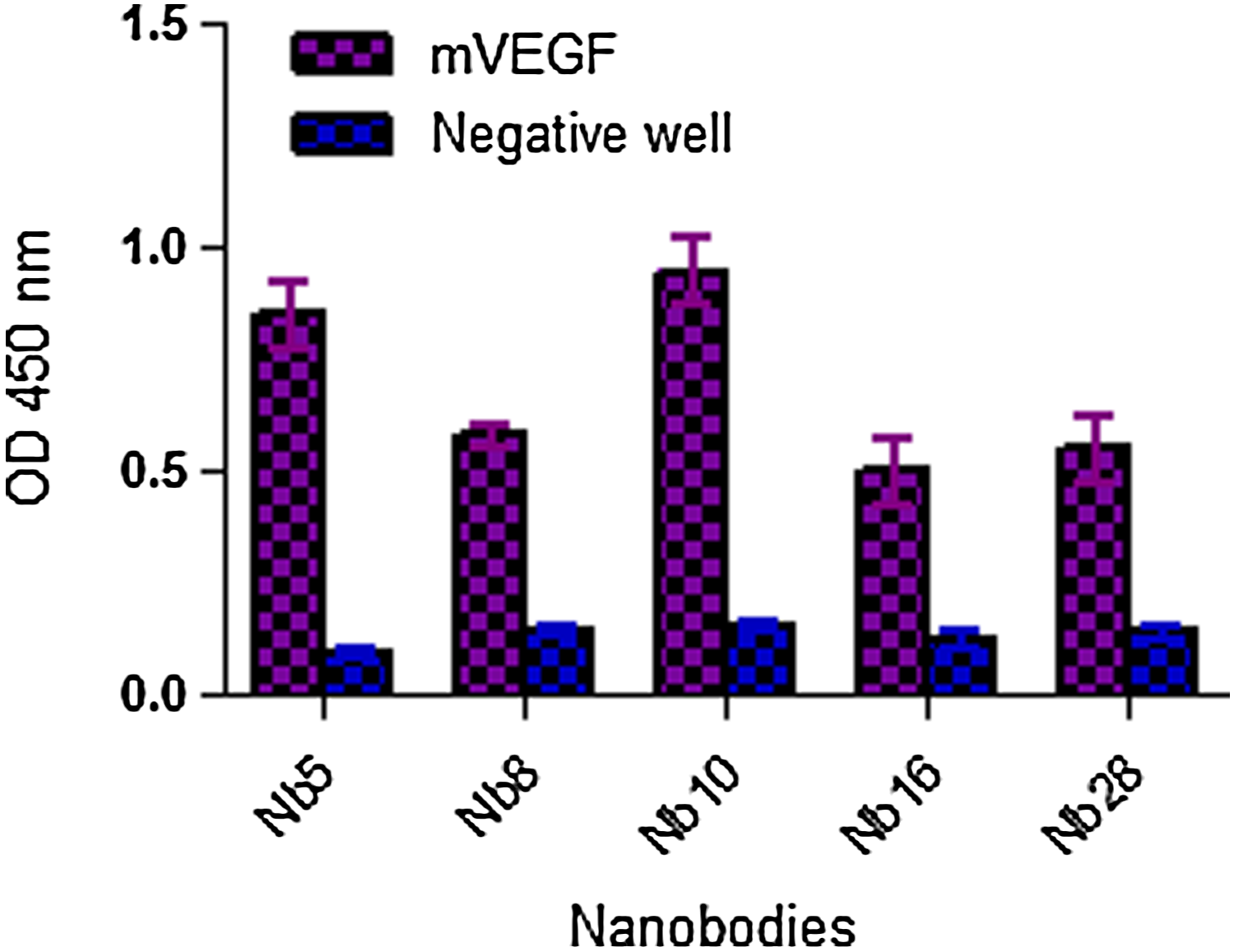
Screening results of Nanobodies. Screening was performed through periplasmic extract ELISA. Five colonies strongly reacted by immobilized mVEGF and were referred as positive clones. Negative well; well containing sodium bicarbonate. The graph represents the mean of triplicate of each assay ± SD.
Expression and purification results of mVEGF-specific Nanobodies
Specific Nanobodies were subcloned into pHEN6c vector and expressed as His-tag fusion proteins in WK6 E. coli cells, and expression was induced by 1 mM IPTG. Purification was performed on periplasmic proteins through nickel affinity chromatography. Purification was confirmed by SDS-PAGE 15% (Fig. 3a) and Western blot analysis (Fig. 3b). A band of ∼15 kDa refers to Nanobody in SDS-PAGE and Western blot. The anti-His HRP conjugated antibody was used for detection of Nanobody in Western blot.
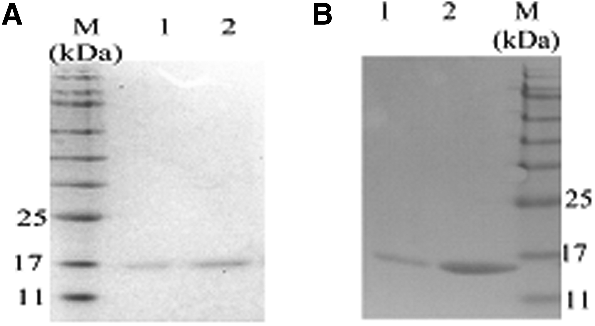
Purification results of Nanobodies.
Characterization of mVEGF-specific Nanobodies
Binding specificity of selected Nanobodies was evaluated using various antigens, and results revealed that both Nanobodies specifically react with mVEGF (Standard, R&D) and recombinant mVEGF (expressed in this study) and don't show cross-reactivity with other antigens (Fig. 4). Affinity of Nanobodies was determined according to the Beatty method (Table 2). Determined affinity for Nb5 and Nb10 was 1.3 and 9 nM, respectively. Affinity graphs are shown in Figure 5.
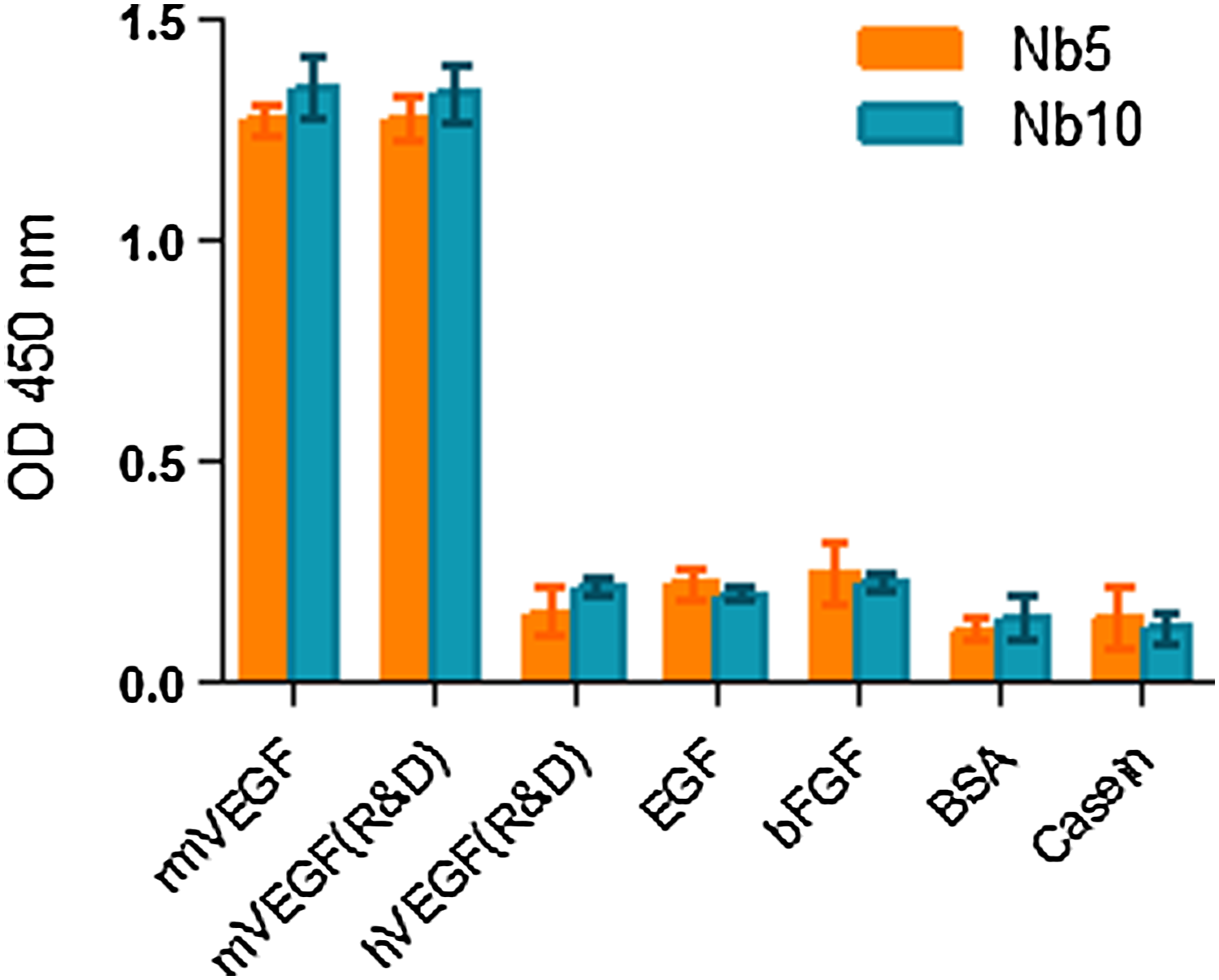
Binding specificity results. Selected Nanobodies were specific to rmVEGF (recombinant mVEGF expressed in this study) and mVEGF(R&D). Data are expressed as triplicate mean ± SD.
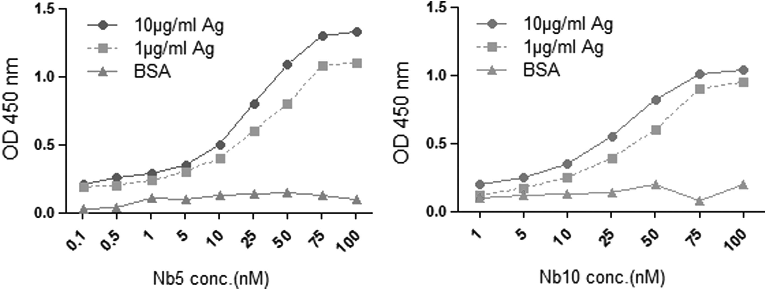
Affinity graphs. Affinity was calculated according to Beatty method. Affinity of Nb5 and Nb10 was 1.3 and 9 nM, respectively. Bovine serum albumin was used as negative control. Graphs are representative for mean of triplicate assay.
Discussion
Currently, there are two approaches for angiogenesis inhibition as follows: therapeutics targeting VEGF(21,22) and VEGF receptors. Many researchers are trying to develop novel therapeutics to efficiently target VEGF. The main aim of the current study was to develop and characterize specific Nanobodies against mVEGF. For this, we used a Camelus dromedarius hyperimmunized Nanobody library and isolated mVEGF-specific Nanobodies through four consecutive rounds of biopanning on immobilized mVEGF. There are two ways for increasing the stringency of biopanning as follows: 1- increasing the washing step and 2- increasing the concentration of Tween 20 in washing buffer. To isolate high affinity and also the specificity of Nanobodies to immobilized mVEGF, we increased the stringency of biopanning conditions by increasing the Tween 20 concentration in washing buffer. Enrichment estimation of the Nanobody library was performed by output phage titration and polyclonal phage ELISA. Results of phage titration and polyclonal phage ELISA revealed the progress of the biopanning procedure on immobilized mVEGF. According to the observed signal intensity in the polyclonal phage ELISA, the phage of the fourth round was screened for specific binding to immobilized mVEGF through periplasmic extract monoclonal phage ELISA. Five positive clones (clones with high signal value in periplasmic extract ELISA) were detected. Two of the five positive clones were randomly selected and sequenced. Sequencing results revealed that selected Nanobodies (Nb5 and Nb10) have different CRD3 sequences, indicating that different lymphocytic clones were stimulated.(17,18) For expression, these Nanobodies were subcloned into pHEN6c expression vector. Purification of Nanobodies was performed using nickel affinity chromatography. Binding specificity results showed that the selected Nanobodies (Nb5 and Nb10) specifically detected and reacted with mVEGF, but not other antigens. The measured affinity for these Nanobodies was in agreement with the affinity achieved in other related Nanobody studies.(13,17–19,23) Single domain entity and having small molecular weight make Nanobodies promising tools in diagnostic and therapeutic applications. Several studies have been reported describing the isolation of human VEGF-specific Nanobody,(24,25) including our previous study,(18) but not mouse VEGF. Therefore, this is the first report on the development of Nanobodies against mVEGF. In fact, Nanobodies are of interest for our research team and have been developed for diagnostic or therapeutic purposes.(13,17–19) We developed an anti-mVEGF-specific Nanobody to evaluate its antiangiogenic effect in an animal model in further studies. In conclusion, as need of research to animal model evaluation, we developed and characterized two specific Nanobodies (Nb5 and Nb10) against mVEGF using phage display. Nanobody library was panned through four consecutive rounds of biopanning against immobilized mVEGF. The selected Nanobodies specifically detected mVEGF in cross-reactivity assay and showed high affinity to immobilized mVEGF. The developed anti-mVEGF Nanobodies promise to be potential tools for diagnostic or therapeutic research purposes.
Footnotes
Acknowledgment
This work was financially supported by Pasteur Institute of Iran, Tehran, Iran.
Author Disclosure Statement
No competing financial interests exist.