
Abstract
Podoplanin (PDPN)/Aggrus is a type I transmembrane O-glycoprotein, which is expressed in several normal tissues including podocytes of kidney and lymphatic endothelial cells. PDPN activates platelet aggregation by binding to C-type lectin-like receptor-2 (CLEC-2) on platelet; however, only bovine PDPN (bovPDPN) does not possess the platelet-aggregating activity. Although many monoclonal antibodies (mAbs) against human PDPN, mouse PDPN, rat PDPN, and rabbit PDPN have been established, anti-bovPDPN mAbs have not been developed. In this study, we immunized mice with the recombinant proteins of bovPDPN and developed anti-bovPDPN mAbs, which are useful in immunohistochemical analysis. One of the clones, PMab-44, is useful for detecting podocytes and lymphatic endothelial cells in normal bovine tissues. PMab-44 also detected bovPDPN specifically in flow cytometry. PMab-44 is expected to be useful for investigating the function of bovPDPN.
Introduction
P
Materials and Methods
Cell lines and animals
Chinese hamster ovary (CHO)-K1, Madin–Darby bovine kidney (MDBK), and P3U1 were purchased from the American Type Culture Collection (ATCC, Manassas, VA). CHO-K1, stable CHO transfectants, and P3U1 were cultured in RPMI 1640 medium (Nacalai Tesque, Inc. (Nacalai), Kyoto, Japan), and MDBK was cultured in Dulbecco's Modified Eagle's Medium (Nacalai), supplemented with 10% heat-inactivated fetal bovine serum (Thermo Fisher Scientific, Inc. (Thermo), Waltham, MA), 100 U/mL of penicillin, 100 μg/mL of streptomycin, and 25 μg/mL of amphotericin B (Nacalai) at 37°C in a humidified atmosphere of 5% CO2 and 95% air. Female BALB/c mice (four weeks old) were purchased from CLEA Japan (Tokyo, Japan). The animals were housed under pathogen-free conditions. The Animal Care and Use Committee of Tohoku University approved the animal experiments described herein.
Hybridoma production
Bovine PDPN with N-terminal PA tag and C-terminal RAP tag-MAP tag (PA-bovPDPN-RAP-MAP) was inserted into pCAG-zeo vector (Wako Pure Chemical Industries Ltd. (Wako), Osaka, Japan). RAP tag consists of 12 amino acids (DMVNPGLEDRIE) and MAP tag consists of 12 amino acids (GDGMVPPGIEDK). CHO-K1 was transfected with pCAG-zeo/PA-bovPDPN-RAP-MAP using Gene Pulser Xcell electroporation system (Bio-Rad Laboratories, Inc. (Bio-Rad), Berkeley, CA). For the purification of PA-bovPDPN-RAP-MAP from cell membrane, we used the PA tag system.(7,8) BALB/c mice were immunized by intraperitoneal (i.p.) injection of 100 μg of recombinant PA-bovPDPN-RAP-MAP together with Imject Alum (Thermo). After several additional immunizations of 50 μg, a booster injection of 100 μg was given i.p. 2 days before spleen cells were harvested. The spleen cells were fused with P3U1 cells using PEG1500 (Roche Diagnostics, Indianapolis, IN). The hybridomas were grown in RPMI medium with hypoxanthine, aminopterin, and thymidine selection medium supplement (Thermo). Recombinant proteins of PA-bovPDPN-RAP-MAP were immobilized on Nunc Maxisorp 96-well immunoplates (Thermo) at 1 μg/mL for 30 minutes. After blocking with 1% BSA in 0.05% Tween20/phosphate-buffered saline (PBS), the plates were incubated with culture supernatant followed by 1:3000 diluted peroxidase-conjugated anti-mouse IgG (Dako; Agilent Technologies, Inc., Santa Clara, CA). The enzymatic reaction was conducted with a 1-Step Ultra TMB-ELISA (Thermo). The optical density was measured at 655 nm using an iMark microplate reader (Bio-Rad).
Immunohistochemical analyses
Four micrometer-thick histologic sections were deparaffinized in xylene and rehydrated, and were autoclaved in citrate buffer (pH 6.0; Dako) for 20 minutes. Sections were incubated with 1 μg/mL of primary mAbs for 1 hour at room temperature followed by treatment with Envision+ kit for 30 minutes (Dako). Color was developed using 3,3-diaminobenzidine tetrahydrochloride (DAB; Dako) for 1 minute, and then the sections were counterstained with hematoxylin (Wako).
Flow cytometry
The pcDNA3/bovPDPN-FLAG(6) was transfected into CHO-K1 cells for flow cytometry. Stable transfectant of CHO/bovPDPN-FLAG was established using SH800 (Sony Corp., Tokyo, Japan). The other stable transfectants (CHO/hPDPN-FLAG, CHO/mPDPN-FLAG, CHO/rPDPN-His, and CHO/PA-rabPDPN) were previously established.(9–11) Cells were harvested by brief exposure to 0.25% Trypsin/1 mM EDTA (Nacalai). After washing with 0.1% BSA/PBS, the cells were treated with primary mAbs (1 μg/mL) for 30 minutes at 4°C followed by treatment with Oregon green-conjugated anti-mouse IgG or anti-rat IgG (1:1000 diluted; Thermo). Fluorescence data were collected using a Cell Analyzer EC800 (Sony Corp.).
Results
Production of mAbs against bovPDPN
We first immunized mice with the recombinant proteins, which were purified from CHO/PA-bovPDPN-RAP-MAP cells, and the ELISA screening was performed. Among ELISA-positive wells, supernatants from four wells reacted with bovPDPN in immunohistochemical analysis (data not shown). These hybridomas were cloned by limiting dilution. After purification using protein G, we further investigated the sensitivity of those mAbs in immunohistochemistry. One of the clones, PMab-44 (IgG1, kappa), reacted with bovPDPN of lymphatic endothelial cells in kidney or colon and with podocytes and Bowman's capsule in kidney the most sensitively compared with PMab-43 (IgG1, kappa), PMab-45 (IgG1, kappa), and PMab-46 (IgG1, kappa) (Fig. 1). Furthermore, PMab-44 does not need the antigen retrieval procedure to detect bovPDPN in all normal tissues; in contrast, the other anti-bovPDPN mAbs need the antigen retrieval (data not shown). These data demonstrate that PMab-44 is useful for immunohistochemistry using paraffin-embedded tissues.
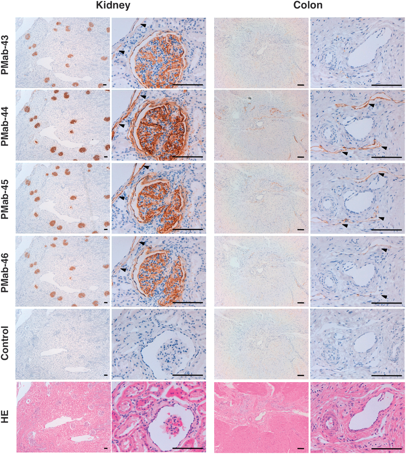
Immunohistochemical analysis by anti-bovPDPN mAbs. Sections of bovine kidney and colon were autoclaved in citrate buffer (pH 6.0). After blocking, they were incubated with 1 μg/mL of PMab-43, PMab-44, PMab-45, and PMab-46, followed by EnVision+ kit, and color was developed using DAB and counterstained with hematoxylin. PBS was used as negative control. HE staining was performed against serial sections. Arrow heads, lymphatic vessels. Scale bar: 100 μm. HE, hematoxylin and eosin; mAbs, monoclonal antibodies; PBS, phosphate-buffered saline.
Specificity of PMab-44 against bovine PDPN in flow cytometry
PMab-44 reacted with CHO/bovPDPN-FLAG, not with CHO/hPDPN-FLAG, CHO/mPDPN-FLAG, CHO/rPDPN-His, CHO/PA-rabPDPN, and CHO-K1 cells in flow cytometry (Fig. 2). LpMab-12 (anti-hPDPN mAb; mouse IgG1),(12) PMab-1 (anti-mPDPN mAb; rat IgG2a),(13) PMab-2 (anti-rPDPN mAb; mouse IgG1),(10) and PMab-32 (anti-rabPDPN mAb; mouse IgG1)(11) specifically reacted with CHO/hPDPN-FLAG, CHO/mPDPN-FLAG, CHO/rPDPN-His, and CHO/PA-rabPDPN, respectively. LpMab-12 reacted with CHO/rPDPN-His and CHO/PA-rabPDPN very weakly. Furthermore, PMab-44 reacted with MDBK cells, which express endogenous bovPDPN; in contrast, the other anti-PDPN mAbs did not. These results indicate that PMab-44 detects not only exogenous but also endogenous bovPDPN, and is very specific against bovPDPN.
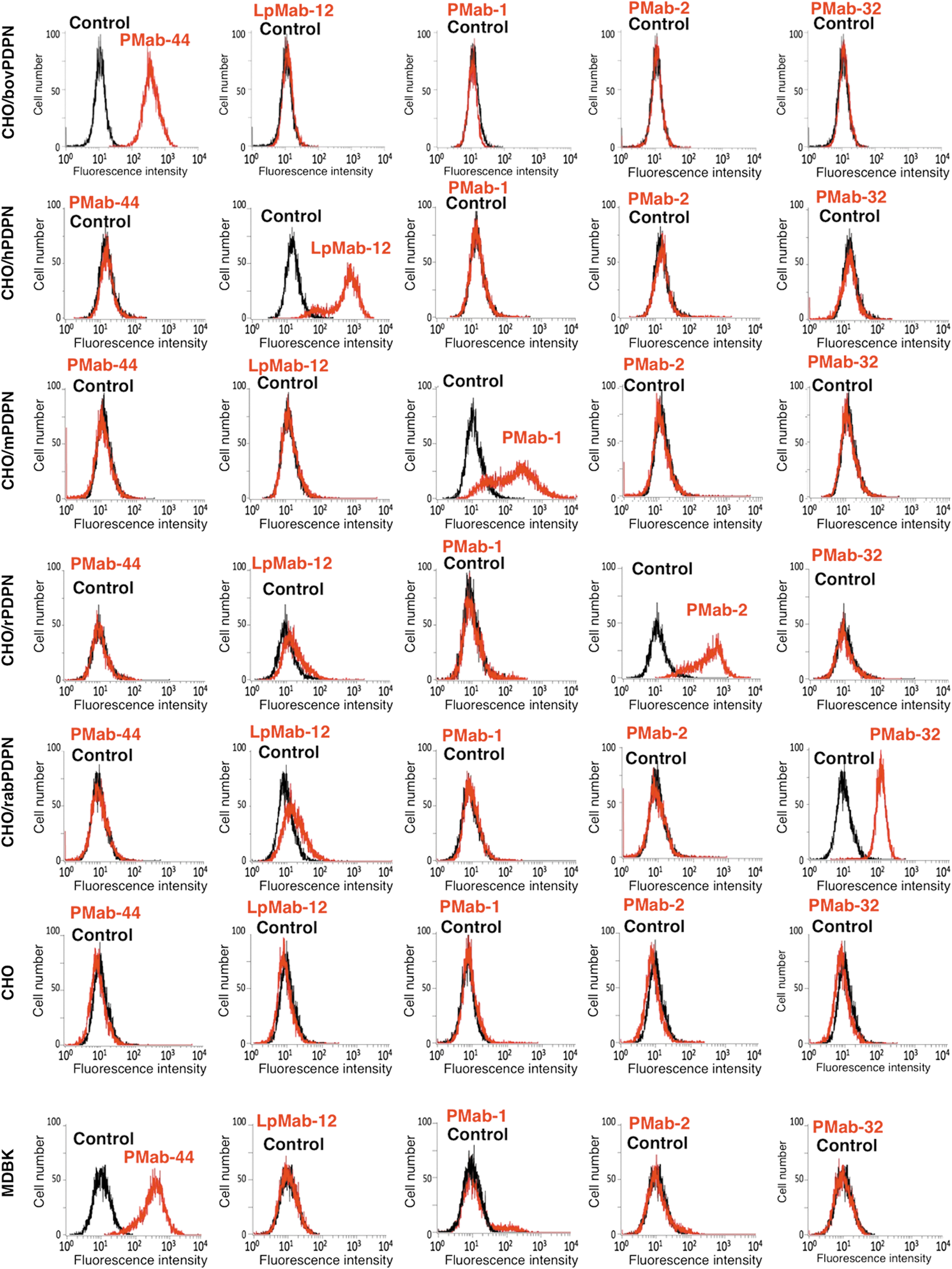
Flow cytometric analysis by anti-PDPN mAbs. CHO/bovPDPN-FLAG, CHO/hPDPN-FLAG, CHO/mPDPN-FLAG, CHO/rPDPN-His, CHO/PA-rabPDPN, CHO-K1, and MDBK were treated with PMab-44, LpMab-12, PMab-1, PMab-2, and PMab-32 followed by treatment with Oregon green-conjugated anti-mouse IgG against PMab-44, LpMab-12, PMab-2, and PMab-32 or anti-rat IgG against PMab-1. Red line: PMab-44, LpMab-12, PMab-1, PMab-2, and PMab-32. Black line: negative control.
Discussion
Expression of hPDPN has been reported in many malignant tumors such as osteosarcomas,(14) malignant brain tumors,(15–18) oral squamous cell carcinomas,(19) lung cancers,(20) esophageal cancers,(21) malignant mesotheliomas,(22) and testicular tumors.(23) Therefore, many mAbs against hPDPN have been established.(14,18,24–29) Several mAbs against mPDPN,(13) rPDPN,(10) and rabPDPN(11,30) have also been established. In contrast, useful mAbs against bovPDPN have not been reported. Because we recently established specific mAbs against rabPDPN by immunizing mice with membranous rabPDPN, we purified membranous bovPDPN from CHO/PA-bovPDPN-RAP-MAP cells and immunized mice with those recombinant proteins. We used several original epitope tags such as PA tag, MAP tag, and RAP tag in this study. All anti-tag mAbs detected recombinant proteins of PA-bovPDPN-RAP-MAP (data not shown). The combination of several tag systems is very useful not only for purification but also for establishing mAbs efficiently.
We previously constructed a phylogenetic tree of six mammalian PDPN amino acid sequences: hPDPN, mPDPN, rPDPN, bovPDPN, hamster PDPN (hamPDPN), and dog PDPN. The rodents (mPDPN, rPDPN, hamPDPN) and the bovine lineages showed longer branches in contrast with the human and the dog lineages.(6) It is likely that some kinds of functional changes are related to higher rates of amino acid substitution in the bovine lineages. In fact, bovPDPN did not induce platelet aggregation probably because of a sporadic deletion mutation in amino acid sequence of the PLAG domain.(6) The platelet aggregation by PDPN is known to be associated with blood/lymphatic vessel separation in mice(2); therefore, there might be the other mechanism in bovine blood/lymphatic vessel separation. PMab-44 could be useful to uncover the pathophysiological function of bovPDPN in the near future.
Footnotes
Acknowledgments
We thank Noriko Saidoh and Kanae Yoshida for their excellent technical assistance. This work was supported, in part, by the Regional Innovation Strategy Support Program from the Ministry of Education, Culture, Sports, Science and Technology (MEXT) of Japan (Y.K.), by JSPS KAKENHI Grant Number 26440019 (M.K.K.), Grant Number 25462242 (Y.K.), and Grant Number 16K10748 (Y.K.) by Takeda Science Foundation (S.O.), by the Platform for Drug Discovery, Informatics, and Structural Life Science (PDIS) from Japan Agency for Medical Research and development, AMED (Y.K.), and by the Basic Science and Platform Technology Program for Innovative Biological Medicine from AMED (Y.K.). This work was performed, in part, under the Cooperative Research Program of Institute for Protein Research, Osaka University, CR-15-05 and CR-16-05.
Author Disclosure Statement
No competing financial interests exist.