
Abstract
Podoplanin (PDPN) is expressed in several normal tissues including podocytes of renal glomerulus, lymphatic endothelial cells (LECs), and type I alveolar cells of lung. PDPN activates platelet aggregation by binding to C-type lectin-like receptor-2 (CLEC-2) on platelets. Many monoclonal antibodies (mAbs) against human PDPN, mouse PDPN, rat PDPN, rabbit PDPN, and bovine PDPN have been established; antidog PDPN (dPDPN) mAbs have not been developed. Herein, we immunized mice with the recombinant proteins of dPDPN and developed anti-dPDPN mAbs. One of the clones, PMab-38, is useful for detecting podocytes in immunohistochemical analysis; in contrast, it did not react with LECs or type I alveolar cells. PMab-38 also detected dPDPN specifically in flow cytometry and Western blot analysis. PMab-38 is expected to be useful for investigating the function of dPDPN, which is expressed in podocytes.
Introduction
P
PDPN activates platelet aggregation by binding to C-type lectin-like receptor-2 (CLEC-2) on platelets.(21,23–25) The interaction between PDPN and CLEC-2 facilitates blood/lymphatic vessel separation.(26) PDPN is also expressed in human fetal rib and chondrocytes of the proliferative and hypertrophic regions of the growth plate.(27)
Herein, we immunized mice with the recombinant proteins of dPDPN and established anti-dPDPN monoclonal antibodies (mAbs).
Materials and Methods
Cell lines, dog tissues, and animals
Chinese hamster ovary (CHO)-K1 and P3U1 were purchased from the American Type Culture Collection (ATCC, Manassas, VA). CHO-K1, stable CHO transfectants, and P3U1 were cultured in RPMI 1640 medium (Nacalai Tesque, Inc., Kyoto, Japan) supplemented with 10% heat-inactivated fetal bovine serum (Thermo Fisher Scientific, Inc., Waltham, MA), 100 U/mL of penicillin, 100 μg/mL of streptomycin, and 25 μg/mL of amphotericin B (Nacalai Tesque, Inc.) at 37°C in a humidified atmosphere of 5% CO2 and 95% air. Female BALB/c mice (4 weeks old) were purchased from CLEA Japan (Tokyo, Japan). Normal dog tissues were obtained from North lab (Hokkaido, Japan). Animals were housed under pathogen-free conditions. The Animal Care and Use Committee of Tohoku University approved the animal experiments described herein.
Hybridoma production
The dPDPN with N-terminal PA tag and C-terminal RAP tag-MAP tag (PA-dPDPN-RAP-MAP) was inserted into pCAG-zeo vector (Wako Pure Chemical Industries Ltd., Osaka, Japan). RAP tag consists of 12 amino acids (DMVNPGLEDRIE) and MAP tag consists of 12 amino acids (GDGMVPPGIEDK). CHO-K1 was transfected with pCAG-zeo/PA-dPDPN-RAP-MAP using Gene Pulser Xcell electroporation system (Bio-Rad Laboratories, Inc., Berkeley, CA). For the purification of PA-dPDPN-RAP-MAP from cell membrane, we used the PA tag system.(28,29) BALB/c mice were immunized by intraperitoneal (i.p.) injection of 100 μg of recombinant PA-dPDPN-RAP-MAP together with Imject Alum (Thermo Fisher Scientific, Inc.). After several additional immunizations of 50 μg, a booster injection of 100 μg was given i.p. 2 days before spleen cells were harvested. The spleen cells were fused with P3U1 cells using PEG1500 (Roche Diagnostics, Indianapolis, IN). The hybridomas were grown in RPMI medium with hypoxanthine, aminopterin, and thymidine selection medium supplement (Thermo Fisher Scientific, Inc.).
The culture supernatants were screened using enzyme-linked immunosorbent assay (ELISA) for binding to purified recombinant PA-dPDPN-RAP-MAP. Recombinant proteins of PA-dPDPN-RAP-MAP were immobilized on Nunc Maxisorp 96-well immunoplates (Thermo Fisher Scientific, Inc.) at 1 μg/mL for 30 minutes. After blocking with 1% BSA in 0.05% Tween20/phosphate-buffered saline (PBS; Nacalai Tesque, Inc.), the plates were incubated with culture supernatant followed by 1:3000 diluted peroxidase-conjugated antimouse IgG (Dako; Agilent Technologies, Inc., Santa Clara, CA). The enzymatic reaction was conducted with a 1-Step Ultra TMB-ELISA (Thermo Fisher Scientific, Inc.). The optical density was measured at 655 nm using an iMark microplate reader (Bio-Rad Laboratories, Inc.).
Immunohistochemical analyses
Four-micrometer-thick histologic sections were deparaffinized in xylene and rehydrated and were autoclaved in citrate buffer (pH 6.0; Dako) for 20 minutes. Sections were incubated with 10 μg/mL of primary mAbs for 1 hour at room temperature followed by treatment with Envision+ kit for 30 minutes (Dako). Color was developed using 3,3-diaminobenzidine tetrahydrochloride (DAB; Dako) for 1 minute, and then the sections were counterstained with hematoxylin (Wako Pure Chemical Industries Ltd.).
Flow cytometry
Stable transfectant of CHO/PA-dPDPN-RAP-MAP was established using SH800 (Sony Corp., Tokyo, Japan). The other stable transfectants [CHO/PA-bovine PDPN (bovPDPN)-RAP-MAP, CHO/hPDPN-FLAG, CHO/mouse PDPN (mPDPN)-FLAG, CHO/rat PDPN (rPDPN)-His, and CHO/PA-rabbit PDPN (rabPDPN)] were previously established.(1,30–32) Cells were harvested by brief exposure to 0.25% trypsin/1 mM EDTA (Nacalai Tesque, Inc.). After washing with 0.1% BSA/PBS, the cells were treated with primary mAbs (1 μg/mL) for 30 minutes at 4°C followed by treatment with Oregon green-conjugated antimouse IgG (1:1000 diluted; Thermo Fisher Scientific, Inc.). Fluorescence data were collected using a Cell Analyzer EC800 (Sony Corp.).
Western blot analysis
Cell lysates (10 μg) of CHO/PA-dPDPN-RAP-MAP, CHO/bovPDPN-FLAG, CHO/hPDPN-FLAG, CHO/mPDPN-FLAG, CHO/rPDPN-His, CHO/PA-rabPDPN, and CHO-K1 were boiled in SDS sample buffer (Nacalai Tesque, Inc.). The proteins were electrophoresed on 5%–20% polyacrylamide gels (Wako Pure Chemical Industries Ltd.) and were transferred onto a polyvinylidene difluoride (PVDF) membrane (EMD Millipore Corp., Billerica, MA). After blocking with 4% skim milk (Nacalai Tesque, Inc.), the membrane was incubated with PMab-38 and anti-β-actin (clone AC-15; Sigma-Aldrich Corp., St. Louis, MO) and then with peroxidase-conjugated antimouse IgG (1:1000 diluted; Dako) and developed with the Pierce Western Blotting Substrate Plus (Thermo Fisher Scientific, Inc.) using a Sayaca-Imager (DRC Co. Ltd., Tokyo, Japan).
Results
Production of mAbs against dPDPN
We immunized mice with the recombinant proteins, which were purified from CHO/PA-dPDPN-RAP-MAP cells, and the ELISA screening was performed. Anti-PA tag mAb (clone: NZ-1), anti-MAP tag mAb (clone: PMab-1), and anti-RAP tag mAb (clone: PMab-2) detected the PA-dPDPN-RAP-MAP protein in Western blot analysis (data not shown). Among ELISA-positive wells, the second screening was performed by flow cytometry. Finally, a positive wells were selected in immunohistochemical analysis. After limiting dilution, one of the clones, PMab-38 (IgG1, kappa), was established. PMab-38 reacted with podocytes of renal glomerulus (Fig. 1A), whereas it did not react with other normal cells such as LECs (Fig. 1B) or type I alveolar cells of lung (Fig. 1C), in which PDPN expression has been reported in other species.(3,7) These data indicate that PMab-38 is useful for immunohistochemistry using paraffin-embedded tissues.
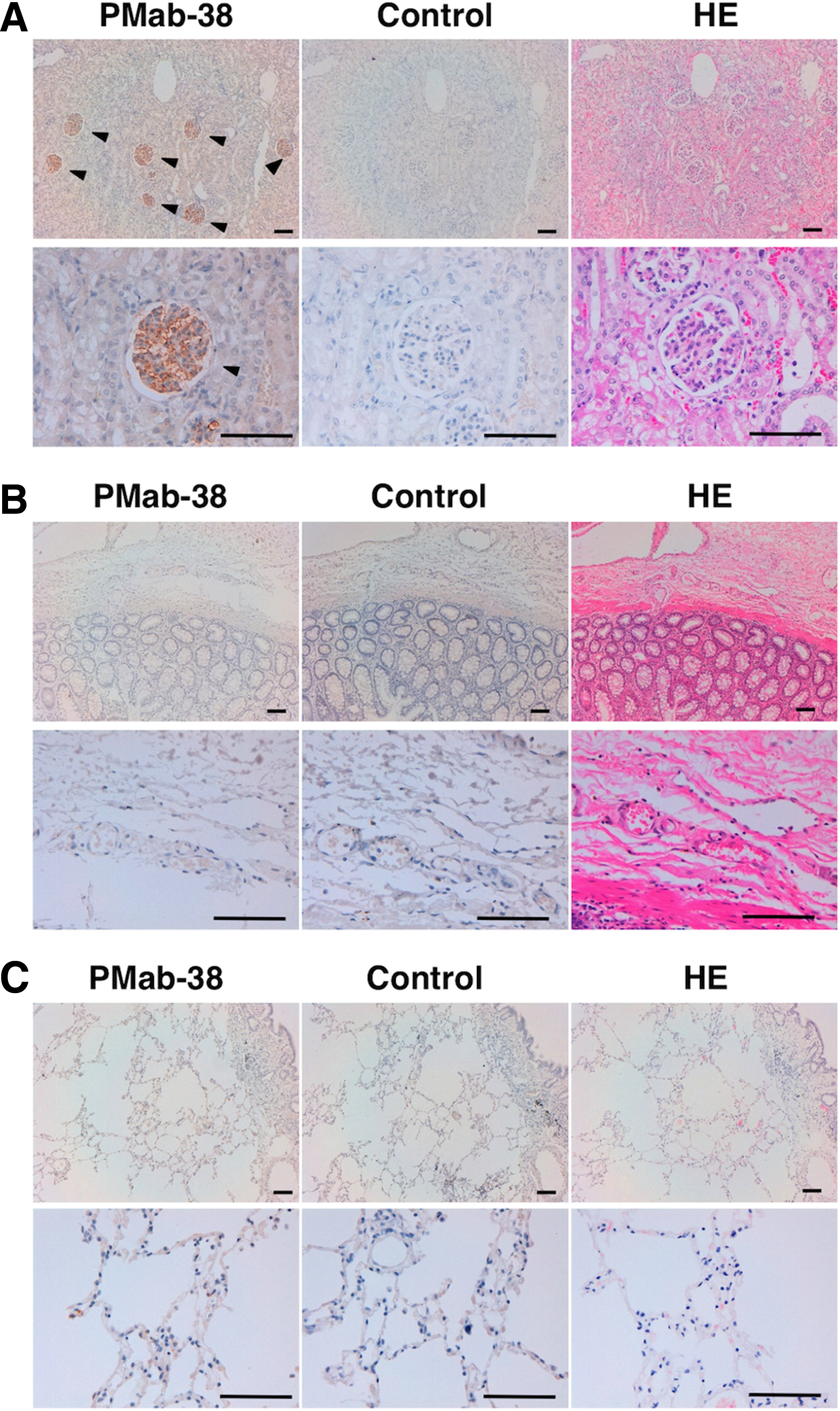
Immunohistochemical analysis by PMab-38. Sections of dog kidney
Specificity of PMab-38 against dPDPN in flow cytometry and Western blot
PMab-38 reacted with CHO/PA-dPDPN-RAP-MAP, not with CHO/PA-bovPDPN-RAP-MAP, CHO/hPDPN-FLAG, CHO/mPDPN-FLAG, CHO/rPDPN-His, CHO/PA-rabPDPN, and CHO-K1 cells in flow cytometry (Fig. 2A). In Western blot analysis, PMab-38 recognized only dPDPN of CHO/PA-dPDPN-RAP-MAP, and did not react with PDPNs of CHO/bovPDPN-FLAG, CHO/hPDPN-FLAG, CHO/mPDPN-FLAG, CHO/rPDPN-His, CHO/PA-rabPDPN, or CHO-K1 cells (Fig. 2B). These results indicate that PMab-38 detects dPDPN specifically.
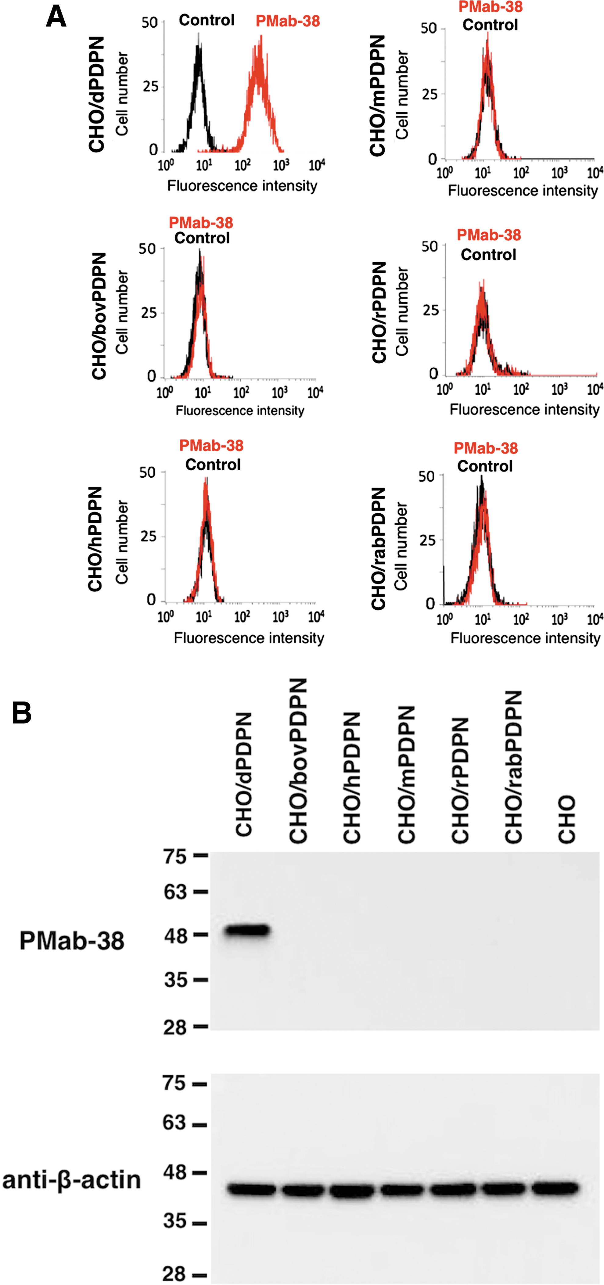
Flow cytometric and Western blot analyses by PMab-38.
Discussion
Although there are many mAbs and polyclonal Abs against PDPN have been produced, anti-dPDPN mAbs have not been developed. Our previous reports revealed that PMab-44 (anti-bovPDPN mAb; mouse IgG1),(32) LpMab-12 (anti-hPDPN mAb; mouse IgG1),(33) PMab-1 (anti-mPDPN mAb; rat IgG2a),(34) PMab-2 (anti-rPDPN mAb; mouse IgG1),(30) and PMab-32 (anti-rabPDPN mAb; mouse IgG1)(31,35) specifically react with CHO/bovPDPN, CHO/hPDPN, CHO/mPDPN, CHO/rPDPN, and CHO/rabPDPN, respectively. Many other anti-hPDPN mAbs also detect hPDPN specifically.(12,13,17,19,36–47) Herein, we immunized mice with the recombinant proteins of dPDPN, and developed several anti-dPDPN mAbs including PMab-38. When PMab-38 was incubated for 18 hours at 4°C in immunohistochemistry, the stronger staining was observed in renal glomerulus, and Bowman's capsule was also stained by PMab-38, whereas LECs or type I alveolar cells were not stained (data not shown). These data demonstrate that PMab-38 is useful for immunohistochemistry using paraffin-embedded tissues, but the reaction of PMab-38 is limited to podocytes of renal glomerulus.
Taken together, PMab-38 could be useful to uncover the pathophysiological function of dPDPN in podocytes of renal glomerulus. In the future, we should determine the epitope of PMab-38 and clarify the reason for the specificity of PMab-38 against podocytes of renal glomerulus in immunohistochemistry.
Footnotes
Acknowledgments
We thank Takuro Nakamura, Noriko Saidoh, and Kanae Yoshida for their excellent technical assistance. This work was supported in part by the Regional Innovation Strategy Support Program from the Ministry of Education, Culture, Sports, Science, and Technology (MEXT) of Japan (Y.K.), by JSPS KAKENHI Grant Number 26440019 (M.K.K.) and Grant Number 25462242 and 16K10748 (Y.K.), by Takeda Science Foundation (S.O.), by the Platform for Drug Discovery, Informatics, and Structural Life Science (PDIS) from Japan Agency for Medical Research and Development, AMED (Y.K.), and by the Basic Science and Platform Technology Program for Innovative Biological Medicine from AMED (Y.K.). This work was performed, in part, under the Cooperative Research Program of Institute for Protein Research, Osaka University, CR-15-05 and CR-16-05.
Author Disclosure Statement
No competing financial interests exist.