
Abstract
Human transferrin receptor (hTfR1), a type II homodimeric transmembrane protein, is extensively expressed at low levels in most normal human tissues. However, the expression level of hTfR1 in some tumor tissues and the blood–brain barrier (BBB) is comparatively higher. Therefore, we developed an hTfR1 monoclonal antibody 2C9, which may be utilized to deliver drugs across the BBB through receptor-mediated endocytosis in the future. 2C9 was obtained by hybridoma cells fused by the SP2/0-Ag14 cell line and splenic B cells from hTfR1-immunized BALB/c mice. In this study, we used indirect ELISA, Western blot, and immunofluorescence to explore the characterization and application of MAb 2C9. The results showed that the affinity constant (Kaff) of the MAb 2C9 produced from ascites was 2.85 × 10⁻⁸ M and its isotype is IgG2a (Kappa chain). Above all, this report provides a more comprehensive protocol to produce monoclonal antibodies against the extracellular domain of hTfR1.
Introduction
T
It is well known that iron is essential for a number of cellular functions, including DNA synthesis and electron transport.(4) The available data suggest that the transferrin (Tf)/TfR pathway is the main route of iron transport across the BBB.(5) Tf-bound iron is internalized in the endothelial cells through TfR-mediated endocytosis. Once inside the cell, iron is dissociated from the Tf-TfR complex due to acidification of the endosome and is released into the cytosol before being transported to the brain parenchyma.(6)
The BBB is constituted by specialized vascular endothelium that interacts directly with astrocytes, neurons, and pericytes. It also has an ensemble of enzymes, receptors, transporters, and efflux pumps. Thus, the BBB serves as a major impediment to effective delivery of many therapies from the bloodstream into the brain parenchyma.(2) The biggest challenge for the treatment of brain diseases is the limitation caused by the inadequacy of its delivery system.(7) Currently, chemical modifications of drugs, such as increasing their lipophilicity, make them easier to be internalized in the brain. Other mechanisms include the use of molecular tools to bind the drugs such as immunoglobulins, peptides, liposomes, or nanoparticles that will act as Trojan horses favoring the drug delivery in brain.(8,9)
In recent years, TfR1 has been recognized as a target receptor for drug delivery across the BBB to treat some brain disorders such as Alzheimer's disease, Parkinson's disease, and stroke.(10) In this article, we prepared the monoclonal antibody against the extracellular domain of the TfR1and the monoclonal antibody possesses potent targeted activity and antigen specificity to BBB. Collectively, the MAb 2C9 would be a potential delivery tool for drugs across the BBB through receptor-mediated transcytosis.
Materials and Methods
Antigen, animals, and reagent
hTfR1 was prepared and served as antigen.(11) Six-week-old female BALB/c mice were purchased from Experimental Animals of Yangzhou University (College of Animal Science and Technology, Yangzhou, China). Freund's adjuvant, bovine serum albumin (BSA), polyethylene glycol (PEG4000, 50%w/v), hypoxanthine/aminopterin/thymidine (HAT), hypoxanthine/thymidine (HT), and O-phenylenediamine were purchased from Sigma-Aldrich (St. Louis, MO). Dulbecco's modified Eagle's medium (DMEM) was purchased from Gibco (Invitrogen, Carlsbad, CA). Fetal bovine serum was purchased from Clark. Peroxidase-labeled goat anti-mouse IgG (H+L) (HRP-IgG) was purchased from ZSGB-BIO (Beijing, China), Mouse monoclonal antibody subtype identification kit was purchased from Proteintech (Wuhan, China).
Immunization of BALB/c mice
Five female 6-week-old BALB/c mice were immunized with 200 μL emulsion containing 100 μg of recombinant TfR1 protein and Freund's complete adjuvant by subcutaneous injection. At 3-week intervals, two boosters of 200 μL emulsion were administered by intraperitoneal injection, and serum from the immunized mice was monitored for antibody titers against hTfR1 by indirect ELISA. Mice with sustained antibody titers above 1.6 × 10⁵ were selected and intravenously injected with 200 μg TfR1 without Freund's adjuvant 3 days before cell fusion.
ELISA
Washes with 1× PBST (NaCl 8.0 g, KH2PO4 0.2 g, Na2HPO4·12H2O 0.89 g, KCl 0.2 g, Tween 20 0.5 mL, 1000 mL, pH7.4) were performed between each step. Ninety six-well microplates were coated with 100 μL purified complete hTfR1 (1 μg/mL) in 0.1 M carbonate coating buffer (pH 9.5) at 37°C for 2 hours. The plates were subsequently blocked with 200 μL 3% BSA at 37°C for 2 hours. Samples of immune serum were diluted in 1% BSA. Incubation took place for 2 hours at 37°C. Negative control was incubated with serum from nonimmunized mice. After incubation with primary antibodies, the plates were exposed to 100 μL HRP (horseradish peroxidase)-conjugated goat anti-mouse IgG (1:5000) at 37°C for 1 hour. Finally, 100 μL TMB was added into the plates with incubation for 30 minutes, and the reaction was stopped by adding 100 μL 2 M H2SO4. The results were measured by Multiscan Spectrum at 450 nm wavelength.(12)
Cell fusion
Preparation of hybridoma was performed by the marine method of Kohler and Milstein.(13) Spleens of the positive mice were removed and dispersed 72 hours after the last booster; 1 × 10⁸ spleen cells were suspended with DMEM and then mixed with 1 × 10⁷ SP2/0-Ag14 myeloma cells. Then, the mixture was centrifuged twice with DMEM at 1000 rpm for 5 minutes, followed by the addition of 1 mL 50% PEG 1450 solution for 1 minute, and then 3 mL DMEM for 3 minutes. Finally, 10 mL DMEM was added with gentle stirring for 1 minute. The fused cells were centrifuged and suspended in HAT medium and then seeded in 96-well plates, which had been seeded with feeder cells the day before. Hybridoma cells were cultured in HT medium 10 days later. Positive hybridoma clones were selected by indirect ELISA.
Production and purification of MAb
After being selected three times with indirect ELISA, a cell line 2C9, which can secrete high-affinity antibody, was obtained and massively cultured in high-glucose DMEM containing 10% fetal bovine serum (FBS). Before the hybridoma injection, several adult female BALB/c mice were intraperitoneally injected with sterile paraffin oil (0.5 mL per mouse). After 7 days, each mouse was injected with 1 × 10⁶ hybridoma cells 2C9. Within 1–2 weeks, ascites were collected and centrifuged at 12,000 rpm for 5 minutes to acquire the supernatant.
The ascites collected were diluted at least 50 times with binding buffer (1.409 g NaH2PO42H2O; 4.372 g Na2HPO4·12H2O) and then were purified by HiTrap protein A column (GE Healthcare) according to the manufacturer's protocol to obtain purified monoclonal antibody named TfRMAb. To assess the purity, the purified TfRMAb was subjected to sodium salt–polyacrylamide gel electrophoresis (SDS-PAGE) and analyzed by Quantity One software (Bio-Rad).
Isotype analysis
The TfRMAb was diluted into 20 ng/mL, and then the class and subclass of the TfRMAb were identified by a mouse monoclonal antibody isotyping reagent (Proteintech) according to the manufacturer's directions. Each strip contains IgG1, IgG2a, IgG2b, IgG2c, IgG3, and IgM heavy chain, as well as kappa and lambda light chains.
Monoclonal antibody affinity constant determination
To determine the affinity constant (Kaff) of TfRMAb, a noncompetitive enzyme immunoassay was performed. Briefly, two different concentrations of recombinant hTfR1, including [Ag′] = 0.50 μg/mL and [Ag] = 1.00 μg/mL, were coated in a 96-well plate. After incubation at 37°C for 2 hours, the antibody was serially diluted to 50, 25, 12.5, 6.25, 3.12, 1.56, 0.78, and 0.39 μg/mL and added to the microplate. Then, the process was performed as previously described in the ELISA. Sigmoid curves were plotted to represent the relationship of OD450 value versus MAb concentration in two different antigen concentrations. The half maximum OD (OD-50) was assigned for all selected curves from which the corresponding antibody concentration ([Ab]t and [Ab′]t) was extrapolated. Accordingly, [Ab]t and [Ab′]t are the measurable total antibody concentrations at OD-50 and OD′-50 for plates coated with [Ag] and [Ag′], respectively. The affinity constant was finally determined using the following equation:
Western blot analysis
The concentration of TfRMAb was determined by bicinchoninic acid assays. Sixteen microliters hTfR was mixed with 4 μL loading buffer and directly loaded for SDS-PAGE analysis. Then, proteins were electrophoretically transferred to 0.22 μm polyvinylidene fluoride (PVDF) membranes by Western blot transfer system (Bio-Rad) for 1 hour at 200 mV. The PVDF membranes were washed with TBST (Trsi-buffered saline containing 0.5% Tween 20) and blocked with 5% BSA overnight at 4°C. Thereafter, the membranes were washed with TBST five times and incubated with anti-TfR1 monoclonal antibody for 1 hour at 37°C, then washed with TBST five times and incubated with HRP-labeled goat anti-mouse IgG (1:5000) for 1 hour at 37°C. Proteins of interest were visualized with enhanced chemiluminescence in Gel Doc EZ Image Lab (Bio-Rad).(14)
Cell ELISA
The cell ELISA was similar to indirect ELISA. First, 5 × 10⁴ hepatoma carcinoma cells (HepG-2) and MCF-7 cells were seeded in 96-well plates and cultured at 37°C, 5% CO2, until it reached 80% cell density. The plates were washed three times with PBST. Subsequently, the cells were fixed with 4% paraformaldehyde for 20 minutes. The rest of the steps were the same as the ELISA. Both the TfRMAb harvested from supernatants and ascites were used as the primary antibody. The results were measured by Multiscan Spectrum at 450 nm wavelength.
Immunocytochemistry
To analyze the specificity of TfRMAb to TfR expressed on cells, one tumor cell line HepG2 that overexpressed TfR was grown on a six-well plate in complete RPMI 1640 medium with 10% FBS until the cells reached 80%–90% confluence. Then, cells were washed twice with cold phosphate-buffered saline (PBS) and fixed with cold 4% paraformaldehyde. After washing three times with PBS and blocking with 5% BSA-PBST for 1 hour, fixed cells were incubated with purified anti-TfR1 monoclonal antibody (2C9) and negative controls at 37°C for 1 h. After washing five times with PBS, fluorescein isothiocyanate (FITC)-conjugated goat-anti-mouse IgG (H+L) (SAB) was added and incubated with the cells for 1 hour at RT. The coverslips were rinsed with PBS and examined under a fluorescence microscope (ZEISS, Germany).(15)
Results
Immunized BALB/c mice antiserum samples
After immunization three times, blood from four mice was harvested and centrifuged to separate serum from other cells. The serum was diluted into a series of concentrations (1:10000; 1:20000; 1:40000; 1:80000; 1:160000; 1:320000; 1:640000; 1:1280000) and tested by indirect ELISA. The results (Fig. 1) showed that the titers of four immunized mice are up to 1:640000 or 1:1280000. Relatively, the first mouse had the highest titer with 1:1280000, which means it was suitable for cell fusion.
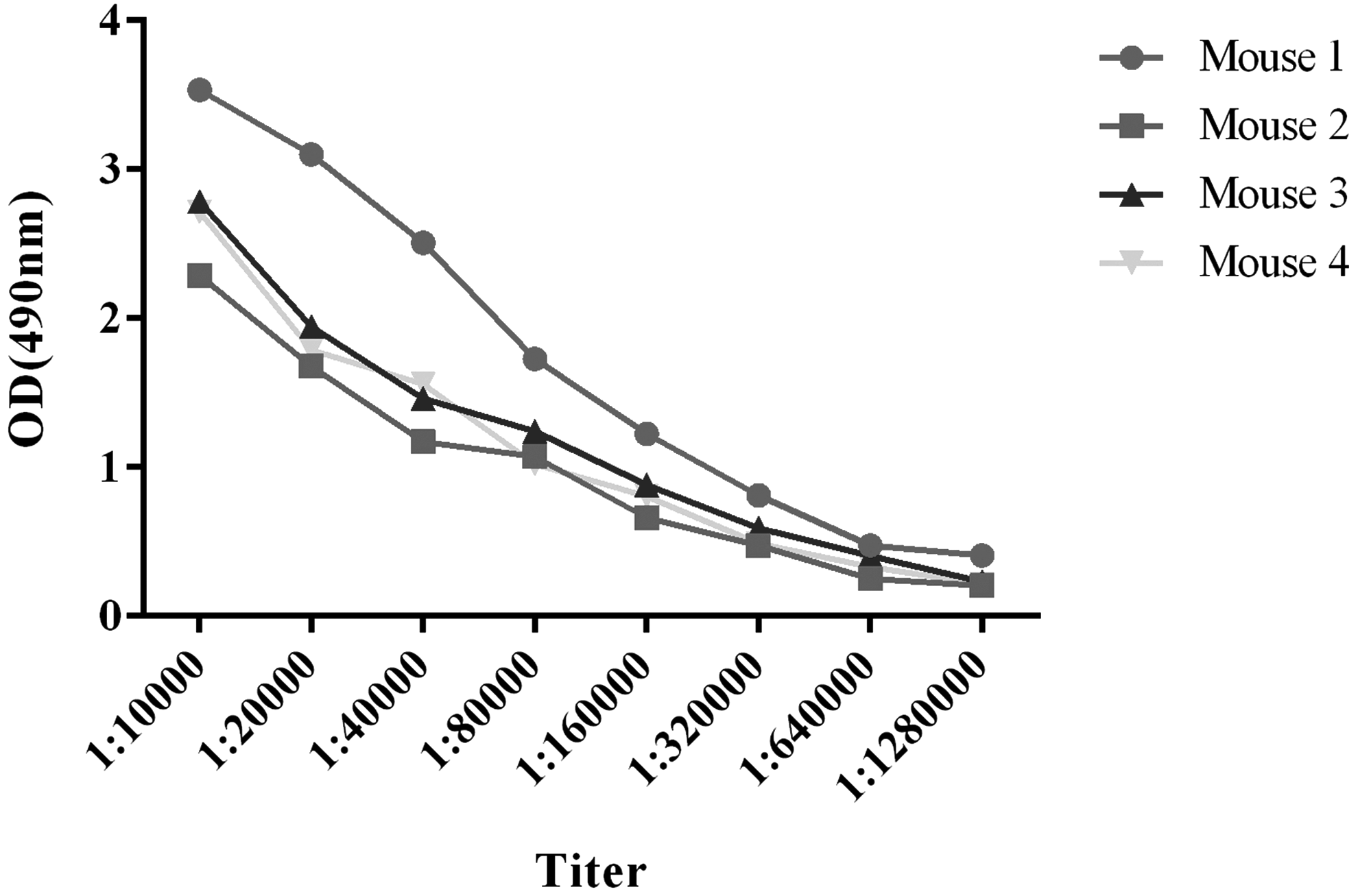
The titers of antiserum samples from immunized mice. The titer of mouse 1 was 1:1280000, which was higher than the others.
Production and purification of TfRMAb
Clones were obtained 7 days after cell fusion. Supernatants from hybridoma cells were detected by indirect ELISA. The specific high-affinity hybridoma cells were harvested after subcloning three times. Among them, one hybridoma, named 2C9, exhibited a relatively higher binding activity to hTfR1. Therefore, all subsequent tests were carried out with 2C9.
Both supernatants from the 2C9 culture and ascites from BALB/c mice immunized with 2C9 were purified using HiTrap protein A column. SDS-PAGE and Western blot were carried out to identify the purity and specificity of TfRMAb against hTfR1. Nonreduced electrophoresis results explicitly exhibited that integrated TfRMAb was about 170 kD (Fig. 2A). Reduced electrophoresis results showed that TfRMAb was divided into heavy chain (∼55kD) and light chain (∼28kD) (Fig. 2A). The purity was about 95%, which was analyzed by Quantity One software (Bio-Rad, Tokyo, Japan). Western blot results demonstrated that TfRMAb can specifically combine with the recombinant protein hTfR1 and the hTfR1 without Trx-tag (Fig. 2B).

The purity and specificity analysis of TfRMAb.
Isotype and affinity constant of TfRMAb
The isotype of TfRMAb was analyzed by anti-isotype reagents, and the results indicated that the isotype is IgG2a, as shown in Figure 3. The type of light chain is Kappa (K), as shown in Figure 3.
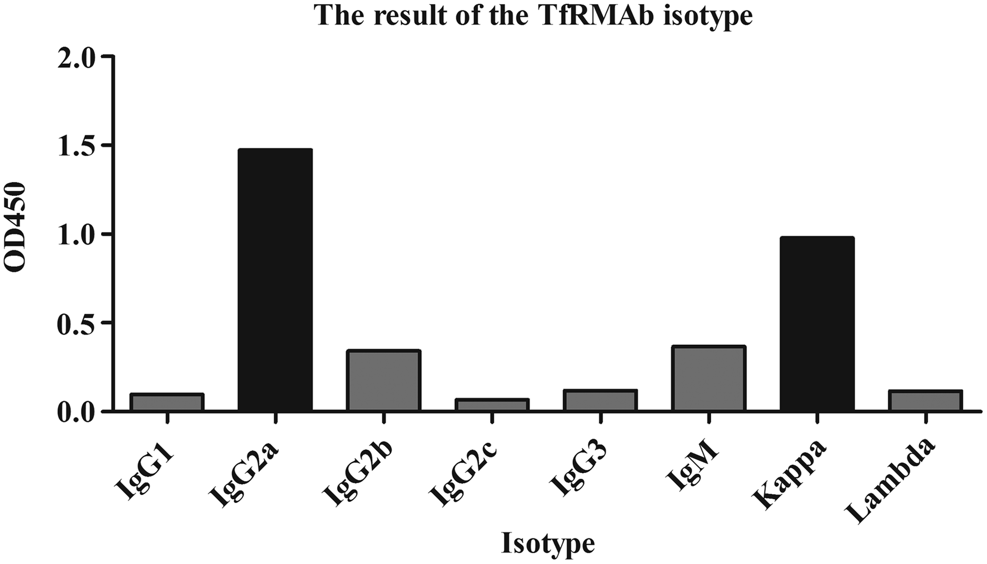
The result of the TfRMAb isotype. The OD450 nm values in different subtypes. The type of heavy chain is IgG2a as shown. OD450 nm values in IgG2a were much higher than OD450 nm values in IgG1, IgG2b, IgG2c, IgG3, and IgM. The type of light chain is Kappa (K), OD450 nm values in Kappa were significantly higher than OD450 nm values in Lambda.
Sigmoid curves were plotted to represent the relationship of OD450 nm values versus TfRMAb concentration in two different antigen concentrations. According to the Beatty method, the Kaff of TfRMAb was calculated to be 2.85 × 10⁻⁸M (Table 1).
[Ab] = 8.514 μg/mL is the measurable antibody concentration at OD-50 when plates are coated with [Ag] = 1 μg/mL, [Ab′] = 14.967 μg/mL is the measurable total antibody concentration at OD′-50 when plates are coated with [Ag′] = 0.5 μg/mL, respectively. The affinity constant was 2.85 × 10⁻⁸ M, which was finally determined by Beatty equation.
Antigen specificity of TfRMAb
The Western blot result of TfRMAb against recombinant hTfR1 has been described above. As antigen used in immunization was expressed in prokaryotic cells, its structure might be somewhat distinct from the hTfR1 expressed in eukaryotic cells. Therefore, human liver cancer cells HepG-2 and human breast cancer cells MCF-7 were used in cell ELISA as antigen to examine the specificity of TfRMAb against hTfR1. The results proved that TfRMAb purified from ascites had the specific binding capacity to the natural hTfR1 expressed on HepG-2 and MCF-7 (Fig. 4). In addition, the TfRMAb could significantly combine with HepG-2 (Fig. 4A, B). Conversely, the TfRMAb purified from supernatants has no antigen specificity capacity.

The antigen specificity of TfRMAb.
To explore the reason for this binding difference, circular dichroism (CD), a kind of fast, simple, and accurate method to study the protein conformation in diluted solution, which is relatively similar with its physiological state, was used here to determine the secondary structure of proteins. The secondary structures of the TfRMAb purified from supernatant and ascites are shown in Figure 5A, B, respectively. The waveforms and data analysis results showed that the TfRMAb from different sources (supernatant and ascites) exhibited various secondary structures (Tables 2, 3). The TfRMAb obtained from supernatants only had α-helix and turn structure, while from ascites, they had one more structure—random coil. Meanwhile, the ratio of helix and turn in TfRMAb from supernatants was higher than relevant structures in TfRMAb purified from ascites. We tried to explain the difference in various ways. First, by adjusting the percentage of serum from 5 to 10% FBS, we found that the activity of TfRMAb purified from the medium with 10% FBS was twofold than that from the medium with 5% FBS. Second, we adjusted the pH of culture medium and found that the bioactivity of the TfRMAb purified from pH = 7.4 medium was 1.2 times higher than that from pH = 7.0 medium. Therefore, the percent of FBS and the medium pH probably led to the change of TfRMAb bioactivity.

The circular dichroism (CD) spectrum analysis of TfRMAb purified from two sources.
The experiment was set in the condition at 37°C, range from 190 to 260 nm, scanning speed 100 nm/min, data pitch 1 nm, response 1 s, and band width 1 nm.
The experiment was set in the condition at 37°C, range from 190 to 260 nm, scanning speed 100 nm/min, data pitch 1 nm, response 1 s, and band width 1 nm.
Immunofluorescence analysis
The immunofluorescence results of the reaction of HepG-2 to TfRMAb are shown as follows (Fig. 6). TfRMAb purified from ascites was labeled with FITC and served as primary antibody to incubate with HepG-2. The results showed that green fluorescence was mainly gathered around the surface of HepG-2. It meant that purified TfRMAb could specifically combine with hTfR1 expressed on HepG-2.

Immunofluorescence staining results of TfRMAb purified from ascites (200 × ).
Discussion
Although TfR1 is a kind of glycoprotein extensively expressed in different tissues and cell lines, the expression level of TfR1 at the BBB and tumor cells is relatively higher. As is well known, brain tumors are inherently difficult to treat due to the cellular BBB limiting the delivery of therapeutic drugs to the tumor tissue from the systemic circulation.(2) TfR has been widely studied as a molecular Trojan horse to ferry across the BBB large molecule pharmaceuticals, including recombinant proteins, antibodies, RNA interference drugs, or nonviral gene medicines.(16) In this study, our purpose was to prepare an antibody against the extracellular region of hTfR1, which can transport small molecular medicine to the target tissue.
In our research, we developed monoclonal antibodies against hTfR1 using recombinant hTfR1 as antigen. Classical cell fusion method was used to obtain the hybridoma cells, which can express high-affinity TfRMAb. Subsequently, a series of experiments such as ELISA, Western blot, and immunofluorescence assays were used to determine the characteristics of the monoclonal antibody.
The available data suggested that high-affinity monoclonal antibody binding to TfR drives rapid antibody clearance and TfR degradation, thus limiting sustained brain exposure to the antibody. In contrast, very low-affinity binding to TfR results in substantially better peripheral blood antibody exposure as a result of reduced target-mediated clearance; however, maximal brain uptake is also reduced because of the extremely low affinity of antibody for TfR.(5) Therefore, moderate-affinity constant (Kaff) of the TfR monoclonal was important for delivery of drugs across the BBB. The affinity constant (Kaff) of the MAb 2C9 was calculated to be 2.85 × 10⁻⁸ M, which owned medium TfR antigen binding activity compared with others previously reported. Therefore, the exact optimal affinity for TfR leads to sustained brain concentrations of antibody above a therapeutic threshold, which means it has a potential use for delivering drugs across the BBB. The results of ELISA and Western blot showed that TfRMAb exhibited superiority in recognizing recombinant hTfR1. Furthermore, the results of immunofluorescence showed that TfRMAb could specifically combine hTfR1 expressed on HepG-2. In summary, these data demonstrated that the TfRMAb produced by 2C9 cell line will have a potential application for delivering drugs across the BBB in the future.
Footnotes
Acknowledgments
This work was supported by the Fundamental Research Funds for the Central Universities Institutions (Grant No. 201000009), the National Science and Technology Major Project (Grant No. 2013ZX09301303-004), National High Technology Research and Development Program 863 (Grant No. 2012AA02A303), and a project funded by the Priority Academic Program Development of Jiangsu Higher Education Institutions (PAPD).
Author Disclosure Statement
No competing financial interests exist.