
Abstract
The purpose of this study was to detect Streptococcus mutans by using monoclonal antibodies (mAbs) against S. mutans that cause dental caries and compare the levels of the bacterium between the saliva of adolescents undergoing orthodontic treatment (OT) and those not undergoing treatment (NT). Saliva samples, collected from 25 OT adolescents (with a mean age of 12.84 years) and 25 NT adolescents (mean age of 12.4 years), were analyzed by Dentocult-SM and enzyme-linked immunosorbent assay using mAbs against Ag I/II (ckAg I/II) and GTF B (ckGTF B), GTF C (ckGTF C), and GTF D (ckGTF D) of S. mutans. The DMFT index was slightly higher in the OT group (5.12 in OT and 4.96 in NT) and the level of S. mutans (≥105 CFU/mL) was higher in OT (72%) than in NT (56%). The detected levels of ckAg I/II, ckGTF B, ckGTF C, and ckGTF D were slightly higher in OT than in NT. The results of this study indicate that use of mAbs against S. mutans yields sensitive detection for the bacterium in saliva samples and shows that it has a reliable connection to the number of S. mutans and decayed, missing, filled teeth (DMFT), suggesting that the levels of S. mutans in saliva can be defined and compared by the application of the mAbs.
Introduction
S
Orthodontic treatment (OT) is suggested to correct malocclusions that can affect one's quality of life(9–11) and the timing of OT should be decided on a case-by-case basis.(12) However, adolescents who have crowded dentitions often choose to undergo OT even though they lack any serious conditions(13) because adolescents with aligned teeth are considered to appear more attractive and more intelligent than adolescents with crowded teeth.(14,15) OT, however, can have several negative effects as oral bacteria attaches to the surface of teeth. In addition, orthodontic appliances increase the chance of S. mutans colonization(4,16) and plaque accumulation. This is perhaps because it is difficult to maintain adequate oral hygiene during OT.
Considering the widespread use of OT, it would be worthwhile to check and compare the levels of S. mutans in OT and in adolescents not undergoing treatment (NT). Several studies on the levels of oral bacteria in adolescents undergoing OT have been conducted.(17–19) However, no studies have been performed to compare levels of S. mutans between OT and NT using monoclonal antibodies (mAbs). Consequently, the aim of this study was to apply mAbs against virulence factors of S. mutans (ckAg I/II, ckGTF B, ckGTF C, and ckGTF D)(20–23) to investigate and compare the oral bacterial levels in adolescents undergoing OT with those NT.
Materials and Methods
Subjects
Twenty-five adolescents (mean age 12.84 years) who visited the Dental Hospital of Chonbuk National University (Jeonju, Korea) for OT over a 6-month period were randomly selected for the OT group and 25 healthy adolescents (mean age 12.4 years) were gathered for the NT group. All clinical examinations were performed by two dentists. Written consent was obtained from each adolescent and their parents participating in the study. All procedures were approved by the Ethical Committee of Chonbuk National University Hospital (approval number 201107025). A fluoride treatment was applied to all adolescents in the OT group (Topex Topical APF Gel; Sultan Healthcare, Hackensack, NJ) once every 3 months. In addition, they were given specific instructions as to how to brush their teeth.
Saliva sampling and counting the levels of S. mutans in saliva
Subjects spat saliva samples into 50 mL sterile tubes after chewing paraffin wax for 3 or 4 minutes. The samples were kept on ice and analyzed within 24 hours. The colony forming unit (CFU)/mL in saliva was estimated using a Dentocult SM (Orion Diagnostica Co. Ltd, Helsinki, Finland) according to the manufacturer's instructions.
Enzyme-linked immunosorbent assay
Enzyme-linked immunosorbent assay (ELISA) was conducted through the use of mAbs against Ag I/II, GTF B, GTF C, and GTF D to investigate the abundance of S. mutans in each sample. A 96-well, flat-bottomed polystyrene microtiterplate (Nunc, Roskilde, Denmark) was coated with 100 μL of saliva and incubated at 4°C overnight before being blocked with 3% skim milk for 30 minutes at room temperature. After washing three times with phosphate-buffered saline (PBS), 20 ng of mAbs in PBS with 3% skim milk was added and incubated for 1 hour at 37°C and washed three times with PBS. Then, a secondary alkaline phosphatase-labeled goat antimouse immunoglobulin G antibody (Sigma Chemical Co., St. Louis, MO) was added to each well before wells were washed four times with PBS. Color was developed with an alkaline phosphatase substrate and the wells were read at 405 nm using an ELISA reader (Packard Instrument Co., Downers Grove, IL).
Statistical analysis
All experiments were performed in triplicate and mean log titers and their standard errors were calculated. Results are presented as the mean ± standard error. Statistical analyses were performed using SPSS 12 software (SPSS, Inc., Chicago, IL).
Results
In the OT group, 72% of samples contained S. mutans classes 1 to 3, whereas only 56% of the NT group samples contained S. mutans classes 1 to 3 (Table 1). The OT group displayed a slightly higher mean of decayed, missing, filled teeth (DMFT) (mean 5.2 ± 2.82) than the NT group (mean 5.12 ± 4.15). In both groups, however, the most prevalent class was class 1 and there was more class 0 in the NT group (44%) than in the OT group (28%).
CFU, colony forming unit; NT, not undergoing treatment; OT, orthodontic treatment.
Dentocult-SM is considered to be an adequate method and commonly used for the measurement of S. mutans.(24,25) A study reported that the use of specific mAbs against S. mutans is a more accurate method as it allows for the quantification of total S. mutans.(26) Thus, Dentocult-SM and mAbs ckAg I/II, ckGTF B, ckGTF C, and ckGTF D, which have been developed against S. mutans in our laboratory, were used in this experiment to detect S. mutans in the saliva sample of NT group (Fig. 1) and OT group (Fig. 2). As shown in Figure 3, the levels of mAbs in the OT group were higher than in the NT group. The reactions of ckGTF B and ckGTF C with glucosyltransferases B and C were higher in both groups than those of ckGTF D and Ag I/II, indicating that there are more insoluble glucans produced by glucosyltransferases B and C that play an important role in the formation of dental plaque in the saliva (Fig. 3). As shown in Table 1, the DMFT index is lower when the level of S. mutans in saliva is low.
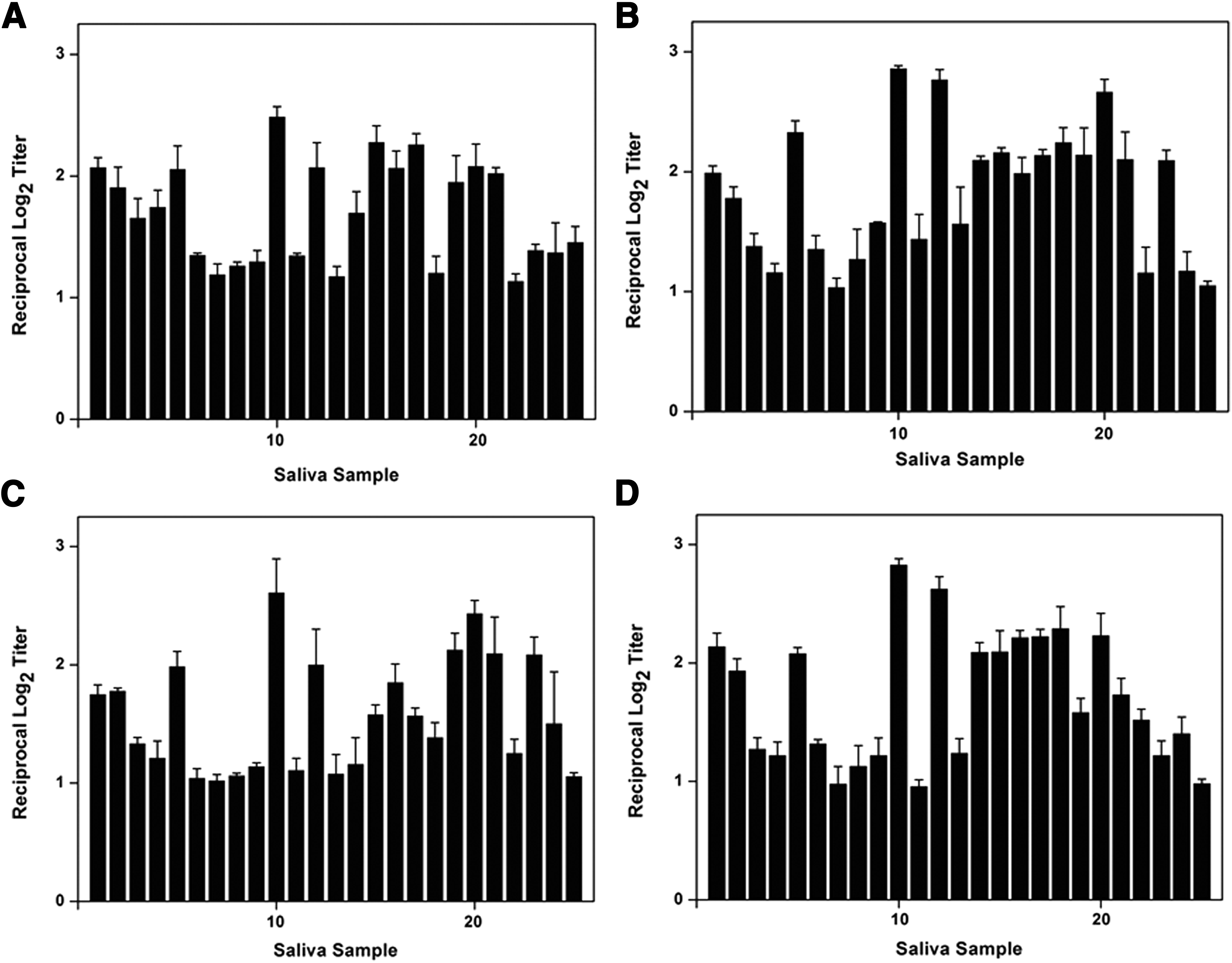
Reaction of mAbs with
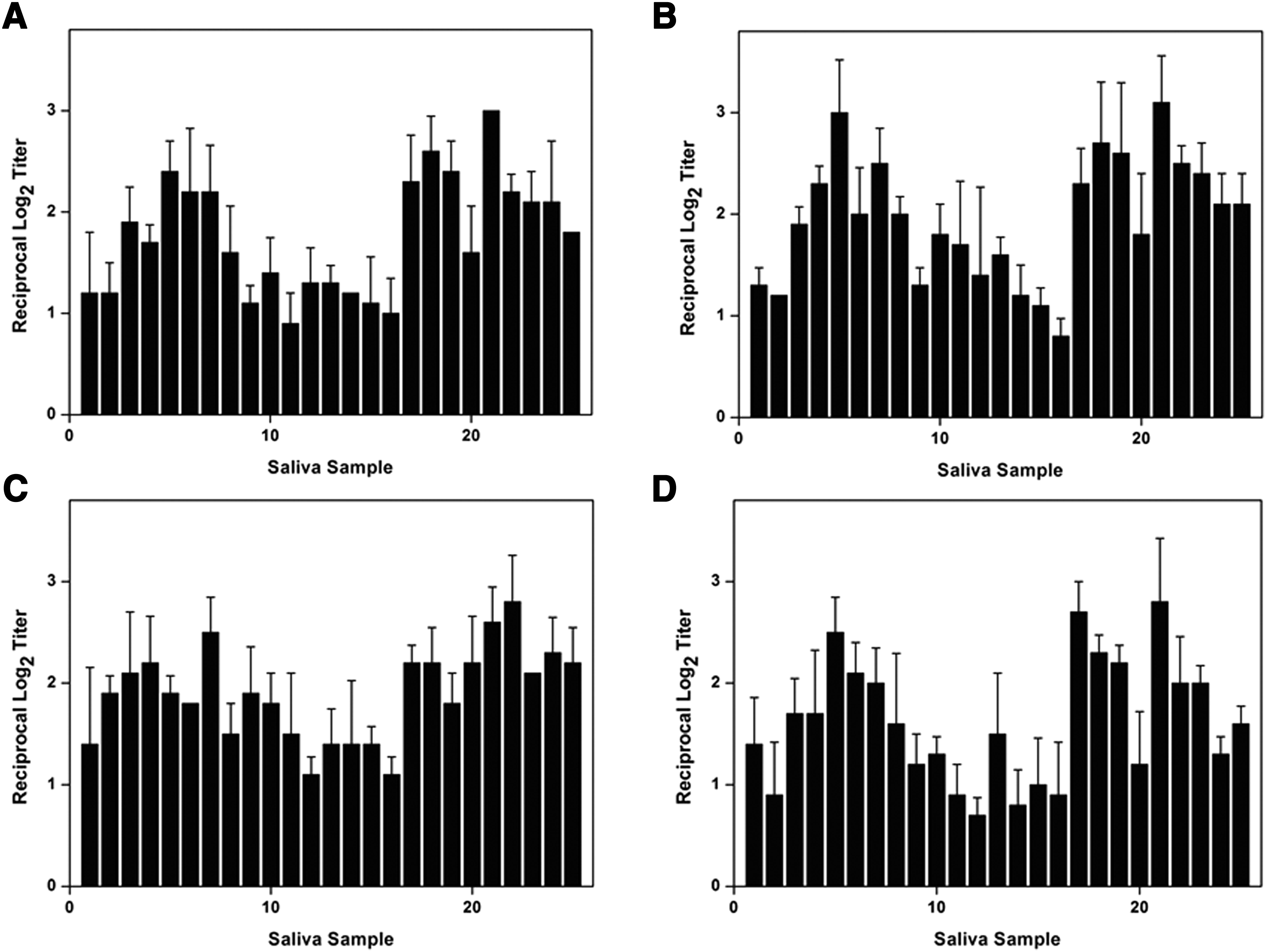
Reaction of mAbs with
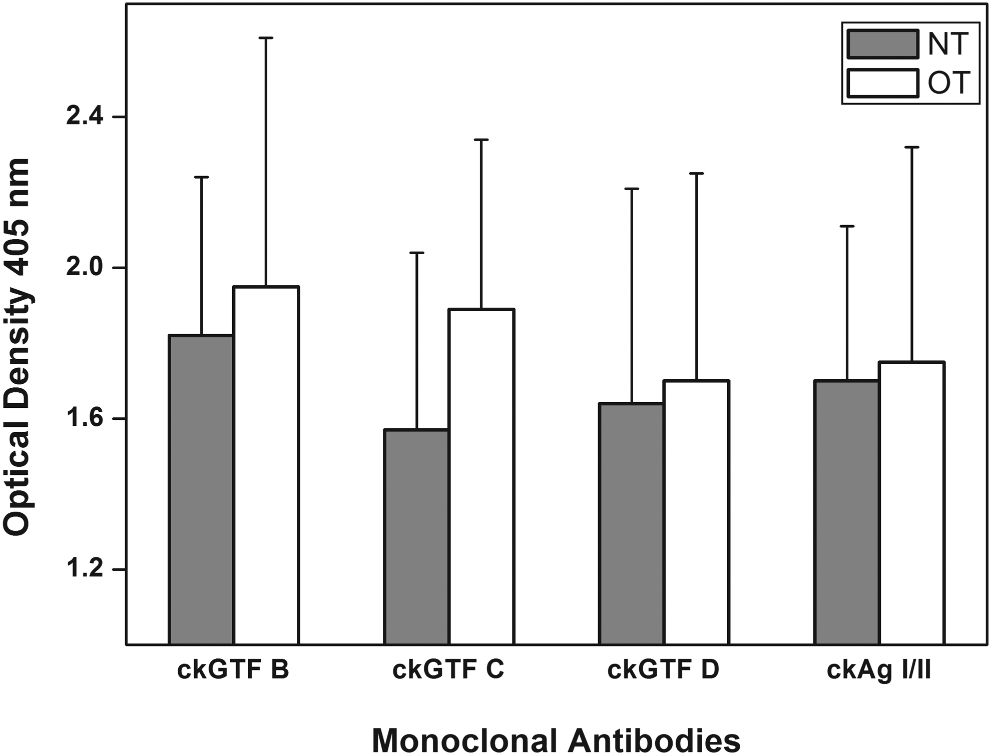
Comparison of the number of S. mutans detected by using mAbs. Saliva samples collected from 25 adolescents undergoing OT and 25 healthy adolescents NT were used. Data represent the mean ± SD of triplicate assays. OT, orthodontic treatment; NT, not undergoing treatment.
Discussion
It has been shown that OT can change a patient's oral ecology,(2,27) increase S. mutans in saliva, and their risk of plaque and dental caries.(3,11,28) Several studies have reported that young patients with OT were especially prone to caries.(1) There is a report that a count of S. mutans alone could not predict the risk for dental caries.(29) Thus, many studies have used CFU counting and other methods such as PCR (polymerase chain reaction) and suggested that there is a positive relationship between the number of mutans streptococci in saliva and the prevalence of dental caries(30,31) and, also between the abundance of S. mutans and the DMFT index.(32,33)
The aim of this study was to investigate the number of S. mutans in OT and NT groups and determine whether there was a link between the OT and the levels of the oral bacterium-causing dental caries. For this purpose, we used mAbs against S. mutans along with Dentocult-SM because a study suggested that mAbs are a valid and reliable method to estimate the levels of S. mutans.(26)
In this study, the results showed that the levels of S. mutans in OT were not significantly higher than those in NT. This can be explained in that the adolescents in the OT group had relatively good oral hygiene because they had undergone OT for over 6 months and, according to several studies, bacterial concentration increased after the first 3 months of OT and decreased after 6 months.(34,35) However, the OT group still had more S. mutans classes 1 to 2 than the NT group (Table 1), suggesting that it is generally more difficult to keep teeth clean during OT even though the patients were educated in oral hygiene and instructed in techniques that eliminate or reduce oral bacteria.
Furthermore, the use of the mAbs against S. mutans provides information on the number of GTFs and Ag I/II in saliva. As shown in Figure 3, the concentration of each enzyme in the groups is different. It is not certain whether the difference of the enzymes affect the incidence of dental caries and needs to be further investigated.
In conclusion, S. mutans in saliva samples were detected by using mAbs and the levels of the bacterium in OT patients were slightly higher than in the NT group. The findings of this study suggest that the use of mAbs against S. mutans could be a useful method to investigate the effect of OT, other dental tools, or agents on the frequency and/or development of dental caries by comparing the quantity of the bacterium.
Footnotes
Acknowledgment
This study was supported by a fund of Biomedical Research Institute, Chonbuk National University Hospital.
Author Disclosure Statement
No competing financial interests exist.