
Abstract
Enzyme-linked immunosorbent assay (ELISA) has been one of the main methods for detecting an antigen in an aqueous sample for more than four decades. Nowadays, one of the biggest concerns for ELISA is still how to improve the sensitivity of the assay, and the luciferase–luciferin reaction system has been noticed as a new detection method with high sensitivity. In this study, a luciferin–luciferase reaction system was used as the detection method for a sandwich ELISA system. It was shown that this new system led to an increase in the detection sensitivity of at least two times when compared with the traditional horseradish peroxidase (HRP) detection method. Lastly, the serum levels of the human extracellular matrix 1 protein of breast cancer patients were determined by the new system, which were overall similar to the HRP chemiluminescent system. Furthermore, this new luciferase reporter can be implemented into other ELISA systems for the purpose of increasing the assay sensitivity.
Introduction
E
The human extracellular matrix 1 (hECM1) protein, which is mapped to chromosome 1q21 outside of the epidermal differentiation complex region, is a secreted glycosylated protein with a molecular weight of 85 kDa.(8–10) The full-length hECM1 contains five different domains, including a signal peptide, an N-terminal cysteine-free domain, two tandem repeats, and a C-terminal domain. The latter three domains have the typical CC-(X7–10)-C arrangement, which generates “double-loop” domains that are involved in important ligand-binding functions.(11) hECM1 is widely expressed in various tissues and has multiple physiological functions,(10,12) such as cell proliferation,(13) chondrocyte development,(14,15) angiogenesis,(16,17) and the regulation of type 2 helper T cells migration.(18) In tumors, ECM1 has higher expression levels than in normal tissues, and was shown to be correlated with microvascular proliferation,(16) tumor grading, lymphatic metastasis, and prognosis of cancers.(19–24) Lee et al. found that high levels of ECM1 in serum of breast cancer patients were directly and inversely related to overall survival.(13) Therefore, rapid and accurate determination of the serum level of ECM1 can be crucial for clinical diagnosis and management.
In this study, we adopted the luciferase detection method to improve the sensitivity of the ELISA system. As a proof of concept, a luciferase-based sandwich ELISA system for the detection of human ECM1 was established. We first ran ELISA to determine the best antibody pair among those domain-specific antihuman ECM1 monoclonal antibodies (MAbs) that we generated previously.(25) We then optimized an HRP-based sandwich ELISA system followed by the integration of a luciferase-based detection system, which led to an increase in sensitivity of at least two times.
Materials and Methods
Purification of MAbs against hECM1
The five hybridomas (1C4, 1G4, 2E3, 2G3, and 2A3) secreting MAbs against hECM1 were produced previously.(25,26) The MAbs were purified preliminarily by saturated ammonium sulfate precipitation followed by protein A/G affinity column according to our previously described method.(25) The purified MAbs were stored at −20°C until use.
Plasmid construction
pGEMT-hECM1 containing full length of ECM1 without stop codon was obtained in our previous study.(25) The full length cDNA encoding avidin or luciferase was generated by polymerase chain reaction (PCR) using the primers shown in Table 1. All PCR products were gel purified and cloned into pGEM-T easy vector (Promega, Madison, WI). The positive clones were confirmed by restriction enzyme digestion and sequencing. The resultant plasmids were named pGEMT-avidin and pGEMT-luciferase. The full length of ECM1 digested from pGEMT-hECM1 was subcloned into adenoviral E1 shuttle vector pAd5-E1-CMV-MCS-(GGGGS)3-6His by restriction sites ClaI and XbaI. The resultant plasmid was named pAd5-E1-CMV-ECM1-(GGGGS)3-6His. The full length of avidin and luciferase digested from pGEMT-avidin and pGEMT-luciferase was subcloned into prokaryotic expression vector pRSET-B (Invitrogen, San Diego, CA) through restriction sites XhoI and ClaI,ClaI and XbaI. The resultant plasmids were named pRSET-B-avidin and pRSET-B-luciferase. Linker of (GGGGS)3 was subcloned into pRSET-B-luciferase through restriction sites XhoI and ClaI and named pRSET-B-(GGGGS)3-luciferase. Then the full length of avidin was subcloned into pRSET-B-(GGGGS)3-luciferase through restriction sites XhoI and ClaI. The resultant plasmid was named pRSET-B-avidin-(GGGGS)3-luciferase.
Expression and purification of recombinant ECM1 protein
HEK 293 cells (human embryonic kidney cell line) were purchased from American Type Culture Collection (ATCC, Manassas, VA) and cultured by Dulbecco's modified Eagle's medium (Gibco, Grand Island, NY) supplemented with 10% fetal bovine serum (Gibco, Grand Island, NY), 1% penicillin–streptomycin, and 1%
Expression and purification of avidin and luciferase proteins
Plasmids of pRSET-B-avidin-(GGGGS)3-luciferase, pRSET-B-avidin, or pRSET-B-luciferase were transformed into Escherichia coli BL21 (DE3). A positive clone was selected and cultured in Luria-Bertani (LB) liquid medium containing ampicillin (Wolsen, Xi'an, China). Then, 1 mM isopropyl-β-
The conjugation of avidin with luciferase
The conjugation of avidin with luciferase was performed using a controlled protein–protein crosslinking kit (Thermo Scientific, Rockford, IL). First, maleimide-avidin was obtained by the reaction of avidin with sulfosuccinimidyl-4-(N-maleimidomethyl)-cyclohexane-1-carboxylate (sulfo-SMCC). Sulfhydryl-luciferase was obtained by the reaction of luciferase with N-succinimidyl-S-acetylthioacetate (SATA). Then the conjugation of avidin with luciferase was achieved by the incubation of maleimide-avidin with sulfhydryl-luciferase at approximately equal molar amounts following the manufacturer's instructions.
Indirect ELISA
Indirect ELISA was performed in 96-well plates (Corning, Inc., Corning, NY), which were coated with 100 μL of 1 μg/mL recombinant ECM1-6His in coating buffer (0.05 M carbonate/bicarbonate buffer, pH 9.6) and incubated overnight at 4°C. After washing three times with PBS containing 0.1% (v/v) Tween-20 (PBST), every well was blocked with 200 μL of PBST containing 10% fetal bovine serum at 37°C for 1 h. Then various dilutions of corresponding anti-ECM1 MAbs or anti-ECM1 MAbs conjugated with HRP or biotin were added. After incubation at 37°C for 1 h, the plate was washed three times and 100 μL of HRP-conjugated sheep antimouse IgG (1:2000, Thermo, Rockford, IL) or avidin-HRP (Thermo, Rockford, IL) was added. Lastly, 100 μL of 0.1 mg/mL 2,2′-azino-bis (3-ethylbenzthiazoline-6-sulfonic acid) (ABTS) (Sigma, St. Louis, MO) dissolved in citrate phosphate buffer (pH 5.0) containing 0.03% (v/v) H2O2 or 3,3′,5,5′- tetramethylbenzidine (TMB) (MP Biomedicals, Solon, OH) was added, and then the light absorbance of each well was determined at 405 or 450 nm.
HRP-based sandwich ELISA
To choose the best antibody pair for the sandwich ELISA system, the five mentioned purified MAbs, 1C4, 1G4, 2E3, 2G3, and 2A3, were labeled with HRP. Anti-hECM1 MAbs (1C4, 1G4, 2E3, 2G3, and 2A3) diluted in coating buffer (0.05 M carbonate/bicarbonate buffer, pH 9.6) were added to each well, respectively, and incubated overnight at 4°C. After washing with PBST, the wells were blocked with 200 μL of PBST containing 10% fetal bovine serum at 37°C for 1 h. After washing, standard ECM1 protein serially diluted with PBST containing 1% fetal bovine serum or serum samples appropriately diluted was added to the wells and incubated at 37°C for 1 h. Then HRP-labeled antibodies (1C4, 1G4, 2E3, 2G3, and 2A3) were added into the wells, respectively. After washing, 100 μL of 0.1 mg/mL ABTS or TMB containing 0.03% (v/v) H2O2 was added. Finally, after incubation at 37°C for 10 min, the light absorbance of each well was determined at 405 or 450 nm after the reaction of TMB was stopped by adding 2 M H2SO4. The optimal antibody pairs were screened based on the light absorbance of each well in the plates. To establish an HRP-based sandwich ELISA kit for detecting hECM1, one antibody of the antibody pairs was used for coating the plate followed by blocking and incubating with samples containing hECM1. Then another biotin-labeled antibody of the antibody pairs was added into the plates followed sequentially by incubating with commercial avidin-HRP. Finally, the light absorbance of each well was determined according to the method already described, and then the ECM1 levels of the samples were quantified.
Patients and normal subjects
The research protocol was approved by the Ethics Committee of Human Experimentation of our University, and was performed in accordance with the principles of the Declaration of Helsinki. Five breast cancer patients were recruited from Fenyang Hospital of Shanxi Province, six normal individuals were provided by Shaanxi Normal University Hospital, and written informed consents were obtained. Serum samples were collected and kept at −80°C until use.
Luciferase-based sandwich ELISA
The experimental procedures were similar to that of HRP-based sandwich ELISA, except 100 μL of avidin-(GGGGS)3-luciferase or the complex of avidin and luciferase conjugate was added and incubated at 37°C for 1 h after incubation with the biotin-labeled antibodies. Finally, 50 μL of 66 μg/mL luciferin was added and the luciferase activity assay of each well was performed at 560 nm by Varioskan Flash (Thermo Fisher scientific, Rockford, IL).
Statistics
All assays were performed in triplicate. A p value less than 0.05 was considered statistically significant; a p value less than 0.01 was considered extremely significant.
Results
Establishment of HRP-based sandwich ELISA system for detecting hECM1
First, to obtain human eukaryotic ECM1 protein to produce the standard curve for sandwich ELISA, the plasmid pAd5-E1-CMV-ECM1-(GGGGS)3-6His expressing human ECM1 protein was constructed. The eukaryotic ECM1 was then purified from the supernatants of HEK 293 cells transfected by the plasmids. Purified ECM1 protein was subjected to 10% SDS-PAGE followed by Coomassie blue staining (line 1 of Fig. 1A) and Western blot using commercial anti-ECM1 antibody (line 2 of Fig. 1A) and anti-His antibody (line 3 of Fig. 1A). The concentration of standard hECM1 protein was 33 μg/mL quantified by BCA assay kit. Second, to obtain an optimal antibody pair, five strains of anti-ECM1 MAbs (1C4, 1G4, 2E3, 2G3, and 2A3) produced in our previous study(25) were purified and tittered (Fig. 1B). The purified MAbs were then used for the screening of the best antibody pair by ELISA using HRP-labeled and unlabeled MAbs. The results indicated that anti-ECM1 MAbs 1C4 and 2E3 made up the best pair (Fig. 1C). Based on this antibody pair, two substrates of HRP, ABTS and TMB, were compared and showed that TMB gave rise to a higher absorbance than ABTS when the same concentration of ECM1 protein was applied (Fig. 1D). Furthermore, biotin–avidin cascade amplifying system was employed to the sandwich ELISA system, which resulted in an increased sensitivity of up to four times as compared with HRP (Fig. 1E). Finally, the dilution of antibody 1C4-biotin, avidin-HRP, and the coating antibody 2E3 was also optimized (Fig. 1F–I).

HRP-based sandwich ELISA system for detecting ECM1.
Establishment of luciferase-based sandwich ELISA system for detecting ECM1
To further improve the sensitivity of the ELISA system we already set up, we introduced the luciferase detection method into the sandwich ELISA system for detecting hECM1. The recombinant avidin-(GGGGS)3-luciferase, avidin, and luciferase fused with 6His peptide were prokaryotically expressed in E. coli BL21 (DE3), and then purified by Ni-NTA purification system. The purified recombinant proteins avidin-(GGGGS)3-luciferase, avidin, and luciferase proteins were subjected to 10% SDS-PAGE followed by Coomassie blue staining (line 1 of Fig. 2A–C). The recombinant avidin and luciferase were further confirmed by Western blot analysis using anti-His antibodies (line 2 of Fig. 2B, C). The fusion protein avidin-(GGGGS)3-luciferase, in combination with the detecting antibody 1C4-biotin, was first utilized for the detection of ECM1. The schematic overview of the strategy is shown in Figure 2D. However, this system failed to improve the detection sensitivity (Fig. 2E). The reason was probably because of the failure of the tetramer formation of avidin protein (23 kDa) caused by the steric hindrance from the large luciferase protein (70 kDa). This problem was not solved by inserting a flexible linker (GGGGS)3 between luciferase and avidin protein. Alternatively, avidin and luciferase were conjugated by protein cross-linking strategy. The resulting conjugate was used to bind 1C4-biotin followed by incubation with luciferase substrate luciferin (Fig. 2F). The results indicated that avidin–luciferase conjugate could significantly improve the system sensitivity by two times (Fig. 2G) when compared with HRP (Fig. 1I).
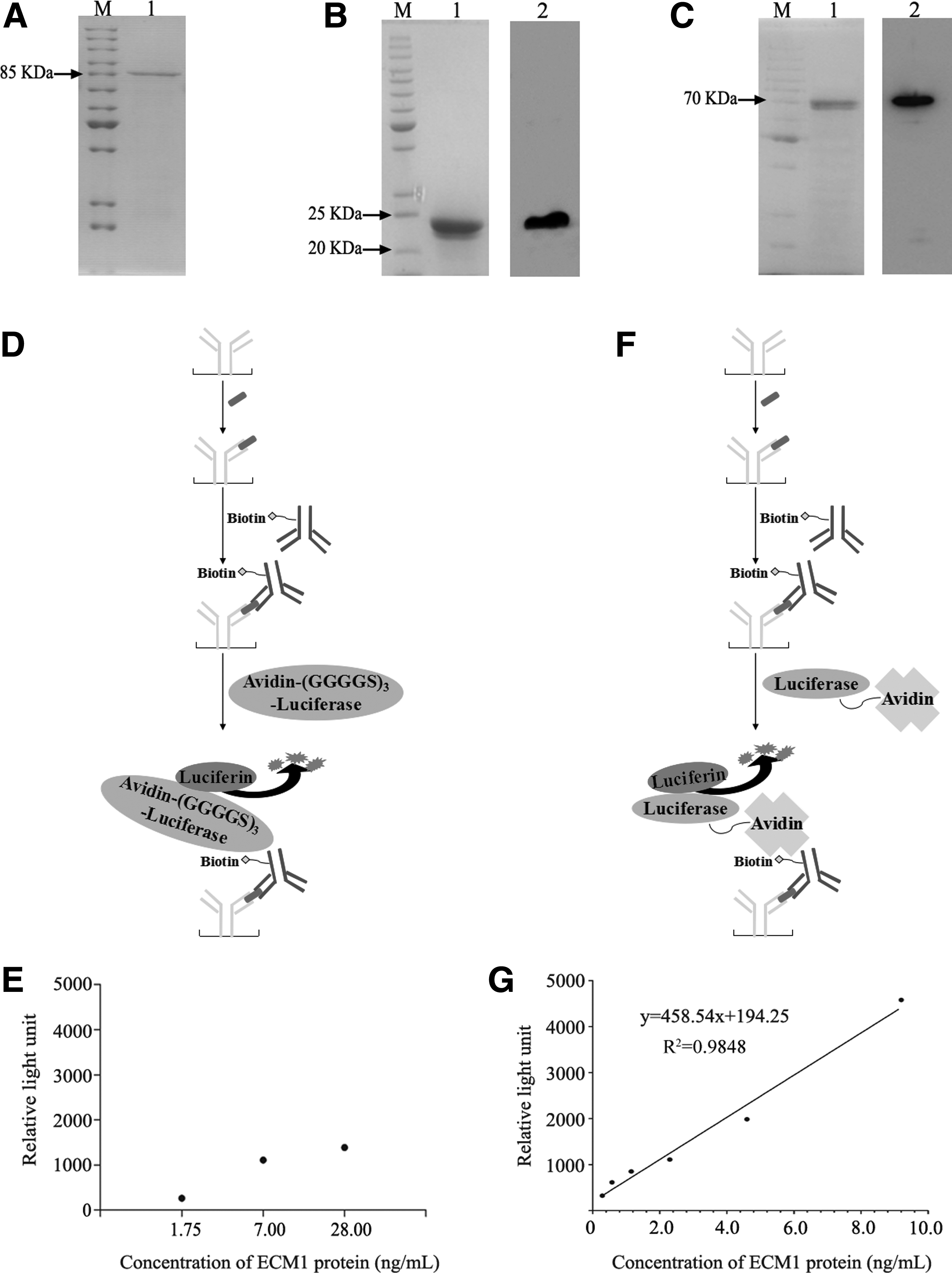
Development of a luciferase-based sandwich ELISA system for detecting hECM1.
Determination of the serum levels of hECM1 in patients with breast cancer
The serum ECM1 levels of patients with breast cancer and normal individuals were determined by using the new system and compared with those of the HRP chemiluminescent system. The results showed that the serum ECM1 levels obtained by the luciferin–luciferase system were overall similar to those obtained by the HRP chemiluminescent system. As expected, breast cancer patients had significantly higher serum ECM1 levels than the normal controls (Fig. 3).

Detection of serum ECM1 levels by HRP-based and luciferase-based sandwich ELISA methods. In normal controls, levels of ECM1 were 5.26 ± 1.85 ng/mL assessed by luciferase-based sandwich ELISA and 5.19 ± 2.43 ng/mL by HRP-based sandwich ELISA; in patients with breast cancer, levels of ECM1 were 11.60 ± 3.77 ng/mL assessed by luciferase-based sandwich ELISA and 10.60 ± 5.19 ng/mL by HRP-based sandwich ELISA. ns, not significant; **p < 0.01 by one-way ANOVA. Error bars represent mean ± standard error of mean.
Discussion
ECM1 is widely expressed in various tissues and involved in multiple physiological functions. ECM1 is over expressed in many kinds of epithelial tumors such as breast tumors and laryngeal carcinoma.(13,16,17,28) High expression level of ECM1 is correlated with microvascular density, lymphatic metastasis, tumor grading, and prognosis.(16,20,28) Therefore, sensitive and accurate determination of the serum level of ECM1 can be crucial for clinical diagnosis and management. For this purpose, a sandwich ELISA system based on HRP for detecting hECM1 was established by the best antibody pair 1C4 and 2E3 (Fig. 1I).
Luciferin–luciferase reaction system has been considered a reporter system with high sensitivity.(5–7,29–32) To improve the sensitivity, we introduced the luciferase detection method into the sandwich ELISA system for detecting hECM1. The fusion protein avidin-(GGGGS)3-luciferase failed to improve the detection sensitivity. This was probably because of the failure of the tetramer formation of avidin protein (23 kDa) caused by the steric hindrance from the large luciferase protein (70 kDa). The problem was not solved by inserting a flexible linker (GGGGS)3 between luciferase and avidin protein. Fortunately, the conjugated protein avidin and luciferase solved the problem effectively and the sensitivity was improved compared with HRP (Fig. 2G).
The data of patients with breast cancer and normal individuals indicated that the new luciferin–luciferase reaction system can accurately detect the serum ECM1 levels. Because the normal serum ECM1 level is much higher than the lowest detection limit of the new system, its advantage over the HRP system of being highly sensitive was not appreciated in this patient cohort. However, the new system would be useful in detecting biomarkers of low serum levels, and hence important in early detection of tumor and/or tumor recurrence.
Footnotes
Acknowledgments
This study was supported by the Innovation Funds of Graduate Programs, Shaanxi Normal University (2013CXB011), and the research grants to H.X. from the National Natural Science Foundation of China (Nos. 81272543 and 81471772) and the Natural Science Foundation of Shaanxi Province, China (No. 2014JM4113).
Author Disclosure Statement
No competing financial interests exist.