




Abstract
Gliomas are the most frequently occurring brain tumors with a heterogeneous molecular background. The molecular subgrouping of gliomas more prognostically stratifies patients into distinct groups compared with conventional histological classification. The most important molecules for the subtype diagnosis of diffuse gliomas are mutations of isocitrate dehydrogenase (IDH), TERT promoter, and α-thalassemia/mental-retardation-syndrome-X-linked (ATRX) and the codeletion of 1p/19q. Among them, IDH and ATRX mutations can be diagnosed using specific monoclonal antibodies (mAbs). We have developed many mAbs against IDH mutants, including HMab-1/HMab-2 against IDH1-R132H and multispecific mAbs MsMab-1/MsMab-2 against IDH1/2 mutations. In contrast, highly sensitive mAbs against ATRX remain to be established. In this study, we immunized mice with recombinant human ATRX and developed a novel mAb, AMab-6. The dissociation constant of AMab-6 was determined to be 9.7 × 10⁻¹⁰ M, indicating that the binding affinity of AMab-6 is very high. Furthermore, AMab-6 sensitively detects ATRX in Western blot and immunohistochemical analyses, indicating that AMab-6 could become the standard marker to determine the ATRX mutation status of gliomas in immunohistochemical analyses.
Introduction
T
ATRX mutation in gliomas results in the loss of ATRX protein expression, which can be diagnosed by immunohistochemistry using anti-ATRX antibodies.(7) Almost all studies of ATRX have used polyclonal antibodies(8) because highly sensitive monoclonal antibodies (mAbs) against human ATRX remain to be established. Herein, we report a novel anti-ATRX mAb, AMab-6, which is very useful in enzyme-linked immunosorbent assay (ELISA), Western blot, and immunohistochemical analyses.
Materials and Methods
Cell lines and animals
P3 U1 was obtained from the American Type Culture Collection (Manassas, VA) and was cultured in the RPMI 1640 medium (Nacalai Tesque, Inc., Kyoto, Japan), supplemented with 10% heat-inactivated fetal bovine serum (Thermo Fisher Scientific, Inc., Waltham, MA) at 37°C in a humidified atmosphere of 5% CO2 and 95% air. Antibiotics, including 100 U/mL of penicillin, 100 μg/mL of streptomycin, and 25 μg/mL of amphotericin B (Nacalai Tesque, Inc.), were added to the media. Female MRL/lpr mice (4 weeks old) were purchased from SLC Japan (Shizuoka, Japan). Animals were housed under pathogen-free conditions. The “Animal Care and Use Committee of Tohoku University” approved the animal experiments described herein.
Hybridoma production
Plasmid preparation and ATRX recombinant protein production are described in the Supplementary Data. MRL/lpr mice were immunized using an intraperitoneal (i.p.) injection of PA-ATRX-RAP-MAP (100 μg) together with Imject Alum (Thermo Fisher Scientific, Inc.). After three additional immunizations, a booster injection was administered by i.p. 2 days before the spleen cells were harvested. The spleen cells were fused with P3 U1 cells using PEG1500 (Roche Diagnostics, Indianapolis, IN). The fused cells were grown in the RPMI medium with hypoxanthine, aminopterin, and thymidine selection medium supplement (Thermo Fisher Scientific, Inc.). Culture supernatants were screened using ELISA for the binding to recombinant MBP-ATRX-MAP-PA or GST-ATRX-MAP-PA.
Enzyme-linked immunosorbent assay
Recombinant MBP-ATRX-MAP-PA or GST-ATRX-MAP-PA was immobilized on Nunc MaxiSorp 96-well immunoplates (Thermo Fisher Scientific, Inc.) at a concentration of 5 μg/mL for 30 minutes. After blocking with 1% bovine serum albumin in 0.05% Tween 20/phosphate-buffered saline, the plates were incubated with culture supernatant followed by 1:3000-diluted peroxidase-conjugated anti-mouse IgG (Dako; Agilent Technologies, Inc., Glostrup, Denmark). The enzymatic reaction was performed with a 1-Step Ultra TMB-ELISA (Thermo Fisher Scientific, Inc.). The optical density was measured at 655 nm using an iMark microplate reader (Bio-Rad Laboratories, Inc., Berkeley, CA).
Determination of binding affinity using ELISA
Purified recombinant protein was immobilized at 5 μg/mL. The plates were incubated with serially diluted antibodies (26 pg/mL–10 μg/mL) followed by 1:1000-diluted peroxidase-conjugated anti-mouse IgG (Dako). The dissociation constant (KD) was obtained by fitting the binding isotherms using the built-in one-site binding models in GraphPad Prism 6 (GraphPad Software, Inc., La Jolla, CA).
Western blot analyses
Recombinant protein (0.1 μg) was boiled in SDS sample buffer (Nacalai Tesque, Inc.). The proteins were electrophoresed on 5%–20% polyacrylamide gels (Wako Pure Chemical Industries Ltd., Osaka, Japan) and were transferred onto a PVDF membrane (EMD Millipore Corp., Billerica, MA). After blocking with 4% skim milk (Nacalai Tesque, Inc.), the membrane was incubated with AMab-6 or anti-MBP (clone: TMab-2; Wako Pure Chemical Industries Ltd.) and then with peroxidase-conjugated anti-mouse or anti-rat antibodies (1:1000 diluted; Dako) and developed with the Pierce Western Blotting Substrate Plus (Thermo Fisher Scientific, Inc.) or the ImmunoStar LD Chemiluminescence Reagent (Wako Pure Chemical Industries Ltd.) using the Sayaca-Imager (DRC Co. Ltd., Tokyo, Japan).
Immunohistochemical analysis
Surgical specimens of oligodendroglioma and diffuse astrocytoma were fixed in 4% paraformaldehyde and embedded in paraffin for routine histopathological and immunohistochemical analysis. Immunohistochemical analysis was performed using avidin-biotin immunoperoxidase technique. In brief, 4-μm-thick tissue sections were deparaffinized using xylene and rehydrated. After antigen retrieval using Target Retrieval Solution, pH 9.0 (Dako), the sections were autoclaved for 10 minutes and then maintained at room temperature for 40 minutes. Sections were quenched using 3% hydrogen peroxide (H2O2) in methanol for 20 minutes and blocked with 5% skim milk in TBST for 30 minutes. Sections were incubated with 3 μg/mL AMab-6 overnight at 4°C. Immunocomplexes were treated with an Envision+ Kit (Dako) for 1 hour. Color was developed using 3, 3-diaminobenzidine tetrahydrochloride (DAB Substrate Kit SK-4100, Vector, Japan) for 5 minutes. Sections were then counterstained with hematoxylin (Wako Pure Chemical Industries Ltd.). Images were acquired with a BZ-X700 microscope (Keyence, Osaka, Japan).
Results and Discussion
The two major methods for identifying these subtypes of gliomas are direct DNA sequencing and immunohistochemical analyses using antibodies.(9) Immunohistochemistry is a robust and widely available method, and immunohistochemical approaches with defined protocols and materials have become essential for assessing molecular genetic changes.(10) Among many molecular parameters, the important molecules for subtype diagnosis of diffuse gliomas are mutations of IDH, TERT promoter, and ATRX and the codeletion of 1p/19q.(11) In particular, mutations of IDH(9) and ATRX(7,8) can be accurately detected by immunohistochemical analysis. We developed many mAbs against IDH mutants, including HMab-1/HMab-2 against IDH1-R132H and multispecific mAbs MsMab-1/MsMab-2 against IDH1/2 mutations.(7,9,12–28) However, highly sensitive mAbs against human ATRX remain to be established.
In this study, the recombinant PA-ATRX-RAP-MAP was captured using NZ-1-Sepharose and efficiently eluted with MES buffer (pH 6.0), containing 3 M MgCl2 (Fig. 1). Mice were then immunized with PA-ATRX-RAP-MAP to develop novel anti-ATRX mAbs. We utilized the C-terminal region of ATRX as immunogen because commercially available polyclonal antibodies (Sigma-Aldrich) against ATRX were also produced against that region.(7) Using ELISA, the culture supernatants of hybridomas were screened for binding to recombinant MBP-ATRX-MAP-PA or GST-ATRX-MAP-PA that was purified from Escherichia coli. As a result, AMab-6 (mouse IgG1, kappa) was established after limiting dilution.
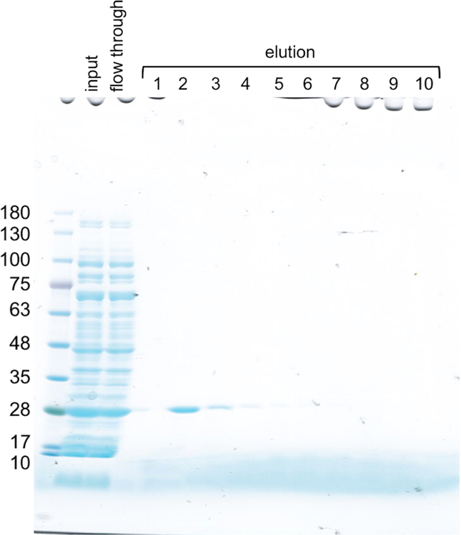
Purification of the recombinant ATRX protein. Cleared lysate (5 μL) from the transformed Escherichia coli before (input) and after (flow through) NZ-1-Sepharose capture, as well as 10 elution fractions (lanes 1–10) during column chromatography, was subjected to 5%–20% gradient SDS-PAGE under a reducing condition and stained with CBB. ATRX, α-thalassemia/mental-retardation-syndrome-X-linked; SDS-PAGE, sodium dodecyl sulfate polyacrylamide gel electrophoresis.
As shown in Figure 2, AMab-6 reacted with MBP-ATRX-MAP-PA in a dose-dependent manner. In contrast, AMab-6 did not react with MBP-IDH1. We further performed a kinetic analysis of the interaction of AMab-6 with MBP-ATRX-MAP-PA (Fig. 2). The KD of AMab-6 was determined to be 9.7 × 10⁻¹⁰ M, indicating that the apparent binding affinity of AMab-6 is very high.
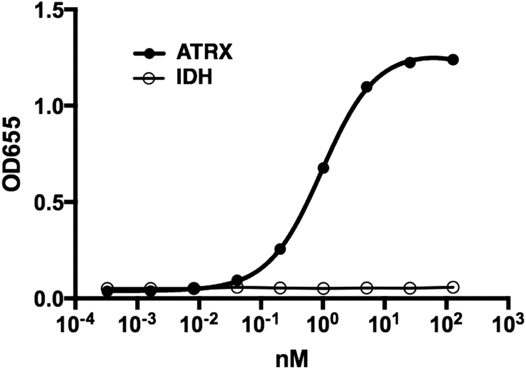
Determination of binding affinity using enzyme-linked immunosorbent assay. Purified recombinant protein (MBP-ATRX-MAP-PA or MBP-IDH1) was immobilized at 5 μg/mL. The plates were incubated with serially diluted antibodies (26 pg/mL–10 μg/mL) followed by 1:1000-diluted peroxidase-conjugated anti-mouse IgG. The dissociation constant (KD) was obtained by fitting the binding isotherms using the built-in one-site binding models in GraphPad Prism 6. IDH, isocitrate dehydrogenase.
Western blot analysis demonstrated that AMab-6 detected MBP-ATRX-MAP-PA, but not MBP-IDH1, indicating that AMab-6 does not detect MBP tag in Western blot analysis (Fig. 3). Immunohistochemical analyses revealed that AMab-6 reacted with the nuclei of oligodendroglioma cells without ATRX mutations (Fig. 4A). ATRX mutations in gliomas result in the loss of ATRX protein expression, which can be diagnosed using immunohistochemical analysis.(7) Likewise, AMab-6 did not recognize the nuclei of diffuse astrocytoma cells, which possess ATRX mutation, but reacted with those of vascular endothelial cells without ATRX mutation (Fig. 4B). These results indicate that AMab-6 is very sensitive against ATRX, which is expressed in gliomas without ATRX mutation.

Western blot analysis. Recombinant proteins (0.1 μg) of MBP-ATRX-MAP-PA and MBP-IDH1 were electrophoresed and transferred onto a PVDF membrane. After blocking, the membrane was incubated with AMab-6, RMab-3 (anti-IDH1), or TMab-2 (anti-MBP).
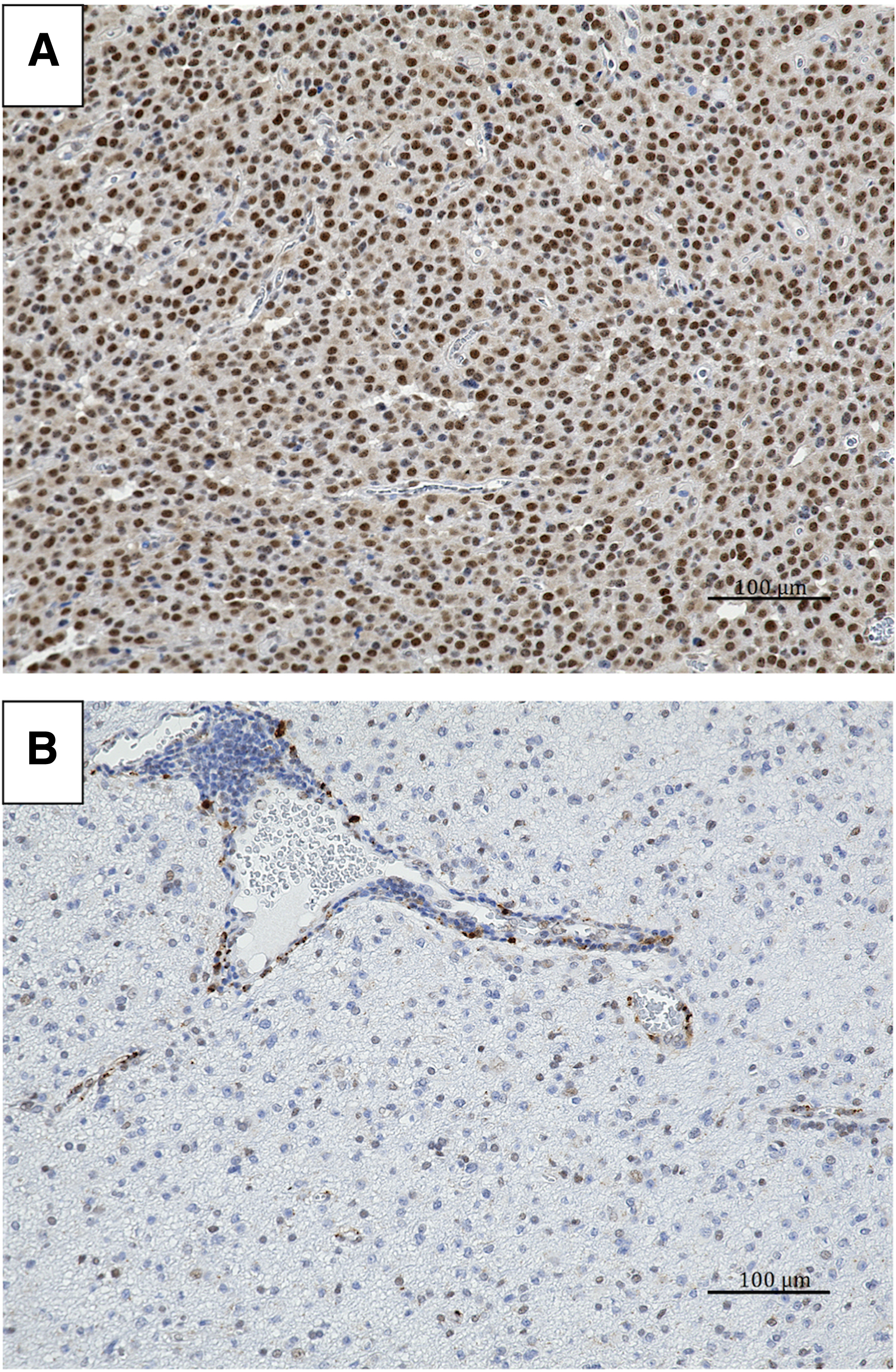
Immunohistochemical analysis.
In conclusion, a novel anti-ATRX mAb, AMab-6, sensitively reacted with human ATRX in ELISA, Western blot, and immunohistochemical analyses. Although many studies have used commercially available polyclonal anti-ATRX antibodies,(7) we confirmed that the reactivity of AMab-6 is compatible with that of polyclonal antibodies (data not shown). Furthermore, we performed immunohistochemical analyses under many other conditions for antigen retrievals (citrate buffer, pH 6.0 or 9.0) or incubation time of AMab-6 (30 minutes, 1 hour, or overnight) and obtained positive staining results as shown in Figure 4. Altogether, AMab-6 could serve as a standard marker to determine the ATRX mutation status of gliomas during immunohistochemical analyses.
Footnotes
Acknowledgments
The authors thank Takuro Nakamura, Noriko Saidoh, and Kanae Yoshida for their excellent technical assistance. This work was supported, in part, by the Practical Research for Innovative Cancer Control from Japan Agency for Medical Research and development, AMED (Y.K.); by JSPS KAKENHI Grant Number 25462242 (Y.K.) and 16K10748 (Y.K.), by the Regional Innovation Strategy Support Program from the Ministry of Education, Culture, Sports, Science and Technology (MEXT) of Japan (Y.K.), by the Platform for Drug Discovery, Informatics, and Structural Life Science (PDIS) from AMED (Y.K.), and by the Basic Science and Platform Technology Program for Innovative Biological Medicine from AMED (Y.K.). This work was performed, in part, under the Cooperative Research Program of Institute for Protein Research, Osaka University, CR-15-05 and CR-16-05.
Author Disclosure Statement
No competing financial interests exist.
References
Supplementary Material
Please find the following supplemental material available below.
For Open Access articles published under a Creative Commons License, all supplemental material carries the same license as the article it is associated with.
For non-Open Access articles published, all supplemental material carries a non-exclusive license, and permission requests for re-use of supplemental material or any part of supplemental material shall be sent directly to the copyright owner as specified in the copyright notice associated with the article.
