
Abstract
AMACR (alpha-methylacyl-CoA racemase) has been recently described as a prostate cancer-specific gene that encodes a protein involved in the beta-oxidation of branched chain fatty acids. Expression of AMACR protein is found in prostatic adenocarcinoma, but not in benign prostatic tissue. Thus, monoclonal antibodies (mAbs) for AMACR detection are an important tool for the diagnosis of AMACR-positive cancers. However, only a few mAbs, especially those applicable for immunohistochemistry (IHC), have been established to date. In this study, we describe the generation of a new hybridoma clone G8 producing anti-AMACR antibodies. G8 mAb specifically binds human AMACR and was successfully used in immunoblotting and immunofluorescence on paraformaldehyde-fixed cells and in IHC of paraffin-embedded tumor specimens. These results indicate that this new anti-AMACR mAb G8 would be useful in the diagnosis of AMACR-related cancers and would be a strong tool in both basic and clinical research on AMACR.
Introduction
A
In immunohistochemistry (IHC), AMACR has been shown to be a specific marker of prostatic adenocarcinoma. AMACR antibody stains the majority of prostate cancers (PCas) and also other types of carcinomas such as hepatomas, breast carcinomas, and pancreatic and islet tumors. PCa is the most frequently diagnosed noncutaneous malignancy in men and the second leading cause of male cancer-related mortality in the United States.(9) Diagnosis of PCa glands can sometimes present a diagnostic challenge for pathologists, since prostate carcinoma can mimic benign prostate glands(10) and the architectural or cytologic clues for the diagnosis of carcinoma may not always be seen in small foci of suspicious glands. In addition, tissue diagnosis of PCa can be difficult in needle biopsies or in a small focus of cancers of radical prostatectomies, presenting one of the major challenges in surgical pathology. Therefore, a PCa specific marker could be of great importance and usefulness as adjunct to facilitate critical diagnostic decisions with high sensitivity and specificity.(11) The following are the International Society of Urological Pathology recommendations for the use of IHC in prostate specimens. Either high-molecular weight cytokeratin (34βE12 or CK5/6 or others) or p63 or a combination of the two with AMACR either in a double or triple cocktail is recommended for the workup of small foci of atypical glands suspicious for adenocarcinoma of the prostate.(12)
In this study, we generated a new monoclonal antibody (mAb) that detects AMACR both in intact cells and in fixed paraffin-embedded sections. We characterized this antibody and demonstrated its usefulness for several applications, including IHC on formalin-fixed paraffin embedded (FFPE) tissue sections. Our results suggest that this new anti-AMACR mAb G8 would be a powerful tool in the diagnostics of AMACR-related cancers and other diseases.
Materials and Methods
Preparation of anti-AMACR mAbs
A synthetic peptide conjugated with recombinant hsp70 from Mycobacterium tuberculosis was purified, mixed with Freund's complete adjuvant (ratio 1:1), and used for immunization. BALB/c mice (16–18 g) were immunized with 50 mkg of immunogen per mouse. Subsequent doses were injected with incomplete Freund's adjuvant at 2-day intervals. After two immunizations, popliteal lymph nodes were collected and fused with sp2/0 myeloma cells in a 50% polyethylene glycol 4000 solution (Wako, Osaka, Japan). The fused cells were plated in 96-well plates in the Dulbecco's Modified Eagle Medium (DMEM) containing 4% fetal calf serum (FCS) (Equitech-Bio, Inc., Kerrville, TX), glutamine, gentamicin, sodium pyruvate, and HAT solution (Invitrogen). After 7 days of incubation at 37°C with 5% carbon dioxide in a humidified environment, culture supernatants were collected and screened for their ability to bind to synthetic peptide immobilized on 96-well plates using indirect enzyme-linked immunosorbent assay (ELISA). Selected positive hybridoma clones were expanded and subcloned by limiting dilution.
Enzyme-linked immunosorbent assay
Peptide was diluted to 5 μg/mL by phosphate-buffered saline (PBS), and the microtiter plate was coated with the protein overnight at 4°C. The plates were washed thrice with PBS and blocked with 5% bovine serum albumin in PBS at 37°C for 2 hours. The plates were then incubated with antiserum or mAbs. Then the plates were washed four times with PBST (PBS containing 0.05% Tween 20) and incubated with HPR labeled goat anti-mouse IgG (1:10,000) at 37°C for 1 hour. Finally, the plates were washed thrice with PBST and incubated with freshly prepared tetramethylbenzidine (TMB) with 30% hydrogen peroxide (TMB/H2O2) at 37°C for 10 minutes. The reaction was stopped by 2 mol/L sulfuric acid. The absorbance was measured by Multiskan (Thermo Scientific, Waltham, MA) at 450 nm.
Antibody subclass identification
The subtype of mAbs was analyzed using the Mouse mAb Isotyping Kit (Genemed, South San Francisco, CA). First, IgA, IgM, IgG1, IgG2a, IgG2b, and IgG3 reagents were diluted by coating buffer (1:1000) and coated overnight at 4°C. The cell culture medium containing mAb was added onto plates and incubated at 37°C for 1 hour. The following procedure was the same as that described in the indirect ELISA.
Plasmid construction
RNA was isolated in PC3-MM2 cells using QIAGEN RNeasy Plus Universal Mini Kit (QIAGEN, Hilden, Germany) according to the recommendations of the manufacturer. Obtained RNA was used for complementary DNA (cDNA) synthesis using a RevertAid RT Reverse Transcription Kit (Thermo Fisher Scientific). Obtained cDNA was used to amplify the coding sequence of human AMACR using polymerase chain reaction (PCR). The following primers were used: forward 5′-TCGAGCGGCCGCGCCAT GGCACTGCAG-3′ and reverse 5′-GCTTGGTACCGAGA CTAGCTTTTAC-3′. The primers contain restriction sites for KpnI and NotI, respectively. The amplified product was purified out of the agarose gel using Wizard® SV Gel and PCR Clean-Up System (Promega) and cloned into the pcDNA3.1(-)myc-His vector between KpnI and NotI restriction sites. Obtained pcDNA3.1(-)AMACR-Myc-His plasmid was propagated in XL1-Blue Escherichia coli (Evrogen). The integrity of the sequence was controlled by sequencing. Plasmid for transfection was isolated using PureYield Plasmid Midiprep system (Promega).
Cell culture and transfection
Human HEK293T and PC3-MM2 cells were maintained in DMEM supplemented with 10% FCS. For transfection, HEK 293T cells were seeded on cover slips in 6-well plates at a density of 6.3 × 10⁵ cells per well. Transfection was performed using Lipofectamine 2000 Transfection Reagent (Thermo Fisher Scientific) according to the manufacturer's instructions using 10 mkl Lipofectamine and 2.5 mkl pcDNA3.1(-)AMACR-Myc-His plasmid DNA. Cells were then washed with PBS and cultivated in complete DMEM for 48 hours. Afterward the cells were fixed and analyzed by immunofluorescence. For the Western blot analysis, the same procedure was repeated without cover slips.
Immunoblotting
Cells were lysed with a cell lysis buffer RIPA (50 mM Tris-HCl [pH: 7.5], 150 mM sodium chloride [NaCl], 2 mM ethylenediaminetetraacetic acid, 1% Triton X-100, 0.1% sodium deoxycholate, and 1/25 protease inhibitor cocktail [Roche]). After incubating on shaker for 20 minutes at 4°C, the samples were centrifuged at 13,200 rpm for 10 minutes. The supernatant was collected, and protein concentrations of the lysates were determined by BCA assay (Bio-Rad). Equal amounts of protein from each cell lysate (15 μg/lane) were subjected to sodium dodecyl sulfate–polyacrylamide gel electrophoresis. Separated proteins were transferred to a nitrocellulose membrane (Bio-Rad), and the membrane was blocked in Tween 20-Tris Buffered Saline (10 mM Tris-HCl pH 7.6, 0.15 M NaCl, 0.05% Tween-20) containing 5% nonfat dry milk (Bio-Rad) for 1 hour, followed by incubation with primary antibodies (anti-AMACR 0.5 mg/mL, 1:1000) at 4°C overnight. After washing, the membrane was incubated with horseradish peroxidase-conjugated secondary antibodies (anti-mouse 1:5000, anti-rabbit 1:65,000) for 1 hour at room temperature. Chemiluminescent detection was carried out with the Novex ECL detection system (Invitrogen) according to the manufacturer's instructions.
Immunofluorescence
To detect AMACR molecules in the cytoplasm, cells were fixed with 3.7% paraformaldehyde in PBS at room temperature for 15 minutes and then incubated with primary mAb (anti-AMACR 0.5 mg/mL, 1:10; anti-myc 1:100) at room temperature for 1 hour. Cells were then stained with the Cy3-conjugated anti-mouse antibody (0.5 mg/mL, 1:300; PrimeBioMed, Moscow, Russia) and fluorescein isothiocyanate (FITC)-conjugated Alexa488-conjugated anti-rabbit antibody (0.5 mg/mL, 1:100; Jackson ImmunoResearch) at room temperature for 1 hour. Images were captured by a fluorescent microscope (Olympus BX53, Tokyo, Japan).
IHC on FFPE section
Immunohistochemical staining was performed on human PCa tissues. Endogenous hydrogen peroxidase was quenched with 3% H2O2 in PBS at room temperature for 7 minutes. Paraffin-embedded sections (4 mm thick) were deparaffinized and then treated with 10 mM citrate buffer (pH 6.0) at 110°C for 10 minutes by Decloaking Chamber (Biocare Medical). Sections were incubated with primary mAb at 5 mkg/mL in PBS at room temperature for 1 hour and with Poly-HRP DAB Kit (Ms+Rb) (Genemed) according to the manufacturer's instructions. Sections were counterstained lightly with Mayer's hematoxylin. Images were captured by light microscopy (Olympus BX53, Tokyo, Japan).
Results and Discussion
Generation and characterization of mouse anti-human AMACR mAbs
For generation of anti-AMACR mAbs, synthetic peptide was used as immunogen. We obtained eight hybridoma clones producing IgGs that recognize AMACR antigen in ELISA. Obtained antibodies were tested for their ability to stain AMACR positive structures in prostatic adenocarcinoma samples. One clone G8 showed a staining pattern similar to the control anti-AMACR antibodies 13H4.
To determine the specificity of anti-AMACR antibody G8, we generated 293T cells transiently transfected with pcDNA3.1(-)AMACR-Myc-His. Obtained cells were stained with G8 antibody and anti-Myc antibody to confirm the identity of the protein detected by G8 antibody. It was found that G8 staining colocalizes with anti-Myc staining (Fig. 1A), indicating that G8 recognizes recombinant Myc-tagged AMACR.

Specificity of G8 monoclonal antibodies for AMACR protein was determined by immunofluorescence
To further confirm the specificity of the obtained antibody, 293T cells transiently transfected with pcDNA3.1(-)AMACR-Myc-His were used for Western blotting with G8 antibody and anti-Myc antibody. It was found that G8 antibody detects protein of the same molecular weight as anti-Myc antibody (Fig. 1B). No protein was detected in mock-transfected cells. These data demonstrate that the newly developed AMACR mAbs are highly specific for detection of AMACR without cross-reaction to other nonspecific proteins.
Comparison of G8 mAb with commercially available anti-AMACR mAb using IHC
To examine whether mAb G8 can detect AMACR on formalin-fixed samples, we first tested staining of paraformaldehyde-fixed PC3-MM2 cells. The cells were grown on cover slips, fixed with paraformaldehyde and stained with G8 or 13H4 antibodies. We observed intensive granular staining with G8 antibody, while no staining was obtained with 13H4 antibody (data not shown).
To test whether the G8 antibody is suitable for IHC on FFPE tissues, clinical specimens were stained with G8 mAb (Fig. 2B, D) and control 13H4 mAb (Fig. 2A, C). Compared with the control mAb 13H4, mAb G8 successfully stained paraffin-embedded sections of prostatic adenocarcinoma. These results indicate that mAb G8 can be successfully used to detect AMACR in FFPE sections.
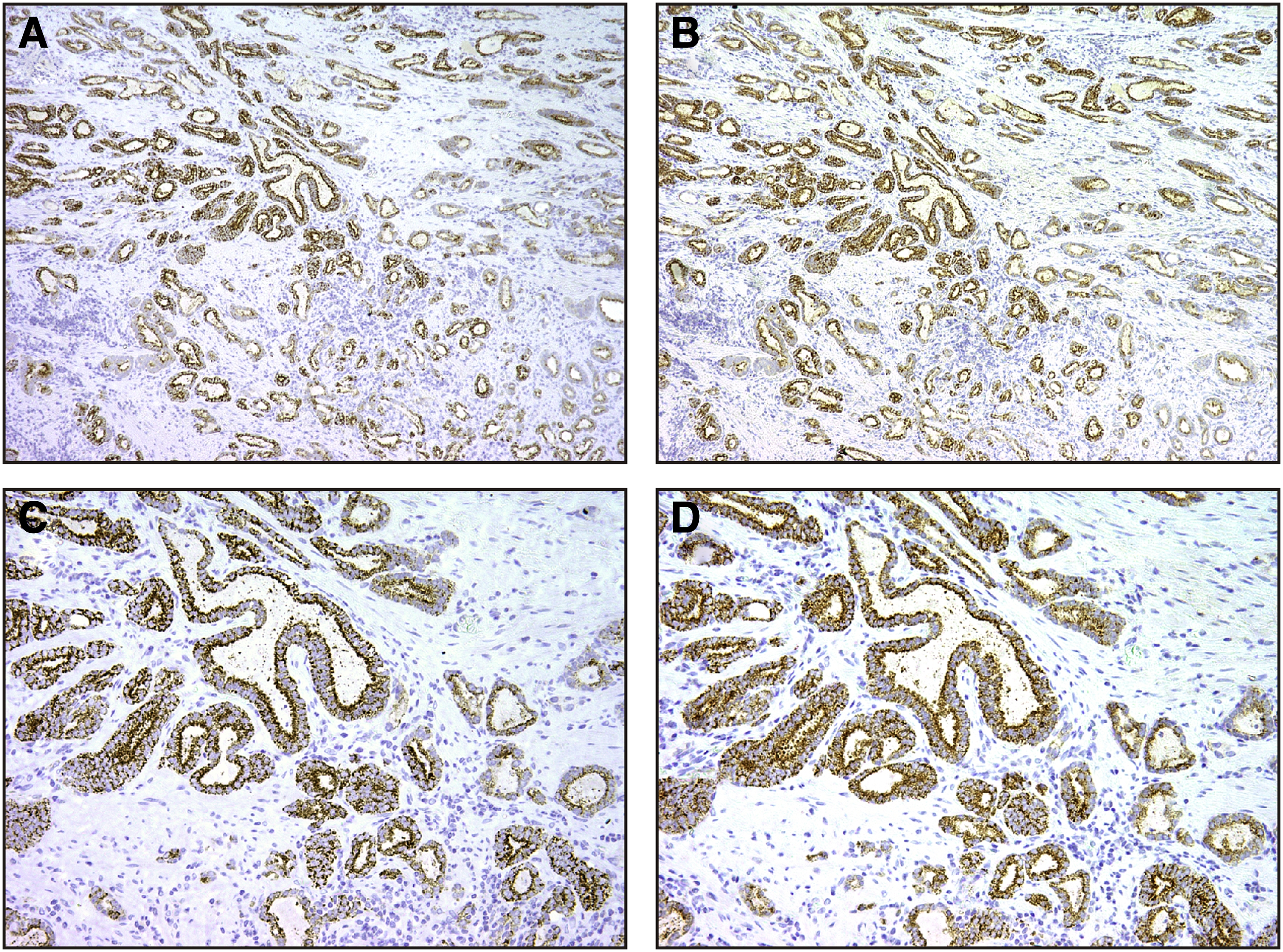
IHC analysis of AMACR in prostatic adenocarcinoma, Gleason score 3 + 3.
In summary, we describe the development and characterization of a novel anti-AMACR mAb, G8, suitable for Western blotting, IHC, and immunofluorescence. The reactivity of this antibody was verified for various types of samples: transfected cells, established cancer cell lines, and PCa tissues. Normal adjacent tissues showed no staining with mAb G8, suggesting that the observed staining was not due to nonspecific binding of mAb G8 to tissues. Thus, our results demonstrate that mAb G8 is the mAb against human AMACR available for IHC of paraffin-embedded sections and suggest that this antibody will be a powerful tool for the study and diagnosis of AMACR-positive cancers and other diseases.
Footnotes
Acknowledgment
This research was supported by Russian Science Foundation grant, project 14-15-00396 (A.G.).
Author Disclosure Statement
No competing financial interests exist.