
Abstract
High mobility group box 1 (HMGB1) is a key member of the “danger associated molecular patterns” (DAMPs), which can localize in various compartments of the cell, and plays important roles in systemic inflammation. In the present study, monoclonal antibodies (MAbs) specifically against chicken HMGB1 were generated. The open reading frame of chicken HMGB1 was amplified by RT-PCR and cloned into the prokaryotic expression vector pET-28a to construct a recombinant plasmid pET-chHMGB1. The recombinant chicken HMGB1 protein was expressed in Escherichia coli Rosetta under IPTG induction and then purified by Ni-NTA Purification System. BALB/c mice were immunized with the purified recombinant HMGB1 protein, and three strains of hybridoma cells named 1F10, 8C11, and 4D8 secreting MAbs of chicken HMGB1 were obtained by hybridoma technique. Western blot and indirect immunofluorescence assays showed that the endogenous HMGB1 in various cell lines and glycosylated HMGB1 could both be specifically recognized by the prepared MAbs. This work indicated that the MAbs against chicken HMGB1 would be a valuable tool for further studies of HMGB1-mediated signaling in virus-infected cells and investigates the role of HMGB1 in avian virus pathogenesis.
Introduction
D
HMGB1 can be expressed in nearly all cell types and is an extremely evolutionarily conserved protein which has 99% identity among all mammals.(19) To facilitate the study of the chicken HMGB1, the nucleotide sequence coding has been established, but specific antibodies are not acquired. Moreover, the function of chicken HMGB1 and the inflammatory signaling pathways activated by HMGB1 are also still not been elucidated. In this study, three monoclonal antibodies (MAbs) for chicken HMGB1 were obtained and have good reactivity with different forms of HMGB1. These MAbs are useful for investigating the functions of chicken HMGB1 in the future.
Materials and Methods
Animals and cell lines
Six-week-old female BALB/c mice (SPF grade) were obtained from the center of SLAC Laboratory Animals of Shanghai (Shanghai, China) and bred in independent ventilation incubators. All the animal protocols used in the study were approved by the Shanghai Veterinary Research Institutional Animal Care Committee. DF1, HeLa, 293T, Vero, MEF, Raw264.7, and SP2/0 cells were purchased from the Shanghai Cell Biology Institutes, Chinese Academy of Sciences (Shanghai, China). MARC-145 cells, PK-15 cells, and porcine alveolar macrophages (PAMs) were kind gifts from Dr. Guangzhi Tong (Shanghai Veterinary Research Institute, Chinese Academy of Agricultural Sciences). Chicken embryo fibroblasts (CEFs) were performed as previously described.(20) Cells were cultured at 37°C in a 5% CO2 atmosphere in Dulbecco's modified Eagle's medium (DMEM; Gibco, Grand Island, NY) or RPMI 1640 medium (Gibco) supplemented with 10% fetal bovine serum (FBS; Gibco).
Molecular cloning and expression vector construction
Based on the mRNA sequence of chicken HMGB1 (GenBank ID no. NM_204902.2), specific primers (forward: 5′-CG
Expression and purification of recombinant chicken HMGB1
The recombinant chicken HMGB1 protein (rchHMGB1) was expressed in Escherichia coli Rosetta transformed with pET28a-chHMGB1. The bacteria were induced with isopropyl-β-D-thiogalactoside (IPTG) at a final concentration of 0.5 mM for 6 hours at 37°C and then harvested by centrifugation and used for ultrasonication. The expression of rchHMGB1 was confirmed by SDS-PAGE and by western blot analysis with anti-His antibody (Sigma-Aldrich, St. Louis, MO). His-tagged rchHMGB1 was purified using the Ni-NTA His•Bind® Resin (Novagen, Madison, WI) according to the manufacturer's instruction. The high purify protein was obtained by ultrafiltration using the tubular ultrafiltration modules (Millipore, Billerica, MA) to remove the imidazole.
Mouse immunization
The mice were immunized with purified rchHMGB1 (100 μg) emulsified with an equal amount of Freund's complete adjuvant using subcutaneous injection at the first immunization. The protein was quantified by RC DC Protein Assay Kit (Bio-Rad, Hercules, CA). At 2-week intervals, a second immunization was given using the purified rchHMGB1 (50 μg) emulsified with an equal amount of Freund's incomplete adjuvant. Then, the immunization was repeated twice with 50 μg of purified rchHMGB1 at 2-week intervals.
Chicken HMGB1 MAb preparation
Cell fusion was performed when the antibody titers reached a certain level. The procedure was performed as previously described with modifications.(21) Briefly, spleen cells obtained from rpHMGB1-immunized BALB/c mice were fused with SP2/0 cells using polyethylene glycol (Sigma-Aldrich, St. Louis, MO). The hybridoma cells were screened for antibody production against HMGB1 using an indirect enzyme linked immunosorbent assay (Indirect ELISA). After subcloning five times, the positive hybridoma cells were inoculated intraperitoneally into pristane-primed BALB/c mice, with a density of 1 × 10⁶ cells for MAb production.
Positive hybridoma clones by Indirect ELISA
The positive hybridoma cells were analyzed by Indirect ELISA assay, according to previously described.(21) Briefly, the ELISA plates were coated with purified rchHMGB1 (1 μg/well) in carbonate bicarbonate buffer (15 mM Na2CO3, 35 mM NaHCO3, pH = 9.6) at 4°C overnight and then blocked with 5% skimmed milk in phosphate buffer with 0.05% Tween-20 (PBST) at 37°C for 2 hours. The plates were incubated with 50 μL cell culture supernatant at 37°C for 1 hour. After washing thrice, horseradish peroxidase-conjugated goat anti-mouse IgG (1:5000 dilution in PBST) was added into the wells for 30 minutes at 37°C. The wells were incubated with TMB liquid (50 μL/well) (Amresco, Solon, OH) and kept in the dark for 10 minutes at RT. After being stopped by 2 M H2SO4, the absorbance was measured at 450 nm. The positive hybridoma cells were subcloned thrice and then extended culture.
Eukaryotic expression vector construction and transfection
Chicken and human eukaryotic expression vector was constructed by cloning the complete CDS of chicken or human HMGB1 into the p3 × FLAG-CMV-7.1 vector (Sigma-Aldrich), primed by FLAG-chHMGB1 primers (forward: 5′-CG
Western blot analysis
To analyze the specificity of the MAbs, HeLa cells overexpressing FLAG-chHMGB1 and FLAG-huHMGB1, CEF, DF1, HeLa, 293T, MARC-145, Vero, MEF, Raw264.7, PAMs, and PK-15 cells were harvested as previously described,(20) and equal amounts of cell lysates were separated by 10% SDS-PAGE and followed by transferring to nitrocellulose (NC) membrane. The membrane was then blocked with 5% skimmed milk in TBST (TBS with 0.1% Tween-20) and probed with anti-FLAG antibody, β-actin antibody (Sigma-Aldrich), or anti-HMGB1 MAbs. The blot was scanned and visualized using an enhanced chemiluminescence detection system (Thermo Fisher Scientific, Waltham, MA).
Indirect immunofluorescence assay
DF1 cells were fixed and then used for the IFA analysis, according to the previously described.(21) Briefly, the cells were fixed with 4% paraformaldehyde for 30 minutes at RT. Next, cells were permeabilized by incubation with 0.25% Triton-100 in PBS for 5 minutes after washing twice with PBS. After being blocked with 3% bovine serum albumin (BSA) in PBS for 30 minutes at 37°C, cells were incubated with the anti-HMGB1 MAbs at 1: 500 in PBS for 1 hour at 37°C. After washing thrice in PBS, Alexa Fluor® 488 donkey anti-mouse IgG (H+L) antibody (Life Technologies, Carlsbad, CA) was added into the wells, and the cells were incubated at 37°C for 30 minutes. Followed by washing, the cells were stained with DAPI for 5 minutes at 37°C. Finally, the cells were washed thrice in PBS, and the fluorescent images were observed on Nikon inverted fluorescence microscope.
Results
Recombinant chicken HMGB1 protein expression and purification
Total RNA of DF1 cells was extracted and used for cDNA synthesis. The 648 bp fragment of chicken HMGB1 gene was amplified by PCR and cloned into pET-28a vector (Fig. 1). Sequencing and restriction enzyme digestion confirmed that the HMGB1 gene inserted into the position is correct. The rchHMGB1 expression was induced by 0.5 mM IPTG at 37°C in E. coli Rosetta. SDS-PAGE analysis showed that the rchHMGB1 expression was achieved at this condition and was expressed at a high level with soluble form. Meanwhile, highly purified rchHMGB1 was purified by Ni-NTA His•Bind® Resin (Fig. 2A). The recombinant chicken HMGB1 has a predicted molecular mass of 27 kDa. As a result of glycosylation, the apparent molecular mass of rchHMGB1 is approximately 34 kDa in SDS-PAGE under reducing conditions. The rpHMGB1 expression was also confirmed by Western blot analysis. The results showed that both rchHMGB1 and glycosylated rchHMGB1 were recognized by anti-His-tag antibody in the predicted size (Fig. 2B).

Amplification of the coding sequence (CDS) of chHMGB1 by RT-PCR from DF1 cDNA. M, DNA marker DL 2,000; lane 1, product of RT-PCR; lane 2, negative control. HMGB1, high mobility group box 1.
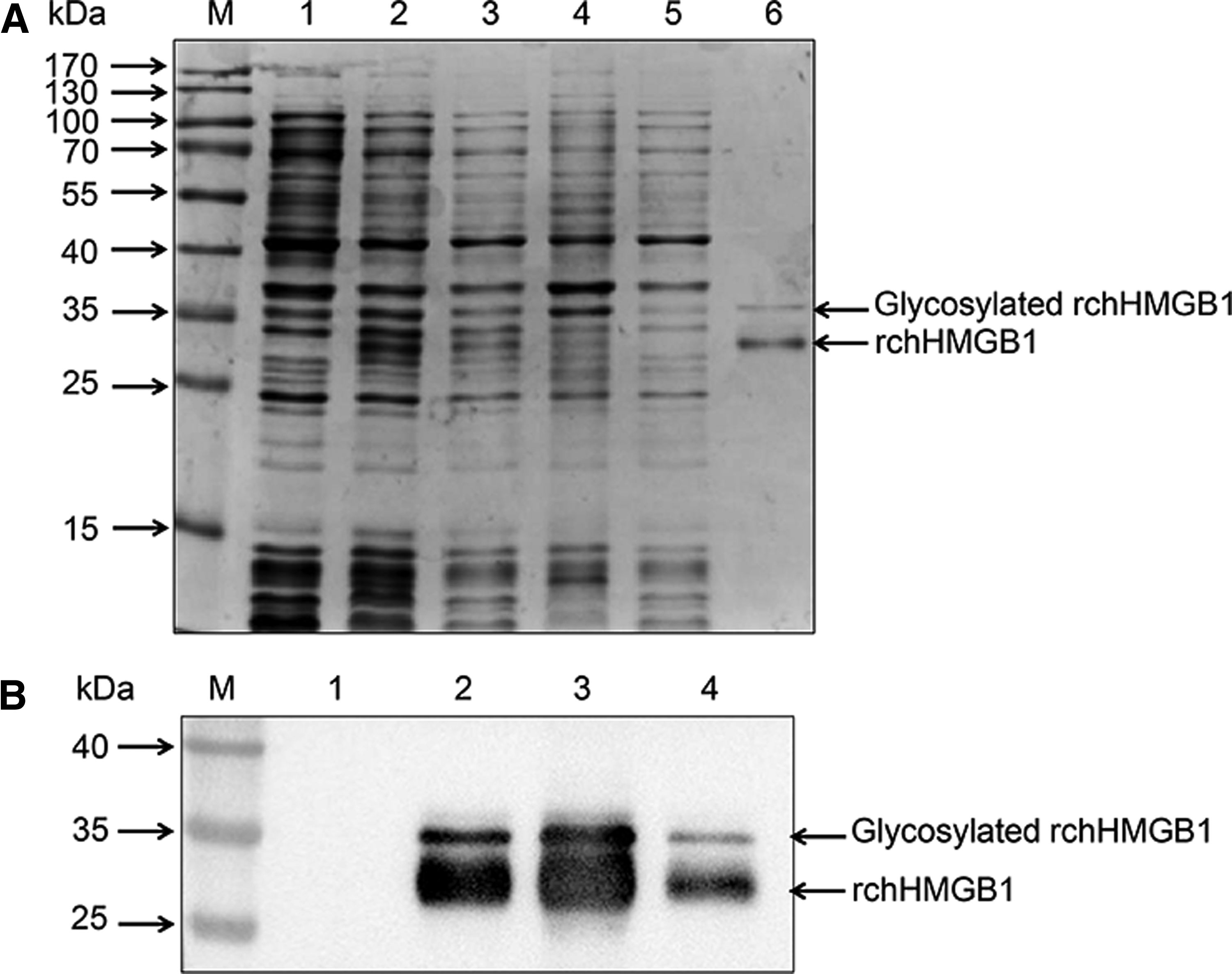
Expression, purification, and identification of rchHMGB1 fusion protein.
Generation of chicken HMGB1 MAbs
After immunization four times, the antisera titers of BALB/c mice were determined by indirect ELISA. Cell fusion was performed using the immunized mice when the antibody titers reached 1:104. Three clones of positive hybridoma cell lines were identified by subcloning and named as clones 1F10, 8C11, and 4D8, respectively. The clones were used to prepare the ascites containing MAbs. The antibody titers of the ascites were up to 1: 2 × 10⁶, 1: 2 × 10⁶, and 1: 1.1 × 10⁶, respectively.
Reactivity and specificity of MAbs
To explore the specificity of the MAbs, endogenous HMGB1, overexpressed chicken HMGB1, and human HMGB1 were detected by the MAbs. As shown in Figure 3A, the overexpressed chicken and human HMGB1 was detected both by anti-FLAG antibody and anti-chicken HMGB1 MAbs. MAb clones 21F10, 8C11, and 4D8 could specifically recognize the endogenous HMGB1 protein in chicken cells (CEF and DF1 cells), human cells (HeLa and 293T cells), and mouse cells (MEF and Raw264.7 cells) (Fig. 3B) and with weak reactivity for monkey cells (MARC-145 and Vero cells) and porcine cells (PAMs and PK-15 cells) (Fig. 3B).

Specificity tests of prepared MAbs by western blot analysis.
Indirect immunofluorescence assay was also performed to further evaluate the reactivity of the MAb to HMGB1. The results showed that specific fluorescence of endogenous HMGB1 protein was detected in the nucleus by the clones 21F10, 8C11, and 4D8 (Fig. 4), suggesting that the MAbs reacted with HMGB1 exclusively and could be further used for IFA analysis.
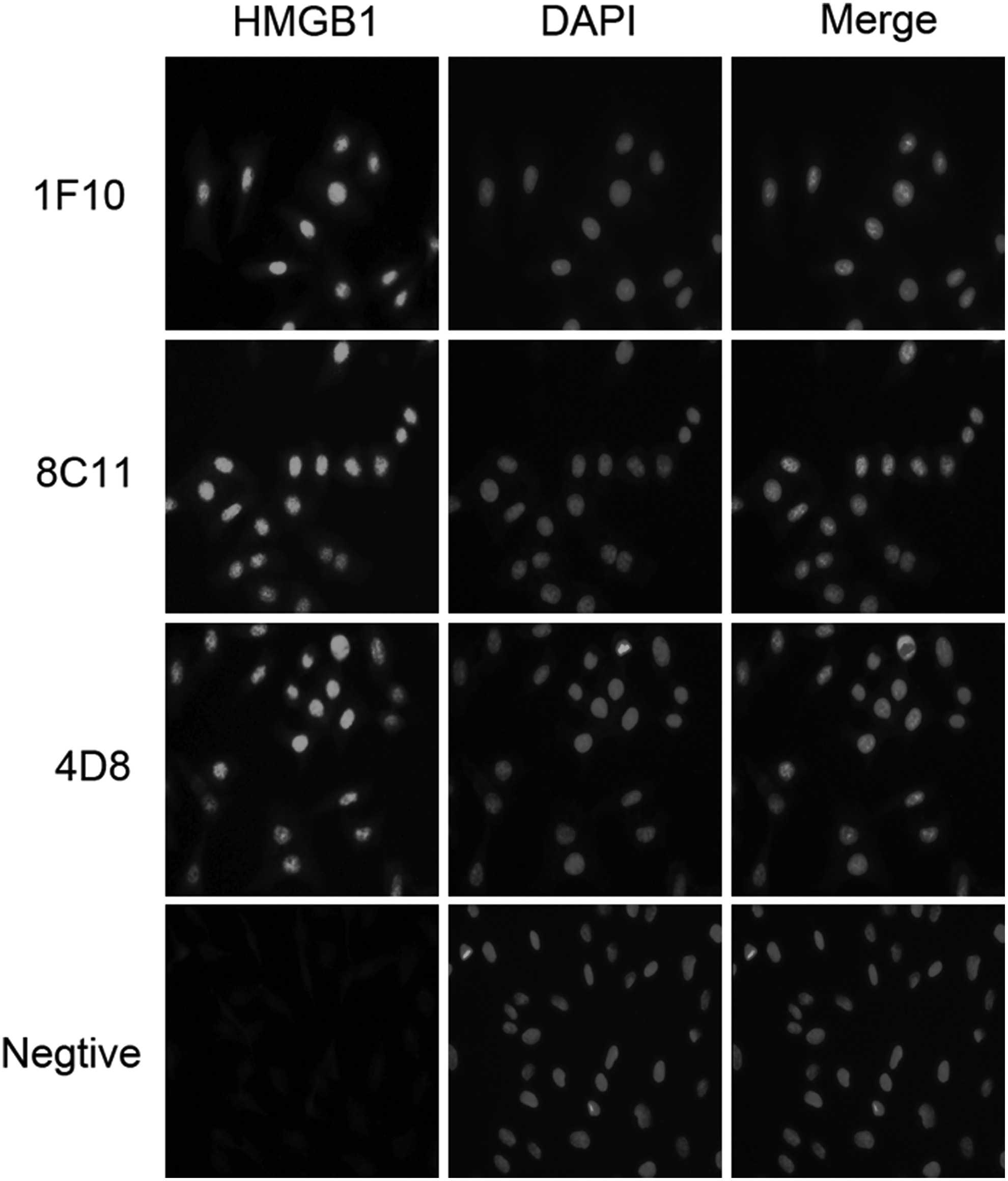
Detection of endogenous chicken HMGB1 expression by indirect immunofluorescence assay. DF1 cells were plated in six-well plate and fixed. The fixed cells were incubated with prepared MAbs (diluted at 1:500) or normal mouse antibody (negative control), followed by Alexa Fluor 488 donkey anti-mouse IgG (H + L) antibody.
Discussion
MAb is a specific biochemical tool for a range of applications and has been applied in the fields of agriculture and medicine, as well as biology. HMGB1 is one of the DAMPs and functions as an extracellular signaling molecule that can induce inflammatory and immune responses during inflammation, cell differentiation, and tumor progression.(22,23) Although there are various monoclonal or polyclonal antibodies against human or mouse HMGB1 that have cross-reactivity with other species, such as monkey, rat, and dog, there are no MAbs that specifically recognize chicken HMGB1. In this study, recombinant chicken HMGB1 protein was expressed prokaryotically and then purified by Ni-NTA Purification System. Three specific MAbs against chicken HMGB1 were prepared using the purified rchHMGB1 as immunogen and were identified by Western blot and IFA. Western blot analysis showed that the MAbs could recognize the chicken HMGB1 and were also able to cross-react with the human, mouse, monkey, and porcine HMGB1. These results indicated that the MAbs against chicken HMGB1 could be used in the study of the function and cell signaling pathways of chicken HMGB1.
There is increasing evidence that HMGB1 plays a potential pathogenic role in viral infectious diseases. The cytoplasmic translocation and passive release of HMGB1 in virus-infected cells have been reported previously following infection with many kinds of virus. WN progeny virus particles can induce necrotic cell death to passive release of HMGB1, and then accumulated HMGB1 will cause an injurious inflammatory response that contributes to the pathogenesis of West Nile encephalitis.(24) HMGB1 also can be passively released by SARS infection-mediated cytolysis. Once released, extracellular HMGB1 may stimulate an injurious pulmonary inflammatory response and, consequently, lead to respiratory failure and death.(25,26) Similarly, PRRSV infection is proved to induce excessive inflammatory response by triggering the secretion of HMGB1 to mediate NF-κB activation.(27) Unfortunately, the relationship between HMGB1 and avian virus infection has not been well determined until now. The deficiency of specific MAbs against chicken HMGB1 could be a main limiting factor for investigations into the role of HMGB1 in avian virus pathogenesis.
Avian influenza virus, Newcastle disease virus, and other viruses pose a serious threat to the poultry industry worldwide, and they are proved to induce an inflammatory response.(28,29) Considering that HMGB1 is associated with a variety of inflammatory diseases and exhibits its cytokine-like function when released from the cell, MAbs against chicken HMGB1 can be used to determine the relationship between HMGB1 and avian virus induced inflammatory cytokines in the future. The cytoplasmic translocation and release of HMGB1 in virus-infected avian cells can be detected by Western blot and IFA assay using the specific MAbs. Since these three MAbs can recognize the endogenous chicken HMGB1, the role of HMGB1 in avian virus replication and virus infection-induced NF-κB activation, as well as subsequent expression of inflammatory cytokines, can be confirmed by knockdown of endogenous HMGB1 with specific siRNAs and blocking extracellular HMGB1 with a HMGB1 neutralizing antibody. If possible, these results will provide new insight into understanding how AIV or other avian viruses induce excessive inflammatory responses. And further investigations into the role of HMGB1 in avian virus pathogenesis will hopefully provide potential therapeutic targets.
In this study, three MAbs against chicken HMGB1 were successfully generated, and they are verified to be suitable for ELISA, western blot, and IFA assay. The prepared MAbs can specifically recognize both prokaryotically and eukaryotically expressed recombinant chicken HMGB1. Endogenous expression of chicken HMGB1 in DF1 cells was also confirmed by these MAbs, which provide a valuable tool for further investigation of the function of chicken HMGB1 and expand the fields of HMGB1 research.
Footnotes
Acknowledgments
This work was funded by National Natural Science Foundation of China (Nos. 31372421 and 31530074) and Special Fund for Agro-scientific Research in the Public Interest (Grant 201303033).
Author Disclosure Statement
The authors have no financial interests to disclose.