
Abstract
Podocalyxin is a CD34-related type I transmembrane protein that is highly glycosylated with N-glycan, O-glycan, and keratan sulfate. Podocalyxin was originally found in the podocytes of rat kidney and is reportedly expressed in many types of tumors, including brain tumors, colorectal cancers, and breast cancers. Overexpression of podocalyxin is an independent predictor of progression, metastasis, and poor outcome. We recently immunized mice with recombinant human podocalyxin, which was produced using LN229 glioblastoma cells, and produced a novel antipodocalyxin monoclonal antibody (mAb), PcMab-47, which reacts with endogenous podocalyxin-expressing cancer cell lines and normal cell lines independent of glycosylation in Western blot, flow cytometry, and immunohistochemical analyses. In this study, we performed immunohistochemical analysis against oral cancers using PcMab-47. PcMab-47-stained oral squamous cell carcinoma cells in a cytoplasmic pattern and detected 26/38 (68.4%) of oral squamous cell carcinoma cells on tissue microarrays. These results indicate that PcMab-47 is useful in detecting podocalyxin of oral cancers for immunohistochemical analysis.
Introduction
P
Several studies have developed antipodocalyxin monoclonal antibodies (mAbs).(12, 13) The antigen of mAbs, TRA-1-60 and TRA-1-81, is keratan sulfate, which is attached to podocalyxin.(3) We recently immunized mice with recombinant human podocalyxin, which was produced using LN229 glioblastoma cells, and produced a novel antipodocalyxin mAb, PcMab-47 (IgG1, kappa).(14) PcMab-47 reacts with endogenous podocalyxin-expressing cancer cell lines and normal cells independently of glycosylation in Western blot, flow cytometry, and immunohistochemical analyses.
In this study, we investigated whether the antipodocalyxin mAb, PcMab-47, is useful for immunohistochemical detection of podocalyxin, which is expressed in oral cancers.
Materials and Methods
Production of hybridoma and purification of mAbs
The development of an antipodocalyxin mAb, PcMab-47, was described previously.(14) In brief, 4-week-old female BALB/c mice (CLEA, Tokyo, Japan) were immunized by intraperitoneal (i.p.) injection of the purified ectodomain of human podocalyxin (100 μg) together with Imject Alum (Thermo Fisher Scientific, Inc., Waltham, MA). After several additional immunizations, a booster i.p. injection of LN229/podocalyxin was given 2 days before the mice were euthanized by cervical dislocation, and spleen cells were harvested. The spleen cells were fused with P3U1 (American Type Culture Collection [ATCC], Manassas, VA) cells using PEG1500 (Roche Diagnostics, Indianapolis, IN). Hybridomas were grown in RPMI 1640 medium including L-glutamine with hypoxanthine, aminopterin, and thymidine selection medium supplement (Thermo Fisher Scientific, Inc.). Culture supernatants were screened using enzyme-linked immunosorbent assay for binding to the purified ectodomain of podocalyxin. MAbs were purified from supernatants of hybridomas cultured in Hybridoma-SFM medium (Thermo Fisher Scientific, Inc.) using Protein G Sepharose 4 Fast Flow (GE Healthcare UK Ltd, Buckinghamshire, England).
Immunohistochemical analyses
Oral cancer tissues were purchased from US Biomax, Inc. (Rockville, MD). Four-micrometer-thick histologic sections were deparaffinized in xylene and rehydrated. After the antigen retrieval procedure (autoclave using citrate buffer, pH 6.0; Agilent Technologies, Inc., Santa Clara, CA), sections were incubated with 5 μg/mL of PcMab-47 for 1 hour at room temperature followed by treatment with Envision+ kit (Agilent Technologies, Inc.) for 30 minutes. Color was developed using 3, 3-diaminobenzidine tetrahydrochloride (Agilent Technologies, Inc.) for 2 minutes, and then the sections were counterstained with hematoxylin (Wako Pure Chemical Industries Ltd., Osaka, Japan).
Results
Until now, podocalyxin expression has not been fully investigated in oral cancers because specific and sensitive mAbs against podocalyxin have not been established. Recently, we developed a sensitive and specific antipodocalyxin mAb, PcMab-47 (mouse IgG1, kappa), which is useful for immunohistochemical analysis.(14) In this study, we performed immunohistochemical analysis against oral cancers using PcMab-47. PcMab-47 stained oral squamous cell carcinoma cells in a cytoplasmic staining pattern (Fig. 1, Supplementary Fig. S1, and Supplementary Fig. S2). In addition, podocalyxin expression was also observed in endothelial cells (ECs) in those cases (Fig. 1). Only ECs were stained by PcMab-47 when no podocalyxin expression was observed in oral cancer cells (Supplementary Fig. S3). Podocalyxin expression was also detected by PcMab-47 in ECs from normal tissues.(14)
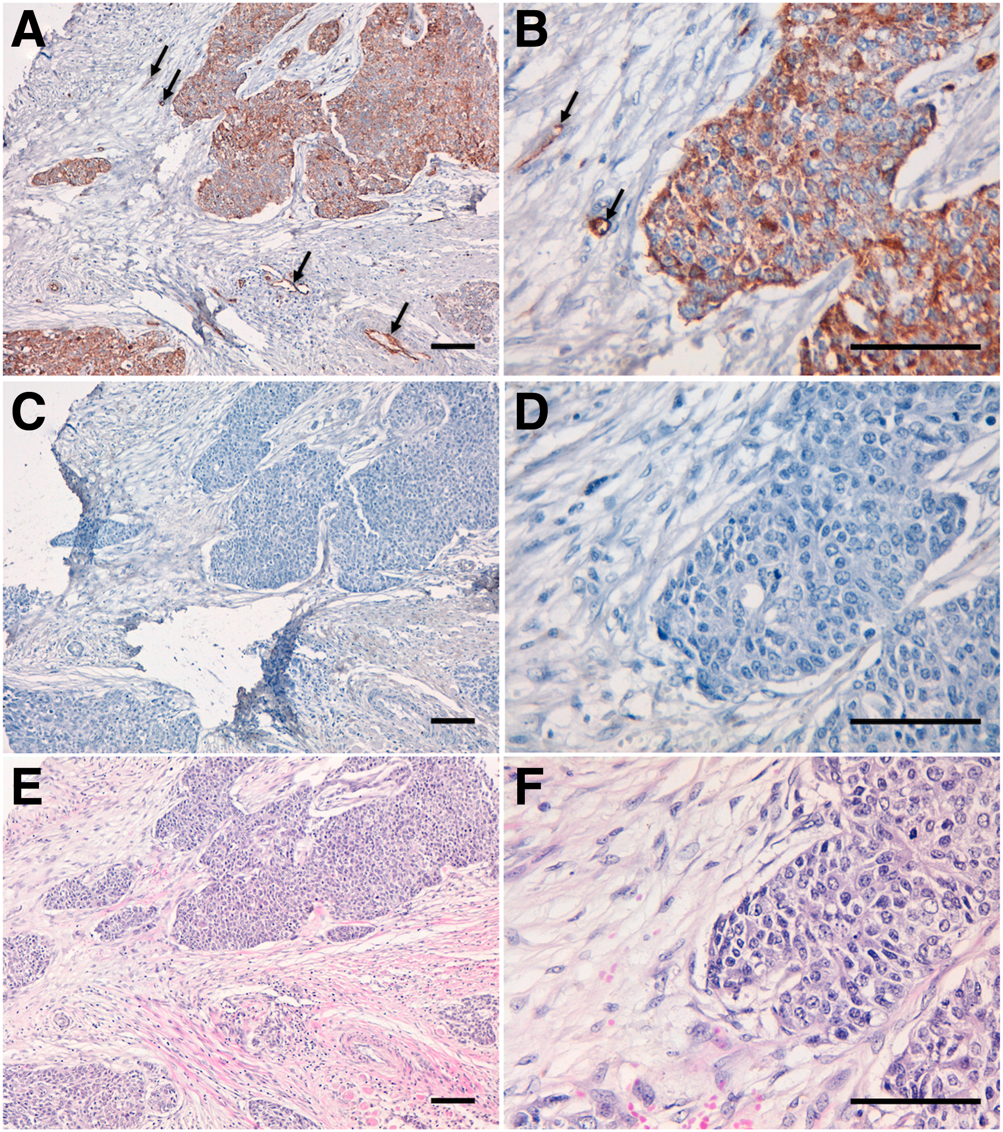
Immunohistochemical analysis of oral cancers by PcMab-47. Sections of oral cancers (Case No. 34) were incubated with 5 μg/mL of PcMab-47, followed by the Envision+ kit. Color was developed using 3, 3-diaminobenzidine tetrahydrochloride (DAB) and counterstained with hematoxylin. PcMab-47
Using oral cancer tissue arrays (Supplementary Table S1), 26/38 (68.4%) of oral squamous cell carcinoma cells were stained by PcMab-47 (Supplementary Table S2). Malignant pleomorphic adenoma and adenolymphoma also showed the positive staining by PcMab-47. In contrast, PcMab-47 did not stain pleomorphic adenoma, adenocystic carcinoma, and mucoepidermoid carcinoma.
Discussion
Podocalyxin is known as a diagnostic marker and prognostic indicator in several cancers, including breast cancers,(15–17) colorectal cancers,(18–21) gastric cancers,(22) prostate cancers,(23) testicular tumors,(2) renal cancers,(24) thyroid cancers,(25) bladder cancers,(26) ovarian cancers,(27) pancreatic cancers,(28, 29) and oral cancers.(30) Previously, we have reported that podocalyxin is associated with the malignant progression in brain tumors.(8, 31) The glycans on podocalyxin bind to E-/P-/L-selectin expressed on endothelium, platelets, and leukocytes, respectively.(32–34) These interactions enhance the formation of leukocyte–tumor–platelet aggregates and tumor cell arrest in the microvasculature.(35) Therefore, podocalyxin overexpression in cancer cells is a potential target for antibody therapy.
We previously developed our original technology for production of cancer-specific monoclonal antibodies (CasMabs) against membrane proteins.(36) Antipodoplanin CasMabs, including LpMab-2 and LpMab-23, specifically recognizes cancer-type PDPN in tumor tissues.(36, 37) In addition, the CasMab technology is useful for generating antiglycopeptide mAbs (GpMabs), such as LpMab-3, LpMab-9, LpMab-12, LpMab-19, and LpMab-21, against podoplanin.(38–40) Moreover, it can generate mAbs, which bind to various novel epitopes of podoplanin, such as LpMab-7, LpMab-10, and LpMab-17.(36,40–42) We further produced a sensitive and specific antipodocalyxin mAb, PcMab-47 (mouse IgG1, kappa).(15) PcMab-47 reacted with not only cancer cell lines, such as Caco-2 (colon adenocarcinoma cell line) and MDA-MB-468 (breast cancer cell line) but also normal cell lines, including HEK-293T (renal epithelial cell line) and vascular endothelial cells. Immunohistochemical studies showed that PcMab-47 not only stained normal cells such as podocytes or ECs of the kidney but also reacted with colon adenocarcinoma, in which a membrane/cytoplasmic-staining pattern was observed.(15) Strong expression of podocalyxin was also observed in ECs in colon adenocarcinoma,(15) indicating that PcMab-47 is very useful for immunohistochemical detection of podocalyxin.
In this study, we demonstrated that PcMab-47 is useful for detecting podocalyxin in oral squamous cell carcinoma cells by immunohistochemical analysis. In addition, we recently produced a mouse-human chimeric PcMab-47 (chPcMab-47) and investigated its antitumor activity against podocalyxin-expressing tumors.(43) chPcMab-47 exerted antitumor activity against mouse xenograft models using Chinese hamster ovary (CHO)/podocalyxin and HCT-15 (a colon cancer cell line),(43) suggesting that chPcMab-47 is useful for antibody therapy against oral squamous cell carcinoma.
Footnotes
Acknowledgments
We thank Takuro Nakamura, Miyuki Yanaka, Noriko Saidoh, Saori Handa, and Yoshimi Nakamura for excellent technical assistance. This work was supported, in part, by the Basic Science and Platform Technology Program for Innovative Biological Medicine from Japan Agency for Medical Research and development, AMED (Y.K.). This work was also supported, in part, by the Platform Project for Supporting Drug Discovery and Life Science Research (Basis for Supporting Innovative Drug Discovery and Life Science Research [BINDS]) from AMED (Y.K.), by project for utilizing glycans in the development of innovative drug discovery technologies from AMED (Y.K.), by the Platform for Drug Discovery, Informatics, and Structural Life Science (PDIS) from AMED (Y.K.), and by JSPS KAKENHI Grant Number 17K07299 (M.K.K.) and 16K10748 (Y.K.).
Author Disclosure Statement
Y.K. received research funding from Ono Pharmaceutical Co., Ltd.
References
Supplementary Material
Please find the following supplemental material available below.
For Open Access articles published under a Creative Commons License, all supplemental material carries the same license as the article it is associated with.
For non-Open Access articles published, all supplemental material carries a non-exclusive license, and permission requests for re-use of supplemental material or any part of supplemental material shall be sent directly to the copyright owner as specified in the copyright notice associated with the article.