
Abstract
Oral squamous cell carcinoma is an aggressive tumor in cats; however, molecular-targeted therapies against this tumor, including antibody therapy, have not been developed. Sensitive and specific monoclonal antibodies (mAbs) against highly expressed membrane proteins are needed to develop antibody therapies. Podoplanin, a type I transmembrane glycoprotein, is expressed in many human malignant tumors, including brain tumor, esophageal cancer, lung cancer, mesothelioma, and oral cancer. Podoplanin binds to C-type lectin-like receptor-2 (CLEC-2) and activates platelet aggregation, which is involved in cancer metastasis. Until now, we have established several mAbs against podoplanin in humans, mice, rats, rabbits, dogs, cattle, and cats. We have reported podoplanin expression in canine melanoma and squamous cell carcinomas using an anti-dog podoplanin mAb PMab-38. In this study, we investigated podoplanin expression in 40 feline squamous cell carcinomas (14 cases of mouth floor, 13 of skin, 9 of ear, and 4 of tongue) by immunohistochemical analysis using an anti-cat podoplanin mAb PMab-52, which we recently developed by cell-based immunization and screening (CBIS) method. Of the total 40 cases, 38 (95%) showed positive staining for PMab-52. In particular, 12 cases (30%) showed a strong membrane-staining pattern of squamous cell carcinoma cells. PMab-52 can be useful for antibody therapy against feline podoplanin-expressing squamous cell carcinomas.
Introduction
O
Podoplanin (PDPN/T1α/Aggrus), a type I transmembrane glycoprotein,(2–6) activates platelet aggregation by binding to the C-type lectin-like receptor-2 (CLEC-2) on the platelets.(7–10) Podoplanin is expressed in normal cells, including renal podocytes, pulmonary type I alveolar cells, and lymphatic endothelial cells.(3) The interaction between podoplanin and CLEC-2 facilitates lymphatic/blood vessel separation.(11) The expression of human podoplanin has been reported in many malignant tumors, including brain tumors,(12–15) oral cancers,(16) esophageal cancers,(17) lung cancers,(18) mesotheliomas,(19,20) testicular tumors,(21) osteosarcomas,(22–24) and chondrosarcomas.(23) Podoplanin expression is associated with cancer metastasis and malignant progression.(8,12,25)
We have established several mAbs against human,(26) mouse,(27) rat,(28) rabbit,(29) dog,(30) bovine,(31) and cat podoplanin.(32) Using these antipodoplanin mAbs, we have investigated podoplanin expression in cancer tissues. PMab-38, an anti-dog podoplanin mAb, reacted with 83% of squamous cell carcinomas (15/18 cases)(33) and 90% of melanomas (9/10 cases).(34) Recently, we have established an anti-cat podoplanin mAb (clone: PMab-52), which is very useful in flow cytometry, Western blot, and immunohistochemical analyses.(32) In this study, we investigated podoplanin expression in feline squamous cell carcinomas by immunohistochemical analysis using PMab-52.
Materials and Methods
Feline tissues
Normal feline tissues were purchased from Zyagen (San Diego, CA). Feline squamous cell carcinoma tissues were obtained from North Lab (Hokkaido, Japan).
Hybridoma production
In brief, BALB/c mice (CLEA Japan, Tokyo, Japan) were immunized by intraperitoneal (i.p.) injection of cat podoplanin-expressing CHO cells (CHO/cat podoplanin), which were produced by transfecting cat podoplanin into CHO-K1 cells(32) [American Type Culture Collection (ATCC), Manassas, VA] along with Imject Alum (Thermo Fisher Scientific, Inc., Waltham, MA). After several additional immunizations of CHO/cat podoplanin cells, a booster injection of CHO/cat podoplanin cells was intraperitoneally administered 2 days before the spleen cells were harvested. The spleen cells were fused with P3U1 cells (ATCC) using PEG1500 (Roche Diagnostics, Indianapolis, IN). The hybridomas were grown in Roswell Park Memorial Institute (RPMI) 1640 medium (Nacalai Tesque, Inc., Kyoto, Japan) supplemented with 10% heat-inactivated fetal bovine serum (Thermo Fisher Scientific, Inc.) and hypoxanthine, aminopterin, and thymidine selection medium supplement (Thermo Fisher Scientific, Inc.). The culture supernatants were screened using flow cytometry for binding to CHO/cat podoplanin and CHO-K1.
Immunohistochemical analyses
First, 4-μm-thick histological sections were deparaffinized in xylene and then rehydrated, followed by autoclaving in citrate buffer (pH 6.0; Agilent Technologies, Inc., Santa Clara, CA) for 20 minutes. The sections were incubated with 1 μg/mL of PMab-52 for 1 hour at room temperature, followed by treatment with Envision+ kit for 30 minutes (Agilent Technologies, Inc.). Color was developed using 3, 3-diaminobenzidine tetrahydrochloride (Agilent Technologies, Inc.) for 2 minutes, and the sections were counterstained with hematoxylin (Wako Pure Chemical Industries, Ltd., Osaka, Japan). The intensity of staining was evaluated as 0, 1+, 2+, and 3+.
Results
In our recent studies, we immunized mice with CHO/cat podoplanin cells and performed flow cytometry as the first screening. Using this cell-based immunization and screening (CBIS) method, we could obtain highly sensitive mAbs, which are useful not only in flow cytometry but also in Western blot and immunohistochemical analyses. Indeed, one of the established clones, PMab-52 (IgM, kappa), was sensitive and specific against cat podoplanin in flow cytometry, Western blot, and immunohistochemical analyses. PMab-52 reacted with only cat podoplanin, whereas it did not react with human, mouse, rat, rabbit, dog, and bovine podoplanin in flow cytometry and Western blot analyses.(32) Although we have investigated podoplanin expression in human, rabbit, and canine cancers, its expression in feline cancer remains to be clarified.
First, we investigated podoplanin expression in normal feline tissues, including the kidney, lung, and rectum, by immunohistochemical analysis. PMab-52 was bound to the Bowman's capsule and glomerular podocytes in feline kidneys (Fig. 1A, C). Also, it was bound to the type I alveolar cells of feline lungs (Fig. 2A, C) and lymphatic endothelial cells (Fig. 3A, C), indicating that PMab-52 can detect cat podoplanin in normal tissues because a similar expression pattern has been reported in other species, including human and mouse.(35)
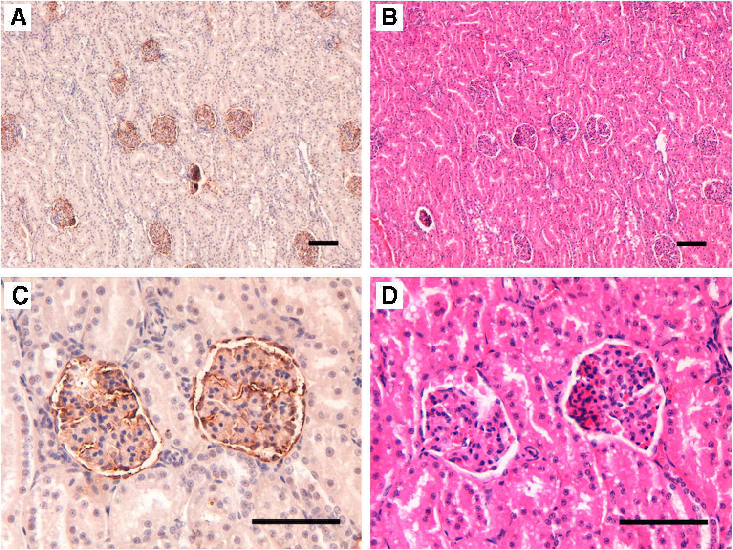
Immunohistochemical analysis of podoplanin using PMab-52 in feline kidney.
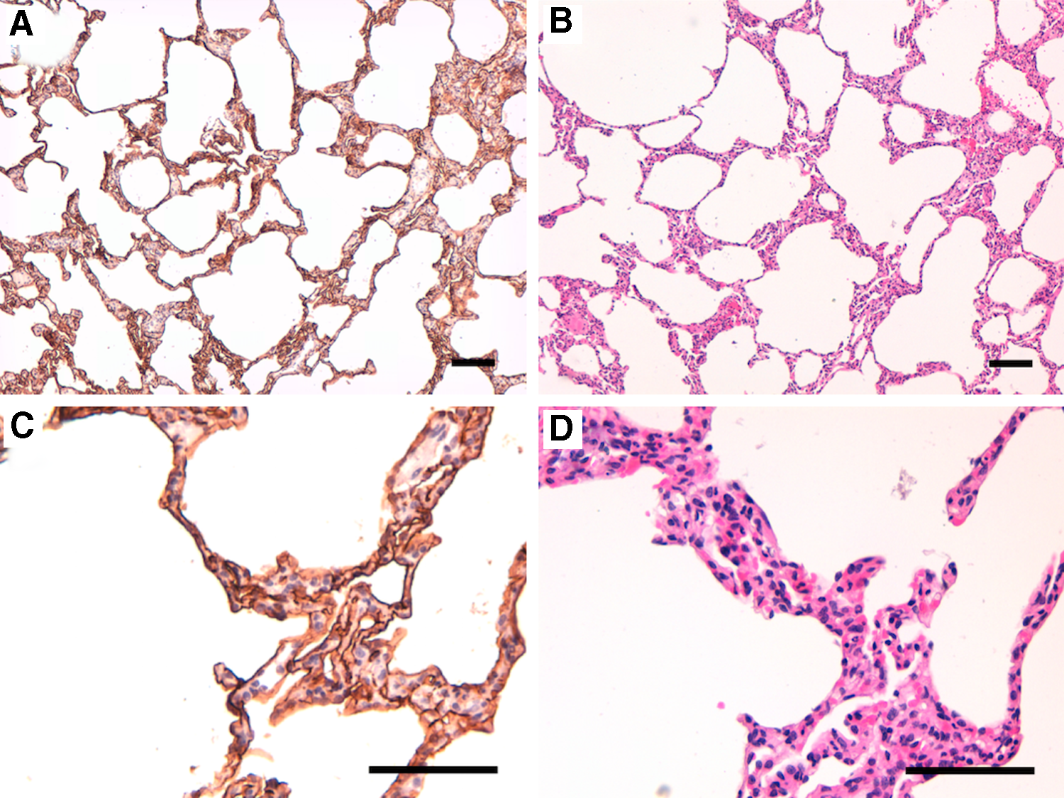
Immunohistochemical analysis of podoplanin using PMab-52 in feline lungs.
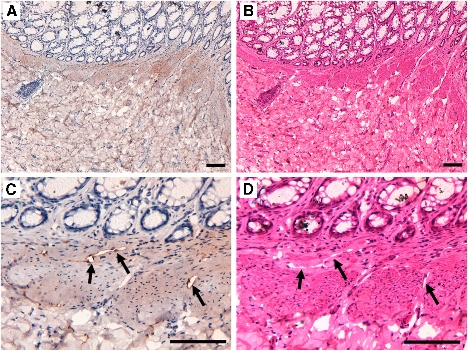
Immunohistochemical analysis of podoplanin using PMab-52 in feline rectum.
Next, we investigated cat podoplanin expression in various squamous cell carcinomas because its expression has been reported in many previous studies with other species.(18,33) All specimens of squamous cell carcinomas used in this study are listed in Table 1. Among the 40 cases analyzed, 14 were from mouth floor, 13 from skin, 9 from ear, and 4 from tongue (Table 2). PMab-52 was observed in squamous cell carcinoma cells in a membrane-staining pattern (Fig. 4). All staining patterns are depicted in Supplementary Figure S1 and S2 (Supplementary Data are available online at
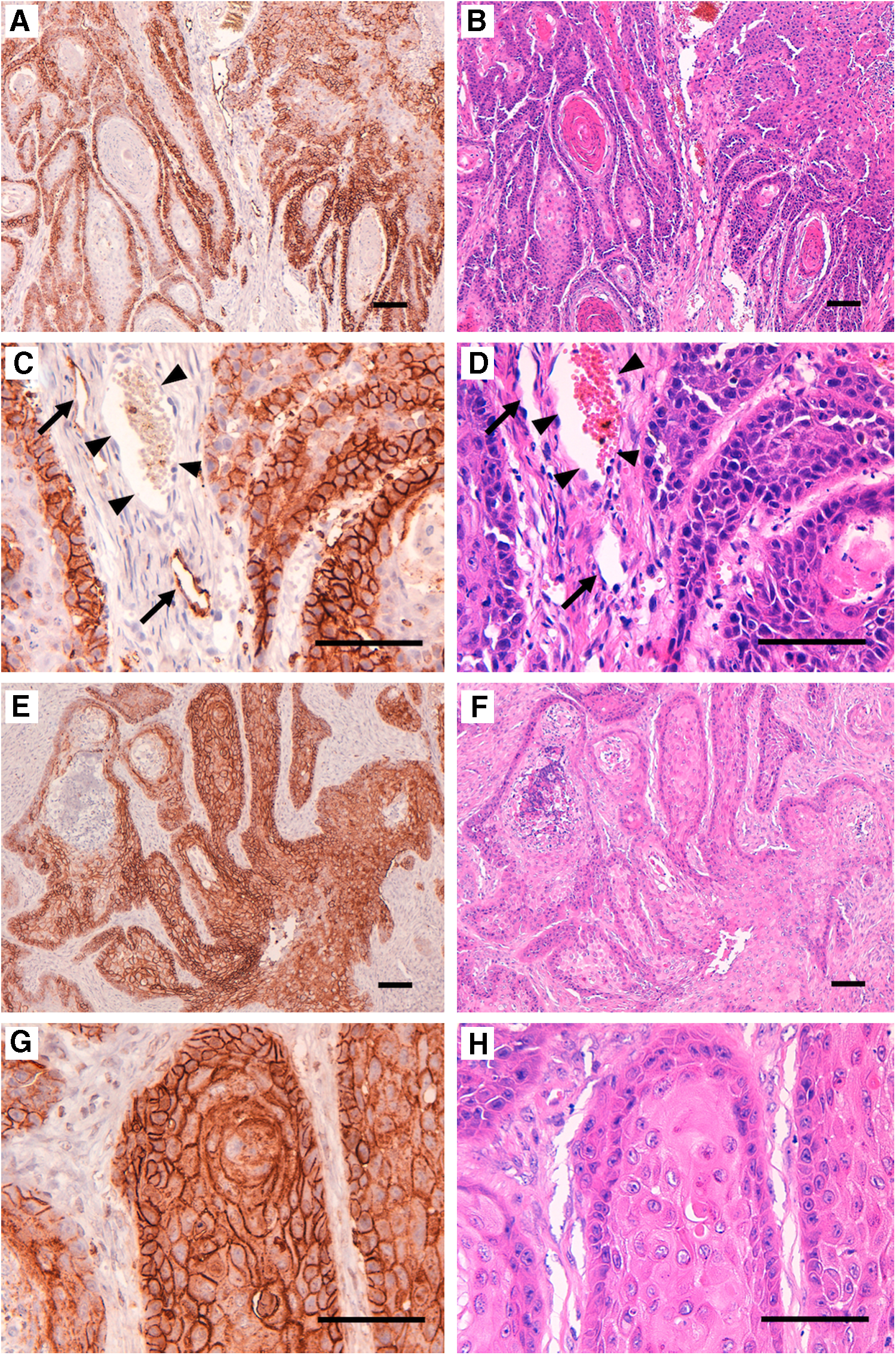
Immunohistochemical analysis of cat podoplanin using PMab-52 in squamous cell carcinomas.
PMab-52 also detected lymphatic endothelial cells around the squamous cell carcinoma cells, but it could not detect vascular endothelial cells (Fig. 4C). PMab-52 very weakly detected normal lymphatic endothelial cells (Fig. 3), indicating that the expression level of podoplanin is different between normal and cancerous tissues.
Discussion
In this study, we first investigated podoplanin expression in feline normal tissues. PMab-52 strongly bound to feline kidneys (Fig. 1) and lungs (Fig. 2). These results are consistent with our previous study regarding podoplanin expression in rabbit normal tissues.(29) In contrast, anti-dog podoplanin mAb PMab-38 reacted with canine kidney, but not with canine lungs.(30) PMab-52 was weakly bound to normal lymphatic endothelial cells (Fig. 3), although podoplanin is known to be a specific marker for lymphatic endothelial cells. Likewise, PMab-38 did not detect normal lymphatic endothelial cells of canine normal tissues.(30) NZ-1, an anti-human podoplanin mAb, detects lymphatic endothelial cells but not those of the lung and kidney.(26) Moreover, cancer-specific mAbs LpMab-2(15) and LpMab-23(36) do not react with human podoplanin in all normal tissues. Taken together, two main reasons can explain the different reaction patterns of anti-podoplanin mAbs: (1) the expression level of podoplanin is different among tissues and (2) the epitopes recognized by antipodoplanin mAbs vary due to many reasons, including tissue-specific posttranslational modification or cancer-specific glycosylation.
We investigated cat podoplanin expression in 40 squamous cell carcinoma tissues. PMab-52 strongly detected lymphatic endothelial cells around squamous cell carcinoma cells (Fig. 4C), whereas it weakly detected normal lymphatic endothelial cells (Fig. 3C), indicating that the expression level of podoplanin differs between normal and cancer tissues. We have previously reported that podoplanin expression can be induced by transforming growth factor-β,(37) which might explain the different expression levels of podoplanin in cancer tissues. Cancer-associated fibroblasts were also detected in some cases (case 14; Supplementary Fig. S1), which have been observed in many previous studies using antipodoplanin mAbs in human cancer tissues(38–40) and using PMab-38 in canine squamous cell carcinomas.(33) Targeted therapy against cancer-associated fibroblasts is also important because cancer-associated fibroblasts have been associated with malignancy.(38–40)
In conclusion, PMab-52 can be useful for clarifying the pathophysiological function of podoplanin in feline squamous cell carcinomas. Furthermore, it is a potential candidate for developing antibody-based therapies against feline squamous cell carcinomas.
Footnotes
Author Disclosure Statement
Y.K. received research funding from Ono Pharmaceutical Co., Ltd.
Acknowledgments
We thank Takuro Nakamura, Miyuki Yanaka, Noriko Saidoh, Saori Handa, and Yoshimi Nakamura for excellent technical assistance. This work was supported, in part, by the Basic Science and Platform Technology Program for Innovative Biological Medicine from Japan Agency for Medical Research and Development, AMED (Y.K.), by Project for utilizing glycans in the development of innovative drug discovery technologies from AMED (Y.K.), and by Platform Project for Supporting Drug Discovery and Life Science Research (Basis for Supporting Innovative Drug Discovery and Life Science Research [BINDS]) from AMED.
References
Supplementary Material
Please find the following supplemental material available below.
For Open Access articles published under a Creative Commons License, all supplemental material carries the same license as the article it is associated with.
For non-Open Access articles published, all supplemental material carries a non-exclusive license, and permission requests for re-use of supplemental material or any part of supplemental material shall be sent directly to the copyright owner as specified in the copyright notice associated with the article.