
Abstract
Human epidermal growth factor receptor 2 (HER2) expression has been reported in several cancers, such as breast, gastric, lung, pancreatic, and colorectal cancers. HER2 is overexpressed in those cancers and is associated with poor clinical outcomes. Trastuzumab, a humanized anti-HER2 antibody, provides significant survival benefits for patients with HER2-overexpressing breast cancers and gastric cancers. In this study, we developed a novel anti-HER2 monoclonal antibody (mAb), H2Mab-139 (IgG1, kappa) and investigated it against colon cancers using flow cytometry, western blot, and immunohistochemical analyses. Flow cytometry analysis revealed that H2Mab-139 reacted with colon cancer cell lines, such as Caco-2, HCT-116, HCT-15, HT-29, LS 174T, COLO 201, COLO 205, HCT-8, SW1116, and DLD-1. Although H2Mab-139 strongly reacted with LN229/HER2 cells on the western blot, we did not observe a specific signal for HER2 in colon cancer cell lines. Immunohistochemical analyses revealed sensitive and specific reactions of H2Mab-139 against colon cancers, indicating that H2Mab-139 is useful in detecting HER2 overexpression in colon cancers using flow cytometry and immunohistochemical analyses.
Introduction
H
Materials and Methods
Cell lines
LN229, Caco-2, HCT-116, HT-29, LS 174T, COLO 201, HCT-8, SW1116, and P3U1 cell lines were obtained from the American Type Culture Collection (ATCC; Manassas, VA). COLO 205, DLD-1, and HCT-15 cell lines were obtained from the Cell Resource Center for Biomedical Research Institute of Development, Aging and Cancer Tohoku University (Miyagi, Japan). LN229/HER2 was produced previously.(12) COLO 205, DLD-1, HCT-15, COLO 201, SW1116, and P3U1 were cultured in RPMI 1640 medium (Nacalai Tesque, Inc., Kyoto, Japan), and LN229, Caco-2, HCT-116, HT-29, LS 174T, and HCT-8 were cultured in DMEM (Dulbecco's modified Eagle's medium) medium (Nacalai Tesque, Inc.), supplemented with 10% heat-inactivated fetal bovine serum (Thermo Fisher Scientific, Inc., Waltham, MA), 100 units/mL of penicillin, 100 μg/mL of streptomycin, and 25 μg/mL of amphotericin B (Nacalai Tesque, Inc.). All cell lines were incubated at 37°C in a humidified atmosphere containing 5% CO2 and 95% air.
Hybridoma production
Female 4-week-old BALB/c mice were purchased from CLEA Japan (Tokyo, Japan). Animals were housed under specific pathogen-free conditions. The Animal Care and Use Committee of Tohoku University approved all the animal experiments described in this study. Anti-HER2 hybridomas were produced, as described previously.(13) Briefly, BALB/c mice were immunized using intraperitoneal (i.p.) injections of 100 μg of recombinant HER2-extracellular domain together with Imject Alum (Thermo Fisher Scientific, Inc.). After several additional immunizations, a booster injection was intraperitoneally administered 2 days before harvesting spleen cells. Spleen cells were then fused with P3U1 cells using PEG1500 (Roche Diagnostics, Indianapolis, IN). The resulting hybridomas were grown in RPMI medium supplemented with hypoxanthine, aminopterin, and thymidine selection medium supplement (Thermo Fisher Scientific, Inc.). Culture supernatants were screened using enzyme-linked immunosorbent assay with recombinant HER2-extracellular domain. MAbs were purified from the supernatants of hybridomas, cultured in Hybridoma-SFM medium (Thermo Fisher Scientific, Inc.) using Protein G Sepharose 4 Fast Flow (GE Healthcare UK, Ltd.).
Flow cytometry
Cells were harvested by brief exposure to 0.25% trypsin/1-mM ethylenediaminetetraacetic acid (EDTA; Nacalai Tesque, Inc.). After washing with 0.1% bovine serum albumin/phosphate-buffered saline, the cells were treated with 10 μg/mL of anti-HER2 (clone H2Mab-139) for 30 minutes at 4°C and subsequently with Alexa Fluor 488-conjugated anti-mouse IgG (1:1000; Cell Signaling Technology, Inc., Danvers, MA). Fluorescence data were collected using EC800 Cell Analyzers (Sony, Corp.).
Western blot analysis
Cell lysates (10 μg) were boiled in sodium dodecyl sulfate (SDS) sample buffer (Nacalai Tesque, Inc.) and proteins were then electrophoresed on 5%–20% polyacrylamide gels (Wako Pure Chemical Industries, Ltd., Osaka, Japan) before being transferred onto polyvinylidene difluoride (PVDF) membranes (Merck KGaA, Darmstadt, Germany). After blocking with 4% skim milk (Nacalai Tesque, Inc.), membranes were incubated with primary antibodies, such as anti-HER2 (10 μg/mL; clone H2Mab-139) and anti-β-actin (1 μg/mL; clone AC-15; Sigma-Aldrich, Corp., St. Louis, MO) and then with peroxidase-conjugated anti-mouse IgG (Agilent Technologies, Inc., Santa Clara, CA; 1:1000 diluted). Finally, membranes were developed using ImmunoStar LD (Wako Pure Chemical Industries, Ltd.) with a Sayaca-Imager (DRC, Co., Ltd., Tokyo, Japan).
Immunohistochemical analyses
Colon cancer tissue arrays were purchased from US Biomax, Inc. (Rockville, MD). Histologic sections of 4-μm thickness were deparaffinized in xylene and subsequently rehydrated and autoclaved in EnVision FLEX Target Retrieval Solution, High pH (Agilent Technologies, Inc.), for 20 minutes. Sections were then incubated with 10 μg/mL of H2Mab-139 for 1 hour at room temperature and treated using an Envision+ kit (Agilent Technologies, Inc.) for 30 minutes. Color was developed using 3,3-diaminobenzidine tetrahydrochloride (DAB; Agilent Technologies, Inc.) for 2 minutes; subsequently, sections were counterstained with hematoxylin (Wako Pure Chemical Industries, Ltd.).
Results and Discussion
We immunized mice with the purified recombinant extracellular domain of HER2. We performed flow cytometry to check reactions with LN229 and LN229/HER2 cells. A stronger reaction against LN229/HER2 was needed compared with LN229 because LN229 cells endogenously express HER2. We obtained one clone: H2Mab-139 (IgG1, kappa). Flow cytometry analysis revealed that H2Mab-139 reacted with LN229/HER2 cells more strongly than with endogenous HER2-expressing LN229 glioblastoma cells (Fig. 1A). H2Mab-139 also reacted with Chinese hamster ovary (CHO)/HER2 cells but did not react with CHO-K1 cells (data not shown), indicating that H2Mab-139 is specific for HER2. Western blot analysis of H2Mab-139 against LN229 and LN229/HER2 cells revealed that the molecular weight of H2Mab-139 was ∼200 kDa in LN229/HER2 cells (Fig. 1B).
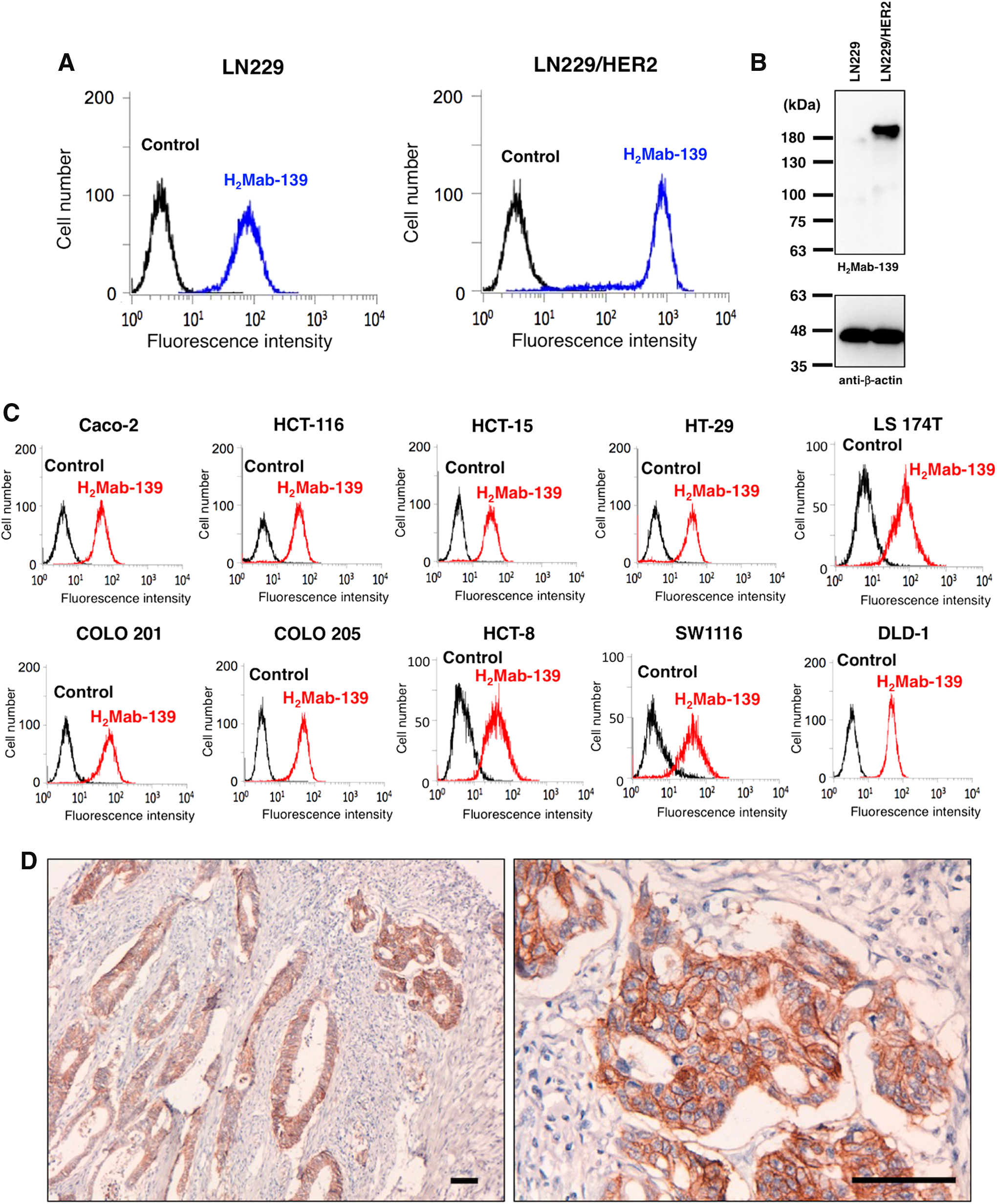
Characterization of H2Mab-139.
H2Mab-139 recognized endogenous HER2 in colon cancer cell lines, such as Caco-2, HCT-116, HCT-15, HT-29, LS 174T, COLO 201, COLO 205, HCT-8, SW1116, and DLD-1 (Fig. 1C). However, H2Mab-139 did not detect HER2 in colon cancer cell lines in western blots (data not shown), indicating that H2Mab-139 is not suitable for western blot analysis of colon cancer cell lines.
We further investigated the immunohistochemical utility of H2Mab-139 in human colon cancers. Figure 1D shows that H2Mab-139 stained the cell membranes of colon cancers that were previously diagnosed as HER2-positive. H2Mab-139 also stained the cell membranes of HER2-positive breast cancer cells (Supplementary Fig. S1), indicating that H2Mab-139 is useful for the immunohistochemical analysis for HER2-positive cancers. In the future, we intend to determine the positive rate of many colon cancer tissues via immunohistochemical analysis using H2Mab-139.
Footnotes
Acknowledgments
This work was supported in part by the Basic Science and Platform Technology Program for Innovative Biological Medicine from Japan Agency for Medical Research and Development, AMED (Y.K.), by Project for utilizing glycans in the development of innovative drug discovery technologies from AMED (Y.K.), and by Platform Project for Supporting Drug Discovery and Life Science Research (Basis for Supporting Innovative Drug Discovery and Life Science Research [BINDS]) from AMED (Y.K.).
Author Disclosure Statement
Y.K. received research funding from Ono Pharmaceutical Co., Ltd. All other authors have nothing to disclose.
References
Supplementary Material
Please find the following supplemental material available below.
For Open Access articles published under a Creative Commons License, all supplemental material carries the same license as the article it is associated with.
For non-Open Access articles published, all supplemental material carries a non-exclusive license, and permission requests for re-use of supplemental material or any part of supplemental material shall be sent directly to the copyright owner as specified in the copyright notice associated with the article.