
Abstract
Firefly luciferase (FLuc) is commonly used as a reporter gene PpyLuc1 in bioanalytical assays. We have produced five mouse-derived monoclonal antibodies (mAbs) that recognize FLuc. The mAbs, DSHB-LUC-2, DSHB-LUC-3, DSHB-LUC-9, DSHB-LUC-16, and DSHB-LUC-24, were generated by immunizing mice with purified 6xHIS-tagged FLuc (6xHis-FLuc) in suspension with an adjuvant. All five were validated by dot blots. Four of the mAbs provided strong signals in western blot analysis, and one a weak signal. All five were validated for immunostaining in fixed cell culture. Only one stained cells embedded in paraffin. The five mAbs are available at cost through the Developmental Studies Hybridoma Bank (DSHB), a nonprofit National Resource created by the National Institutes of Health.
Introduction
L
In this study, we detail the generation and validation of five new mouse anti-FLuc monoclonal antibodies (mAbs). Their utility is demonstrated in dot and western blot hybridization, immunostaining of fixed mammalian cell preparations, and immunostaining of paraformaldehyde-fixed paraffin-embedded cell preparations. Each of the five mAbs is effective for dot blotting and immunostaining of cells in culture. Four of the mAbs (DSHB-LUC-2, DSHB-LUC-9, DSHB-LUC-16, and DSHB-LUC-24) are effective for western blotting. Only one of the mAbs (DSHB-LUC-3), which provided only a weak western blot signal, was highly effective in immunostaining of paraffin-embedded cells. All five mAbs are available for basic research purposes at cost through the Developmental Studies Hybridoma Bank (DSHB), a nonprofit National Resource created by the National Institutes of Health and housed at the University of Iowa.
Materials and Methods
Recombinant protein immunogen production
Production of recombinant 6x histidine-tagged FLuc (6xHis-FLuc) was carried out as previously described by Park et al.(15) for recombinant maltose-binding protein (MBP), with minor modifications. The FLuc-containing vector plasmid, pLIFE-3’UTR, was obtained from the DNASU plasmid repository at the Biodesign Institute of Arizona State University. The FLuc open reading frame (ORF) was amplified from pLIFE-3’UTR by polymerase chain reaction (PCR) with the primers FLuc-F (ATATGGTACCATGGAAGACGCCAAAAACATAAAG) and FLuc-R (TATACTCGAGTTACAATTTGGACTTTCCGCCCTTC). The purified PCR products were digested with KpnI and SacI, and ligated into the plasmid pDB.His.MBP (DNASU), which had been treated with the same restriction enzymes. The resulting plasmid, pDB.His.FLuc, contained the 6xHis-FLuc ORF under the control of the lac operon, allowing the induction of 6xHis-FLuc expression in Escherichia coli in the presence of Isopropryl β-
Mice and immunization
Two 8-week-old female BALB/c mice (Envigo, Indianapolis, IN) were initially immunized by intraperitoneal injection of 50 μg of 6xHis-FLuc, 50 μg of poly(I:C) HMW VacciGrade (InvivoGen, San Diego, CA), and 50 μg of adjuvant anti-CD40 mAb (BioXCell, West Lebanon, NH),(15–17) in 200 μL phosphate-buffered solution (PBS; Gibco, Grand Island, NY). Two weeks after the initial immunization, both mice were boosted with 50 μg of 6xHis-FLuc and 50 μg of poly(I:C) in 200 μL of PBS. Serum was obtained from each mouse 2 weeks after the boost and tested at ≥1:500 dilutions by western blot analysis. Based on the results of the western blot analysis, the mouse with the highest anti-FLuc antibody titer was identified, and that mouse was injected intravascularly with 50 μg of 6xHis-FLuc and 50 μg of poly(I:C) in 100 μL of PBS 3 days before hybridoma fusion.
Hybridoma fusion
Hybridoma fusion was performed as described by Sanchez et al.(17) with minor modification. Spleen cells from the immunized mouse and Sp2/0-Ag14 myeloma cells (CRL-1581; ATCC, Manassas, VA)(18) were washed, passed through a 70 μm filter, resuspended in serum-free RPMI 1640 medium (HyClone, Logan, UT) at a ratio of five nucleated splenocytes to one myeloma cell (5:1), and pelleted. The pelleted cells were dispersed with gentle mixing and warmed to 37°C for 1 minute, followed by the addition of 0.5 mL of prewarmed 50% (v/v) PEG-RPMI 1640 medium. Then the cells were mixed gently at 37°C for an additional 2 minutes. During the subsequent 90 seconds, 15 mL of prewarmed serum-free Iscove's modified Dulbecco's medium (IMDM) (HyClone) was added dropwise to the fusion mixture, and the cells incubated at room temperature for 8 minutes without agitation.
The cells were once again incubated at 37°C for 2 minutes, and then pelleted by centrifugation at 200 g for 5 minutes. After centrifugation, the supernatant was removed and the pellet dispersed by mixing gently and pipetting into 30 mL of hybridoma culture medium IMDM, which contained 15% fetal bovine serum (FBS; HyClone), 20% RPMI 1640 medium conditioned by giant cell tumor (TIB-223; ATCC),(19) and 1× HT-HybriMax. The cell suspension was then transferred to T75 flasks and incubated overnight at 37°C. The following day, 100 μL of the cell suspension was added to each well of 96-well tissue culture plates. After 2 days incubation at 37°C, 100 μL of 2× hypoxanthine–aminopterin–thymidine (HAT; Sigma-Aldrich, St. Louis, MO) medium was added to each well to select for hybridomas. Cells were then weaned from the 2× HAT medium by exchanging 100 μL of the medium with 100 μL of 1× HAT medium twice every 3 days. Once cells had grown to between 25% and 50% confluency, supernatants were tested for the presence of mouse IgG production by a dot blot analysis (Bio-Rad, Hercules, CA). Hybridoma clones that yielded positive results were expanded and the supernatants retested for anti-FLuc activity by western blot analysis. Hybridomas confirmed to be producing anti-FLuc antibodies were expanded and subcloned using fluorescence-activated cell sorting with Propidium Iodide (Life Technologies) staining for viability. The Mouse mAb Isotyping Test Kit (AbD Serotec, Raleigh, NC) was used according to the manufacturer's protocols to determine antibody isotypes from the culture supernatants.
Dot blot
Five microliters of lysate from E. coli strains expressing either the untagged wild-type FLuc protein or the 6xHis-tagged MBP (6xHis-MBP) were applied to dry nitrocellulose membranes. Once the membranes had dried completely, they were subsequently blocked with 5% (w/v) nonfat milk in 1× Tris-buffered saline containing Tween-20 (TBST: 50 mM Tris–HCl, 150 mM NaCl, pH 7.6, 0.005% Tween-20) for 1 hour at room temperature. The membranes were then incubated in blocking buffer containing 0.1 μg/mL of the anti-FLuc mAbs or the rabbit polyclonal anti-FLuc antibody (RBpAB), L0159 (Sigma-Aldrich), for 1 hour at room temperature. The membranes were then washed three times with 1× TBST and incubated at room temperature with IR Dye 800-conjugated goat anti-mouse or goat anti-rabbit secondary antibody (LI-COR Biosciences, Lincoln, NE) at a 1:10,000 dilution for 1 hour. The membranes were washed three times with 1× TBST and scanned in an Odyssey Scanner (LI-COR Biosciences).
Western blot
Samples containing 15 μL of lysate from FLuc-expressing 4T1 murine breast carcinoma cells(20,21) or NTera2 (NT2) human teratocarcinoma cells (ATCC) were denatured and separated in a Tris/Glycine/sodium dodecyl sulfate–polyacrylamide gel electrophoresis (SDS-PAGE) gel (Bio-Rad) with dual color molecular weight markers (Bio-Rad). Proteins were transferred from the gel to nitrocellulose membranes, and the membranes blocked with 5% (w/v) nonfat milk in TBST for 1 hour at room temperature. The blocked membranes were then incubated in a blocking buffer containing 1 μg/mL of anti-FLuc antibodies for 1 hour at room temperature. A rabbit-derived anti-FLuc polyclonal antibody (RBpAB), L0159 (Sigma-Aldrich), was used as a positive control at a dilution of 1:10,000. After incubation with the respective primary antibody, membranes were washed three times with 1× TBST and subsequently incubated for 1 hour, at room temperature in the dark with either IR Dye 800-conjugated goat anti-mouse or goat anti-rabbit secondary antibody (LI-COR Biosciences) at a 1:10,000 dilution. Then, the membranes were once again washed three times with 1× TBST and scanned in an Odyssey Scanner (LI-COR Biosciences).
Immunostaining fixed cells
FLuc-expressing 4T1 cells(20,21) were released from the sides of culture flasks by treating them for 30 minutes with 10 mM EDTA in Dulbecco's PBS without calcium or magnesium (DPBS; Gibco) at 37°C. Released cells were washed and resuspended in IMDM (HyClone), then replated in sterile 12-well Teflon-coated microscope slides (Tekdon, Myakka City, FL) at 2.5 × 10⁴ cells per well. Cells were incubated for up to 48 hours at 37°C to allow cell attachment and spreading. The optimized cell fixation protocol utilized by the Human Cell Atlas was used (Stadler et al.,
22
Paraffin-embedded cells
The 4T1 mouse mammary cell line gently released as above was rinsed in DPBS and resuspended in 4% PF-DPBS for 15 minutes at room temperature. The fixative was removed and the cells were washed by gentle centrifugation in a 1.5-mL Eppendorf tube. The DPBS was removed and the cell pellet was covered with 100 μL of warm 2% agarose–DPBS and the cell pellet disrupted and dispersed by tapping. After the agarose hardened, the sample was cut from the tube and processed for paraffin embedding using standard tissue processing protocols in an RMC Paraffin Tissue Processor 1530. Paraffin sections (7 μm) were placed on Superfrost Plus slides (Fisher Scientific), dried overnight at 37°C, baked for 30 minutes at 65°C, deparaffinized in xylene, and rehydrated through graded alcohols. Sections were blocked with 10% normal goat serum (Sigma-Aldrich) in Tris-buffered saline ([TBS] 50 mM Tris, 150 mM NaCl, pH 8.0) before staining with anti-luciferase-conditioned culture supernatant diluted with TBS to a final concentration of 5 μg/mL at 37°C for 1 hour. After extensive TBS rinsing, preparations were incubated in Alexa Fluor 488-conjugated goat anti-mouse IgG (Jackson ImmunoResearch, West Grove, PA) at 37°C for 45 minutes. Sections were rinsed with TBS, coverslips mounted, and imaged as described above. Control sections were stained using medium containing 5 μg/mL normal mouse IgG (Jackson ImmunoResearch, West Grove, PA).
Results
Generating anti-FLuc monoclonals
To generate anti-FLuc mAbs, mice were immunized against recombinant native 6xHis-FLuc in combination with a water-soluble adjuvant cocktail containing poly(I:C) and anti-CD40 antibody.(17) A booster immunization consisting of 6xHis-FLuc and poly(I:C) was administered 2 weeks after the initial injection. Two weeks after the boost, serum from each mouse was tested and the effects of immunization against FLuc confirmed by immunoblot analysis at ≥1:500 sera dilution. Three days before the fusion process, one mouse was given a final boost with 6xHis-FLuc. The immunization process was completed within 6 weeks, at which point B cells from the spleen of the immunized mouse were fused with Sp2/0-Ag14 myeloma cells.(18,23) After selection with HAT, cloning, and screening hybridomas for anti-FLuc antibody production, five clones (DSHB-LUC-2, DSHB-LUC-3, DSHB-LUC-9, DSHB-LUC-16, and DSHB-LUC-24), exhibiting the most intense levels of anti-FLuc antibody production by western blot analysis, were expanded and the performance of the antibodies they produced tested. The immunoglobulin isotypes of all five hybridomas were demonstrated to be IgG1.
Dot blot
The five mAbs were first tested for their ability to detect native FLuc by a dot blot assay. E. coli cell lysate containing the wild-type FLuc protein was spotted on nitrocellulose membranes. Lysate from an E. coli strain expressing the 6xHis-MBP fusion protein was used as a negate control. The membranes were incubated with either the anti-FLuc mAbs or the anti-FLuc rabbit polyclonal antibody. The polyclonal antiserum RBpAB provided a strong signal against the lysate expressing FLuc and an artificial edge effect against the lysate expressing 6xHis-MBP (Fig. 1A). All five of the generated mAbs produced strong signals with the FLuc-expressing cell lysates, but not with the 6xHis-MBP-expressing cell lysates (Fig. 1B–F). Results indicate that each of the five anti-FLuc mAbs was effective in recognizing the native FLuc protein. These results also rule out the possibility that the five anti-FLuc mAbs recognize the 6xHis tag, which was present on the 6xHis-tagged FLuc immunogen used to generate the mAbs.
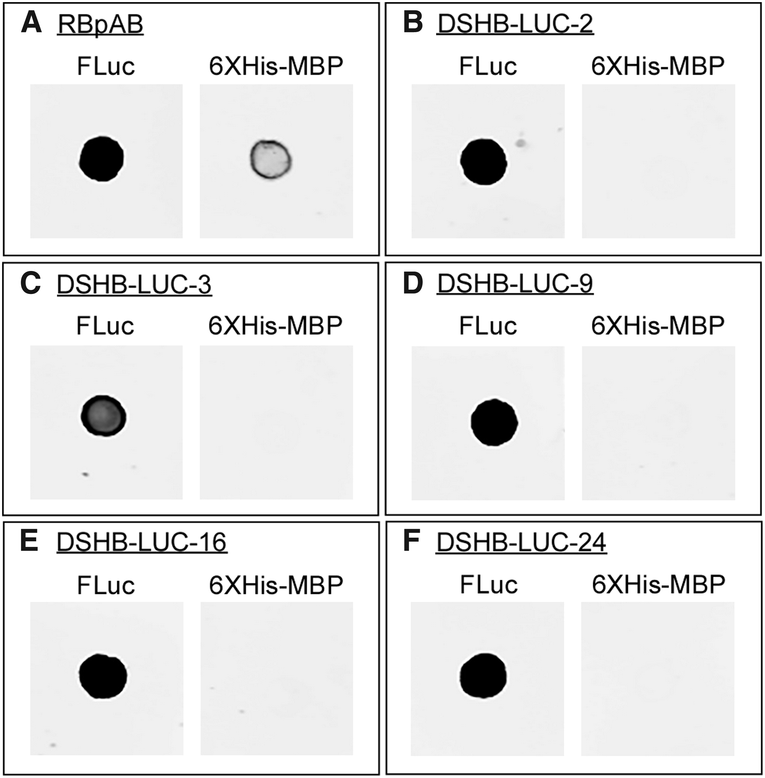
Dot blot analysis of the five anti-FLuc mAbs. Lysates from Escherichia coli strains expressing either the untagged wild-type FLuc or 6xHis-MBP (maltose-binding protein) recombinant proteins were spotted onto nitrocellulose membranes.
Western blot
Each of the five anti-FLuc mAbs was then tested by western blot analysis for the ability to bind denatured FLuc peptides in cell lysates of FLuc-expressing 4T1 cells. The results were also compared with those of the commercially available rabbit polyclonal antibody, (RBpAB), L0159. Each antibody was also tested against the FLuc-free lysate of Ntera2 (NT2) cells. The polyclonal antibody RBpAB recognized in the 4T1 cells a signal protein of 62 kDa, the molecular mass of the FLuc gene product (Fig. 2A). RBpAB recognized no bands in the NT2 cell lysate (Fig. 2A). All of the mAbs we generated (DSHB-LUC-2, DSHB-LUC-3, DSHB-LUC-9, DSHB-LUC-16, and DSHB-LUC-24) also recognized a single band at 62 kDa, contained in the FLuc-expressing 4T1 cell lysate and no bands in the NT2 lysates (Fig. 2B–F). In the case of DSHB-LUC-3, a higher detergent stringency was necessary to remove an increased background signal. The results demonstrate that the five aforementioned anti-FLuc mAbs were able to recognize denatured wild-type FLuc enzyme, although DSHB-LUC-3 exhibited diminished recognition.
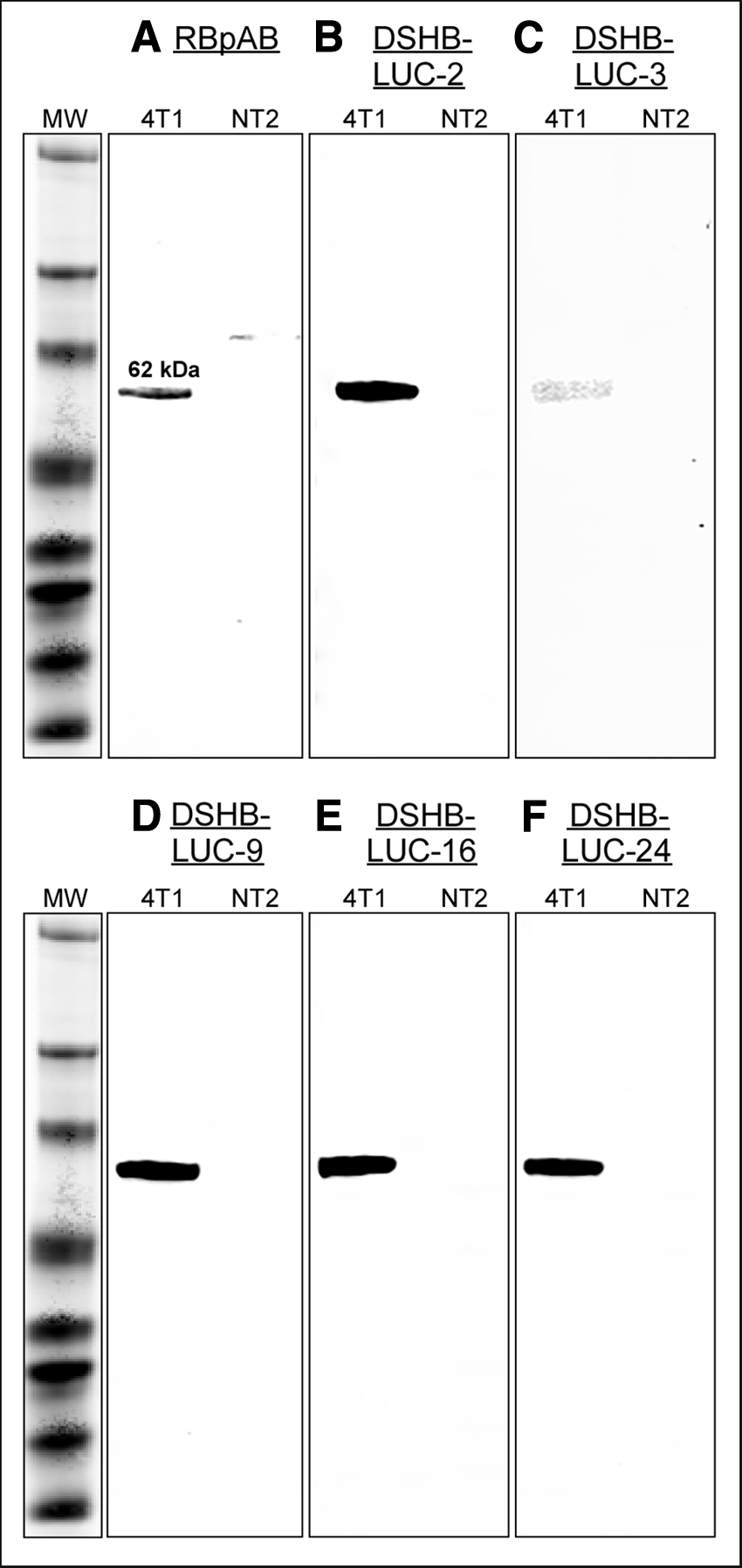
Western blot analysis using whole cell lysates of an
Immunostaining fixed cells
The five anti-FLuc mAbs were tested for immunostaining, using fixed, permeabilized FLuc-expressing 4T1 cells. Cells were treated with each mAb, then with Alexa Fluor 488 goat anti-mouse antibody (green), and then counterstained with the rhodamine-conjugated phalloidin (red). Phalloidin stains F-actin.(24) Each of the five anti-FLuc mAbs stained the cytoplasm of fixed cells (Fig. 3A–E, respectively). Punctate staining was observed within the cytoplasm, as is obvious in enlarged images of stained cells (Fig. 4). Punctate staining reflects localization of FLuc in peroxisomes mediated by a C-terminal peptide-targeting signal.(25) When FLuc expression is higher than the maximum level the peroxisomes can incorporate, as is the case in the FLuc-expressing 4T1 cells, FLuc accumulates in the nonpunctate cytoplasm (Figs. 3 and 4). These results demonstrate that each of the five anti-FLuc mAbs is effective in recognizing FLuc enzyme in cells fixed with paraformaldehyde and permeabilized.

Immunostaining of fixed, permeabilized FLuc-expressing 4T1 murine mammary cell line cells by mAbs. The 4T1 murine breast carcinoma cells constitutively expressing the wild-type FLuc enzyme were fixed with paraformaldehyde, permeabilized, treated with each of the five anti-FLuc mAbs

Increased magnification of FLuc staining of a spread 4T1 cell, in this case by DSHB-LUC-3, reveals both peroxisome staining and diffuse cytoplasmic staining. Fixed, permeabilized cells were stained with the mAb DSHB-LUC-3 (green) and counterstained with rhodamine phalloidin (red) for F-actin localization. Scale bar 10 μM.
Paraffin-embedded cells
FLuc-expressing 4T1 cells were suspended in agarose, fixed with paraformaldehyde, embedded in paraffin, and sectioned for staining. Sections were treated with each mAb and then stained with Alexa Fluor 488 goat anti-mouse antibody. Of the five mAbs, four (DSHB-LUC-2, DSHB-LUC-9, DSHB-LUC-16, and DSHB-LUC-24) either did not stain or very weakly stained paraffin-embedded cell preparations (Fig. 5A, C–E, respectively). Only one mAb, DSHB-LUC-3, provided a strong signal in paraffin-embedded cell preparations (Fig. 5B). Control 4T1 cells not expressing FLuc did not stain in paraffin with DSHB-LUC-3 (Fig. 5F). Control preparations lacking DSHB mAbs did not stain. Therefore, although DSHB-LUC-3 provided only a weak signal in western blot analysis, it provided a strong signal when employed in fixed cell preparations and in cell preparations embedded in paraffin.

Immunofluorescent staining of sections of paraformaldehyde-fixed, paraffin-embedded cells by anti-FLuc mAbs. FLuc-expressing 4T1 mouse mammary cells were suspended in agarose, fixed with paraformaldehyde, embedded in paraffin, sectioned, and treated with the five anti-FLuc mAbs
Discussion
We have generated five mAbs against the purified native FLuc enzyme and characterized their effectiveness in four distinct applications, dot blot staining of native FLuc, western blot analysis of denatured FLuc, and both immunostaining and immunofluorescent staining of paraffin-embedded cells. For immunostaining of fixed cells and immunofluorescent staining of paraffin-embedded cells, we employed a FLuc-expressing mouse cell line derived from the 4T1 mouse mammary carcinoma cell line.(20,21) Table 1 shows a summary of the efficacy of the five mAbs in each of the applications tested. All five mAbs successfully recognized the native protein in dot blot analyses. Four of the five anti-FLuc mAbs provided strong signals in recognizing denatured FLuc in western blotting of cell lysates. All five mAbs were highly effective in staining fixed, permeabilized cells. One of the anti-FLuc mAbs, DSHB-LUC-3, which provided a very weak signal in western blot analyses, was effective in recognizing FLuc in paraffin-embedded cell preparations.
Detergent increased 10-fold to remove nonspecific background binding.
Footnotes
Acknowledgments
This research was funded by the Developmental Studies Hybridoma Bank, a National Resource created by the NIH and housed at the UIOWA. All antibodies can be obtained at cost from the DSHB for use in basic research (
Author Disclosure Statement
No competing financial interests exist.