
Abstract
Receptor tyrosine kinase ROR1 has been introduced as an interesting prognostic cancer marker in histopathology. The aim of this study was to produce a polyclonal antibody (PAb) against recombinant human ROR1 protein to be used as a tool for investigation of ROR1 expression in human cancer tissue blocks. The extracellular part of human ROR1 recombinant protein was expressed using pET-28b(+) plasmid in Escherichia coli Bl21(DE3) host. The recombinant ROR1, as a candidate immunogen, was purified and injected to a New Zealand rabbit. Followed by raising the titration of antibody, polyclonal anti-ROR1 antibody was purified through affinity chromatography column. After determining the purity of PAb anti-ROR1, its specific reactivity was assessed through various assessments. Flow cytometry analysis showed that PAb anti-ROR1 specifically recognizes ROR1 molecule in a number of positive and negative cell lines. Results obtained from detection of ROR1 in paraffin-embedded breast adenocarcinoma tissue blocks (n = 11) also demonstrated that PAb anti-ROR1 can effectively be used in immunohistochemistry. In conclusion, the developed anti-ROR1 PAb can be used as a tool for determining the prognostic value of ROR1 in histopathology of cancer tissues.
Introduction
R
ROR1 is expressed during embryogenesis but not almost all healthy adult tissues.(4–6) The overexpression of ROR1 at gene and protein levels was identified in a number of malignancies, including breast cancer, chronic lymphocytic leukemia, melanoma, and lung adenocarcinoma.(7–9) Moreover, the significant correlation of ROR1 expression with other tumor markers in gastric adenocarcinomas and ovarian cancer introduced ROR1 as a reliable prognostic marker for these types of malignancies.(10,11) Nevertheless, the correlation of ROR1 expression with other tumor markers has remained to be well understood.
In this study, we attempted to develop a specific polyclonal antibody (PAb) against ROR1 to be used as a research tool for further investigation of ROR1 expression in correlation with other tumor markers in human malignancies.
Materials and Methods
PCR amplification of ROR1 gene
The extracellular region of human ROR1 protein, residues 30 through 406, (excluding signal sequence) was amplified from pCMV6-XL6-ROR1 plasmid (OriGene, Rockville, MD), which contains complete cDNA of ROR1. A pair of specific primers was designed according to the published sequence of ROR1 gene (Accession No.: NM_005012). BamHI and XhoI restriction sites were introduced at the 5′ end of the forward and reverse primers, respectively. Forward primer: 5′-
Cloning of ROR1 gene
The PCR product was A-tailed and subsequently ligated in pGEM T-Easy Vector using T4 DNA ligase (Fermentas, Berlin, Germany) according to the manufacture's protocol. Ten microliter of ROR1-pGEM-T Easy construct was transformed in DH5α competent cell. Positive white colonies were screened using colony PCR by T7 and SP6 primers. One of positive colonies was chosen for subcloning in pET-28b(+) after sequencing confirmation. After digestion of ROR1 from pGEM-T Easy vector using BamHI and XhoI, the ROR1 gene was ligated into digested pET-28b(+) vector using T4 DNA ligase. The inserted gene was verified by restriction enzyme digestion and DNA sequencing.
Expression of recombinant ROR1 protein
ROR1-pET-28b(+) plasmid was transformed into Escherichia coli BL21 (DE3) pLysS strain and cultivated in LB agar containing Kanamycin. The expression of ROR1 protein of several colonies under the control of isopropyl-β-
Identity confirmation of purified protein
The identity of purified recombinant ROR1 was confirmed by western blot analysis according to the method described in detail earlier.(13) Horseradish peroxidase (HRP)-conjugated anti-His-tag monoclonal antibody (MAb) (Roche, Basel, Switzerland) at dilution of 1:4000 was used to detect protein. Other staining using anti-ROR1 MAb (ab91187) (Abcam, Cambridge, United Kingdom) at a concentration of 1 μg/mL was performed. HRP sheep anti-mouse immunoglobulin (Avicenna Research Institute, Tehran, Iran) at a dilution of 1:2500 was used for detection of anti-ROR1 MAb.
Rabbit immunization
The rabbit immunization and blood sampling procedures were performed according to the international standard animal welfare guidelines.(14) A female white New Zealand rabbit was immunized 5 times with 2-week intervals for each injection. In the first immunization, 100 μg recombinant ROR1 protein and an equal volume of Freund's complete adjuvant (Sigma, St. Louis, MO) were mixed and injected intramuscularly into the femoral muscle. For subsequent immunizations, 50 μg recombinant ROR1 protein was emulsified in Freund's incomplete adjuvant (Sigma) and injected.
Antibody purification
The antibody purified from rabbit sera by affinity chromatography using an immobilized-recombinant ROR1 protein column. The method of protein coupling to CNBr-activated Sepharose-4B (GE Healthcare, Uppsala, Sweden) and its immobilization to the column was described earlier.(15) The evaluation of antibody reactivity with immunizing protein was examined using enzyme-linked immunosorbent assay (ELISA) as described previously.(16) In brief, 100 ng of recombinant ROR1 protein was coated in each well. Subsequently, the purified anti-ROR1 PAb was titrated from 200 to 3.1 ng. HRP sheep anti-rabbit (1:3000) (Avicenna Research Institute) was used for detection. All values are represented as mean ± standard deviation of two replications.
Cell lines
Cell lines, including chinese hamster ovary (CHO) , A549, HT-29, Raji, and Jurkat (National Cell Bank of Iran, Tehran, Iran), were cultured in RPMI-1640 (Gibco, Grand Island, NY), containing 10% fetal bovine serum (Gibco), 100 U/mL penicillin (ICN Biomedicals, Ohio), and 100 μg/mL streptomycin (Sigma) at 37°C in a humidified incubator with 5% CO2 atmosphere.
Transfection of ROR1 gene to CHO
The human ROR1 gene containing the entire extracellular and transmembrane domains was stably transfected to CHO.(17) In brief, cDNA was extracted from pCMV6-XL5-ROR1 construct (OriGene) and subcloned into pCMV6-neo plasmid (OriGene). The fragment was confirmed by enzyme digestion and DNA sequencing. The constructs of pCMV6-neo-ROR1 or pCMV6-neo-empty vector were transfected to CHO using the JetPEI™ DNA transfection reagent (Polyplus-transfection, New York, NY). After 48 hours, ROR1 expression was analyzed by flow cytometry. Transfected cells were subsequently selected using gradually increasing concentrations of G418 (Gibco) to 850 μg/mL.
Flow cytometry
In brief, 106 cells were washed in phosphate-buffered saline (PBS) and incubated with anti-human ROR1 PAb (5 μg/mL) or anti-ROR1 MAb (ab91187; Abcam) (1 μg/mL) for 1 hour. The purified rabbit immunoglobulin or anti-HIV protein envelope MAb (Avicenna Research Institute) was used at the same concentration as isotype control antibodies. FITC sheep anti-rabbit or anti-mouse immunoglobulin (Avicenna Research Institute) was added at concentration of 1:100 or 1:50, respectively. Subsequently, cells were analyzed by Partec flow cytometer.
Immunocytochemistry
Cells (n = 2 × 10⁴) were centrifuged onto glass slides using a Shandon Cytospin Cytocentrifuge (Thermo Scientific, Rockville, MD). Cells were immediately fixed in cold acetone (−20°C) for 5 minutes. The slides were washed with Tris-buffered saline (pH = 7.4), containing 0.1% bovine serum albumin (BSA) and blocked with 5% sheep serum. Slides were then incubated with 10 μg/mL anti-ROR1 PAb or isotype control, purified rabbit immunoglobulin (Avicenna research Institute) for 1 hour. After quench of endogenous peroxidase by 0.3% H2O2, slides were incubated with HRP-sheep anti-rabbit (Avicenna Research Institute) for 1 hour followed by Diaminobenzidine (DAB) (Roche). Afterward, slides were counterstained with hematoxylin, dehydrated in increasing grades of ethanol, and mounted in Entellan (Merck, Darmstadt, Germany). Finally, cells were examined with a light microscope (Olympus, Center Valley, PA).
Immunohistochemistry
The formalin-fixed paraffin-embedded human breast cancer tissues were collected from pathology department of Khatam Al-Anbia Hospital, Tehran, Iran, based on the Ethics Committee approval with ID No. 90/8047. This study is part of a main project with ID No. 890211-051, which has been carefully reviewed and approved by the Ethics Committee of Avicenna Research Institute.
Subsequently, 4 μm tissue sections were prepared, deparaffinized, and their antigens were retrieved according to the method explained before.(18) The antibody staining was performed according to the method described formerly in immunocytochemistry (ICC) method.
Results
Recombinant human ROR1 protein
The extracellular domain of human ROR1 cDNA without its signal peptide sequence was cloned into the pET-28b(+) vector in frame with a His-tag to facilitate ROR1 protein purification through a nickel charged resin. SDS-PAGE showed the expected band of 43 kDa related to recombinant ROR1 protein in transformed bacteria after IPTG induction (Fig. 1). The integrity of purified ROR1 protein was confirmed by western blotting (Fig. 1). Staining using ab91187 commercial anti-ROR1 MAb and anti-His-tag antibody confirmed the accuracy of protein. Both antibodies detected a protein band of 43 kDa.
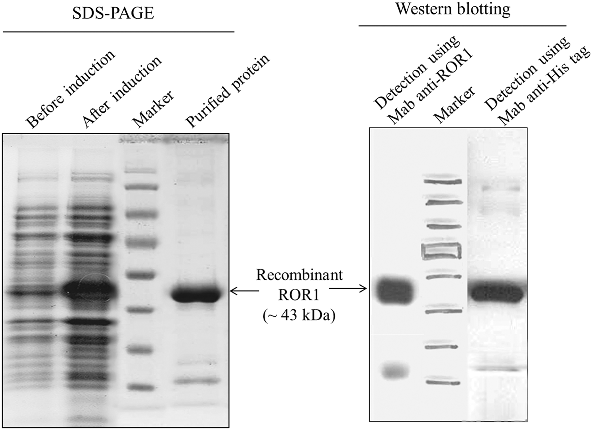
Verification of purified recombinant ROR1 by SDS-PAGE and western blot. Loading the bacteria lysates, before and after induction with IPTG, demonstrated a protein band of 43 kDa related to expression of ROR1 extracellular part (left). Western blot analysis using anti-His-tag and anti-ROR1 MAb (Abcam) confirmed the accuracy of ROR1 protein identity after purification (right). IPTG, isopropyl-β-
Reactivity of anti-ROR1 PAb in ELISA
The anti-human ROR1 PAb was tittered in ELISA (Fig. 2). The excellent immunoreactivity of antibody with its immunizing protein confirmed its functionality after purification. The lack of reactivity of anti-ROR1 PAb with PBS or BSA, as an irrelevant protein, represented its specificity (Fig. 2).

Reactivity of purified rabbit anti-ROR1 PAb in ELISA. Anti-ROR1 PAb was purified by affinity column, and its reactivity with immunizing protein was evaluated by ELISA. In brief, 100 ng of recombinant ROR1 protein was coated in each well. Subsequently, the purified anti-ROR1 PAb was titrated from 200 to 3.1 ng. HRP sheep anti-rabbit (1:3000) (Avicenna Research Institute) was used for detection. All values are represented as mean ± standard deviation of two replications. Lack of reactivity of PAb with bovine serum albumin and phosphate-buffered saline demonstrated the specificity of antibody. ELISA, enzyme-linked immunosorbent assay; HRP, horseradish peroxidase; PAb, polyclonal antibody.
Flow cytometry
The cell surface staining of some cell lines expressing ROR1, including CHO-ROR1 (stably ROR1-transfected cells), A549, and HT-29, using anti-ROR1 PAb, demonstrated that the developed PAb was able to detect the extracellular part of ROR1 (Fig. 3). In contrast, anti-ROR1 PAb did not react with ROR1-nonexpressing cell lines, including CHO-vector (stably pCMV6-Neo-transfected CHO), Raji, and Jurkat, which confirmed its specificity for ROR1 detection in flow cytometry analysis (Fig. 3).

Flow cytometry using anti-ROR1 PAb. The produced PAb detected ROR1 protein on the surfaces of ROR1-expressing cell lines containing CHO-ROR1, A549, and HT-29. In contrast, the antibody did not react with ROR1-nonexpressing cell lines containing CHO-vector, Raji, and Jurkat. To consolidate the results, commercial anti-ROR1 MAb used as positive control. Solid line: produced anti-ROR1 PAb; Dashed line: commercial anti-ROR1 MAb; Gray tinted curve: isotype control antibodies. MAb, monoclonal antibody.
Immunocytochemistry
We prepared cytospin slides of CHO-ROR1 (as positive cell control), CHO-vector (as negative cell control), A549, and HT-29 (which express ROR1 more than 70%) to determine whether anti-ROR1 PAb can identify its target among many other expressed proteins. Figure 4 showed that ROR1 expression obviously was detected by anti-ROR1 PAb in acetone-fixed CHO-ROR1, A549, and HT-29. The location of ROR1 molecule was detected both on cell surface and intracellular in all cell lines. As it was expected, the produced PAb did not detect ROR1 in negative cell line, CHO-vector. The isotype control antibody, rabbit immunoglobulin, did not react with ROR1 in all cell lines.
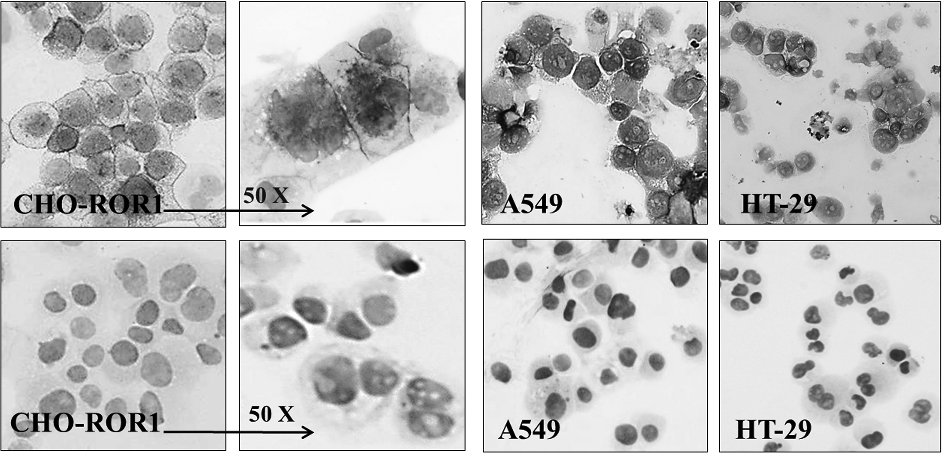
Immunocytochemistry staining using anti-ROR1 PAb. The produced PAb detected ROR1 in a number of ROR1-expressing cell lines, including CHO-ROR1, A549, and HT-29 (upper pictures). The purified rabbit IgG as isotype control did not detect ROR1 in the same cells (lower pictures). CHO, chinese hamster ovary.
Immunohistochemistry
To examine the application of anti-human ROR1 PAb in pathologic study of tumor cells, we immunostained paraffin-embedded tissue blocks that belonged to breast adenocarcinoma patients (n = 11) with median age of 50 years (range 38–60). The patients' clinicopathologic characteristics are summarized in Table 1. Results from immunostaining showed that anti-human ROR1 PAb specifically detected ROR1 in breast adenocarcinoma blocks, while isotype control antibody, rabbit immunoglobulin, did not react (Fig. 5). The figure is reprehensive of 1 from 11 samples. Neoplastic cells of all examined breast adenocarcinoma associated with aggressive disease expressed high-level of ROR1 (score +2 and +3) regardless of the expression level of other breast cancer prognostic markers that is, HER2, estrogen receptor (ER), and progesterone receptor (PR).
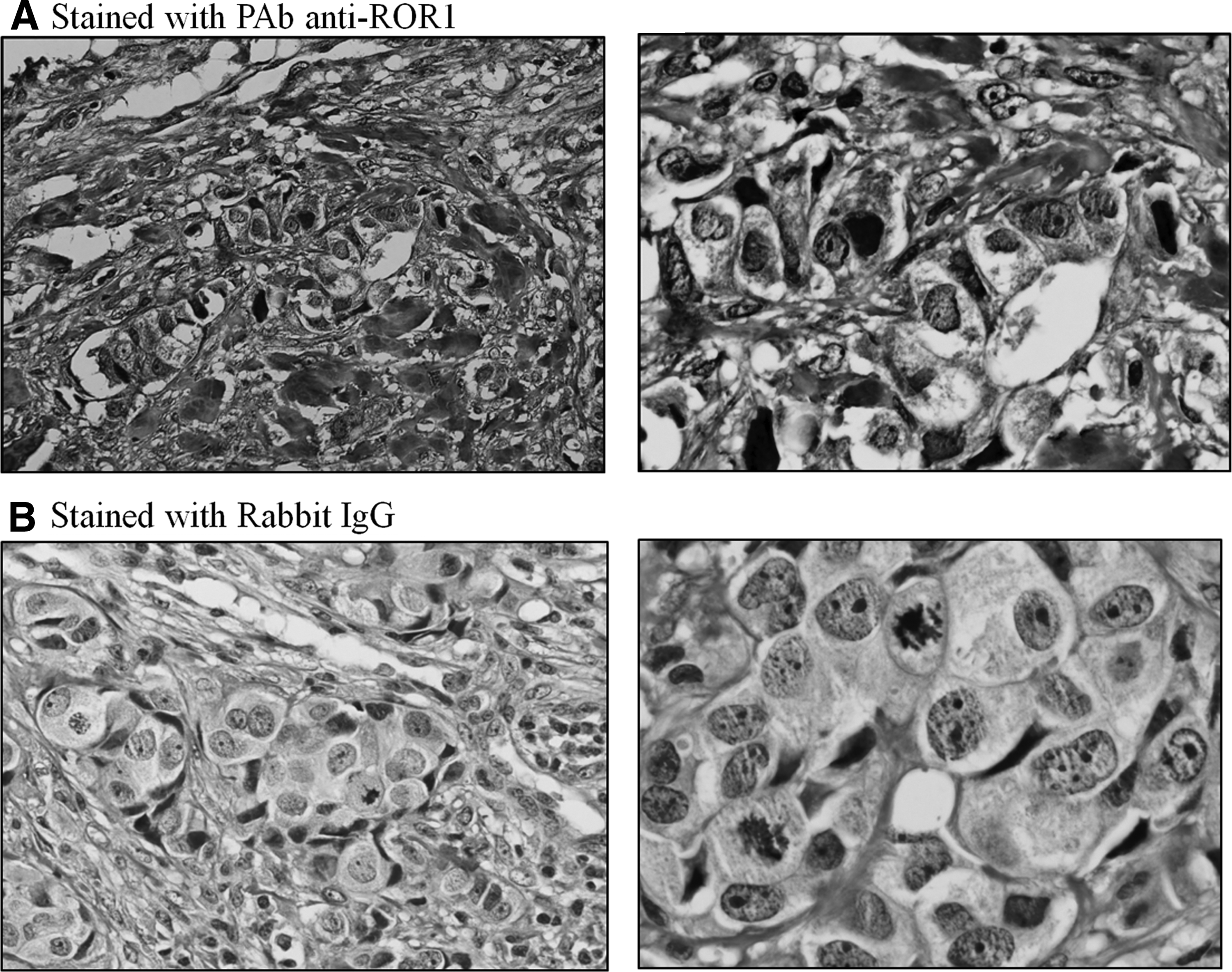
Immunohistochemistry staining using anti-ROR1 PAb in breast adenocarcinoma tissues. The produced PAb detected ROR1 in paraffin-embedded breast cancer tissues
ER, estrogen receptor; IDC, invasive ductal carcinoma; NOS, not otherwise specified; PR, progesterone receptor.
Discussion
Despite the extensive advances in surgery and chemotherapy of human malignancies during the last few decades, therapeutic failure and disease progression are still considerable.(19) Therefore, discovering novel and validated cancer biomarkers with high sensitivity and specificity is important. Recently, ROR1 was introduced as a target of interest for immunotherapy by developing specific anti-ROR1 antibodies due to its tumor-specific expression and its function as tumor survival factor.(20–22)
In this study, we produced and characterized a PAb against recombinant ROR1 protein in rabbit model. Although PAbs do not provide a main supply in therapeutic strategies such as MAbs, there is a great tendency toward its production for application in immunological research purposes.(23) This potency is due to the advantage of PAb obtaining in a relative short time, with little financial investment compared to MAbs.(24) PAbs frequently have higher specificity than Mabs, as they are produced by a large number of B cell clones, each producing antibodies to a specific epitope; and therefore, polyclonal sera are a compound of antibodies with unique specificities.(25) This property causes the PAbs to be considered as interesting tools for specifically detection of antigens in immunohistochemistry (IHC) test at pathological laboratories. The anti-HER2 PAb used in Hercep Test, an FDA-approved IHC test kit, would be an appropriate and distinguished instance for this claim.(26)
In this study, we showed that the produced anti-ROR1 PAb is capable to specifically detect ROR1 in IHC of breast adenocarcinoma tissues. The data were corroborated with Zhang et al., which demonstrated that ROR1 is expressed in high proportions of human breast cancer tissues.(8) Consistent with this finding, our IHC result showed that all 11 breast cancer specimens expressed high level of ROR1, regardless of the expression of other prognostic factors containing Her2, PR, and ER. Data from IHC demonstrated that ROR1 protein localized both intracellularly and on the cell surface of breast adenocarcinoma cells.
Indeed, the produced antibody was capable to specifically detect ROR1 in ICC. Among many available cell lines in our laboratory, A549 (lung adenocarcinoma) and HT-29 (colorectal adenocarcinoma) were chosen due to over 70% expression of ROR1. The high level of ROR1 expression in these cell lines might be attributed to the role of ROR1 as a vital receptor in survival and pathogenesis of lung and colon cancers.(27,28) To consolidate the results, ROR1 and empty vector-transfected CHO provided us the positive and negative cell controls, respectively. We also investigated capability of the developed PAb in detection of cell surface ROR1 in A549 and HT-29 cell lines by flow cytometry. Among the investigated cell lines, the highest expression of ROR1 belonged to CHO-ROR1, due to the insertion of ROR1 gene and the overexpression of ROR1 protein in this cell line. The antibody was able to specifically recognize the extracellular part of ROR1 localized on cell surface.
In conclusion, based on our results, the produced anti-ROR1 PAb is a tool appropriate for IHC, ICC, and flow cytometry analyses. The antibody might be applied in cancer prognosis and indication of the patient outcome by specific detection of ROR1. However, further investigation and methodological standardization are required in the context of application.
Footnotes
Author Disclosure Statement
No competing financial interests exist.