
Abstract
Porcine reproductive and respiratory syndrome virus (PRRSV) is one of the most important viral pathogens that has caused tremendous economic losses to the swine industry worldwide. Although extensive research has been focused on PRRSV, little is known about the structure and biological functions of individual nonstructural viral proteins, especially the nonstructural protein 12 (Nsp12). In this study, we generated and identified the monoclonal antibody (mAb) against PRRSV Nsp12. Six strains of hybridoma cells named 2B10, 2B12, 5E1, 5G6, 5E7, and 8B2 secreting anti-Nsp12 mAbs were obtained by the hybridoma technique. All the mAbs were specifically reacted with PRRSV by indirect immunofluorescence assay and four of them (2B12, 5E1, 5G6, and 5E7) were specifically reacted by Western blot. Furthermore, the 5E7 specifically recognized multiple type 2 PRRSV strains, including highly pathogenic and classical PRRSV strains, but not type 1 PRRSV strain. Taken together, the mAbs against Nsp12 provide a valuable tool to specifically recognize type 2 PRRSV as a diagnostic reagent and study the biological function of Nsp12 in the future.
Introduction
P
Porcine reproductive and respiratory syndrome virus (PRRSV), a member of the arterivirus family, is an enveloped single-strand positive RNA virus..(4,5) Based on their genetic and antigenic differences, PRRSV strains are classified into two distinct genotypes, North American (type 2) and European (type 1), represented by the VR-2332(1) and Lelystad virus,(6) respectively. These two genotypes on two different continents share only ∼60% nucleotide sequence identity.(7,8)
Although extensive research has been focused on PRRSV, little is known about the individual nonstructural viral proteins, except that they exhibit the replicase, protease, and polymerase activities. The replicase-associated genes, ORF1a and ORF1b, are located on the 5′ end of the genome (representing almost 75% of the viral genome) and encode for the polyproteins pp1a and pp1ab.(9) Proteolytic cleavage of pp1ab generates nonstructure protein 9 (Nsp9) through nonstructure protein 12 (Nsp12).(10) The function of Nsp12, which may play essential roles in viral replication and production, still remains unknown.
Herein, we prepared the specific monoclonal antibody (mAb) against PRRSV Nsp12 using the hybridoma technique, which provides a valuable tool to specifically recognize type 2 PRRSV and investigate the functions of Nsp12 in the future.
Materials and Methods
Cells, antibodies, and animals
SP2/0 myeloma cells and MARC-145 cells were grown in Dulbecco's modified Eagle's medium (Invitrogen) with 10% fetal bovine serum (FBS) (Gibco) and were maintained with 2% FBS at 37°C in a humidified 5% CO2 atmosphere as described previously.(11) Horseradish peroxidase (HRP)-conjugated goat antimouse secondary antibody was purchased from Sigma (Shanghai, China). The expression plasmid pCold-I vector was purchased from TaKaRa (Dalian, China). Six-week-old female BALB/c mice were purchased from the Shanghai Slac Laboratory Animal Center (Shanghai). HP-PRRSV strain vHuN4 (GenBank accession No. EF635006) was isolated from the serum of a dying piglet displaying the clinical signs of porcine high-fever disease in 2006,(3) and vF112 is an attenuated vaccine virus strain adapted from vHuN4 strain (GenBank accession No. EF635006).(12) vAPRRS (GenBank accession No. GQ330474)(13) and vSHE(14) were rescued from pAPRRS and pSHE, respectively.
Expression and purification of rNSP12 protein of HP-PRRSV
According to the nucleotide sequence information of HP-PRRSV strain vHuN4, a pair of primers (sense primer 5′-CG
Procedure for immunization of mice
Five 6-week-old female BALB/c mice were immunized with purified rNsp12 (100 μg) plus equal volume of Freund's complete adjuvant through intraperitoneal injection. At 2-week intervals, a second immunization was given using the purified rNsp12 (100 μg) emulsified in Freund's incomplete adjuvant in 1:1 proportion. The immunization was repeated every 2 weeks. Booster immunization was given after 3 days through intraperitoneal injection before cell fusion.
Preparation of anti-rNsp12-specific mAb
The anti-rNsp12 serum was obtained from the immunized mice and the antibody titers were determined by indirect ELISA. Spleen cells of the best immunized mice were isolated and fused with SP2/0 myeloma cells under the action of 50% polyethylene glycol (PEG) as fusion agents. The hybridoma cells were then cultured in 96-well plates in hypoxanthine/aminopterin/thymidine (HAT) screening culture medium with 20% FBS. Positive hybridomas were filtered by indirect ELISA when the cells had covered one-fourth to one-third of the bottoms of 96-well plates. After cloning four times by limiting dilution, the hybridoma cells were injected into pristane-treated BALB/c mice to gain abundant ascetic fluid. The subtype of the mAb was identified using Pierce Rapid ELISA Mouse mAb Isotyping Kit according to the manufacturer's instructions (Thermo Fisher Scientific).
Indirect immunofluorescence assays
Immunofluorescence assays (IFAs) were performed as described previously.(15) MARC-145 cells were infected by PRRSV vHuN4 and fixed with cold methanol followed by blocking with 1% bovine serum albumin and then incubated for 2 hours with the mAbs against Nsp12. After washing with phosphate-buffered saline, the cells were incubated for 1 hour with Alexa Fluor 488-labeled goat antimouse IgG (H+L) antibody (Invitrogen). After the final washing step, cells were visually analyzed using an Olympus inverted fluorescence microscope.
SDS-PAGE and Western blotting
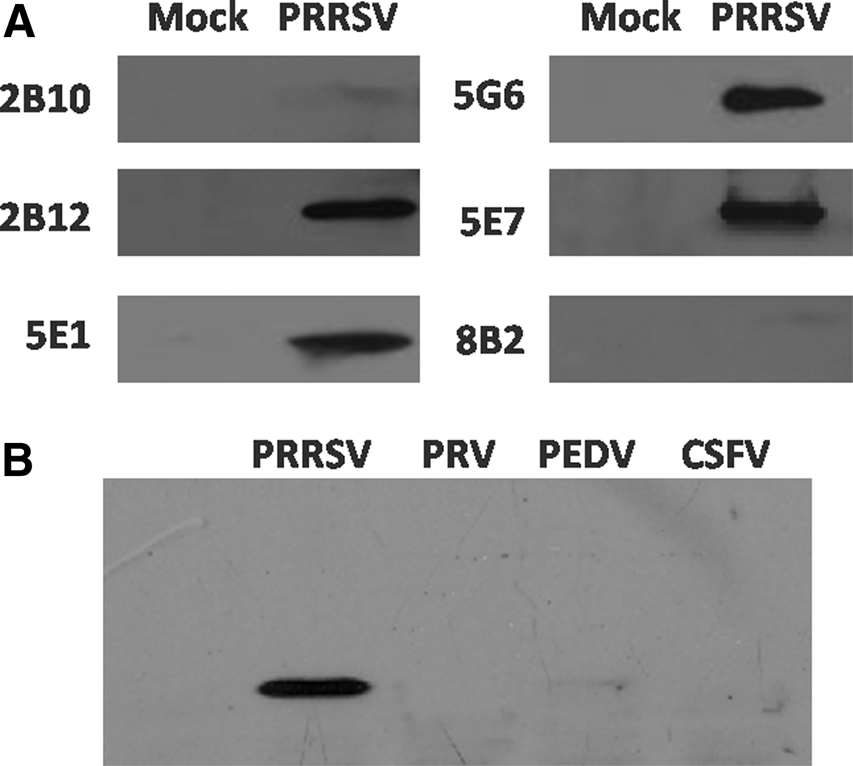
The cell proteins were collected at 36 hours after PRRSV infecting MARC-145 cells. Then, the samples were separated by SDS-PAGE with 15% polyacrylamide gels, and transferred to Hybond-P membranes (Amersham Biosciences). Membranes were blocked with 5% skimmed milk in TBST for 2 hours at room temperature. Membranes were incubated overnight at 4°C with the hybridoma cell supernatants, individually. After washing three times with TBST, blots were incubated with HRP-conjugated goat antimouse secondary antibody (1:6000 dilution in TBST) for 1 hour at room temperature, washed three times again with TBST, and developed by the SuperSignal West Pico or Femto chemiluminescent substrate (Thermo Fisher Scientific). The classical swine fever virus (CSFV), porcine epidemic diarrhea virus (PEDV), and pseudorabies virus (PRV) samples used in this assay were provided by our laboratory.
Results
Expression and purification of rNsp12
Total RNA was extracted from vHuN4-infected MARC-145 cells and was used for cDNA synthesis. The gene fragment of Nsp12 about 462 bp was generated (Fig. 1A). The PCR product was purified and cloned into pCold-I DNA vector, resulting in a recombinant plasmid that was named pCold-Nsp12. The recombinant plasmid was identified by sequencing. For the expression of rNsp12, pCold-Nsp12 was transformed into E. coli BL21 (DE3) and induced by IPTG. As shown in Figure 1B, SDS-PAGE analysis showed that rNsp12 (∼17.6 kDa) was expressed and the highly purified soluble rNsp12 was obtained (Fig. 1B).

Amplification of Nsp12 gene and expression of rNsp12 by prokaryotic expression vector pCold-I-Nsp12.
Generation of mAbs against PRRSV Nsp12
The purified rNsp12 was used to immunize BALB/c mice. The antisera titers were determined by indirect ELISA after four immunizations. The mouse with the highest antibody titers against rNsp12 was selected for isolating splenic cells, which were fused with SP2/0 myeloma cells. After subcloning four times by limiting dilution and screening, six positive mAbs against Nsp12 were identified and named 2B10, 2B12, 5E1, 5G6, 5E7, and 8B2. Using a commercially available isotype classification kit, the subclass of all six mAbs was identified as IgG1. The light chain of 2B12 was λ-type and the light chain of other five mAbs was κ-type.
Reactivity of mAbs against Nsp12
The ascites were purified using NAb Protein G Spin Columns (Thermo Fisher Scientific). Indirect ELISA (Fig. 2) was used to identify the specificity of the mAbs against rNsp12. IFA (Fig. 3) and Western blot analysis (Fig. 4A) were used to identify the specificity of the mAbs against PRRSV Nsp12 in MARC-145 cells. The results suggested that all the mAbs were specifically reacted with PRRSV Nsp12 by indirect IFA and four of them (2B12, 5E1, 5G6, and 5E7) were specifically reacted by Western blot. Furthermore, the mAbs against Nsp12 specifically reacted with PRRSV, whereas it had no reaction with negative control and three other viruses (CSFV, PEDV, and PRV) (Fig. 4B).
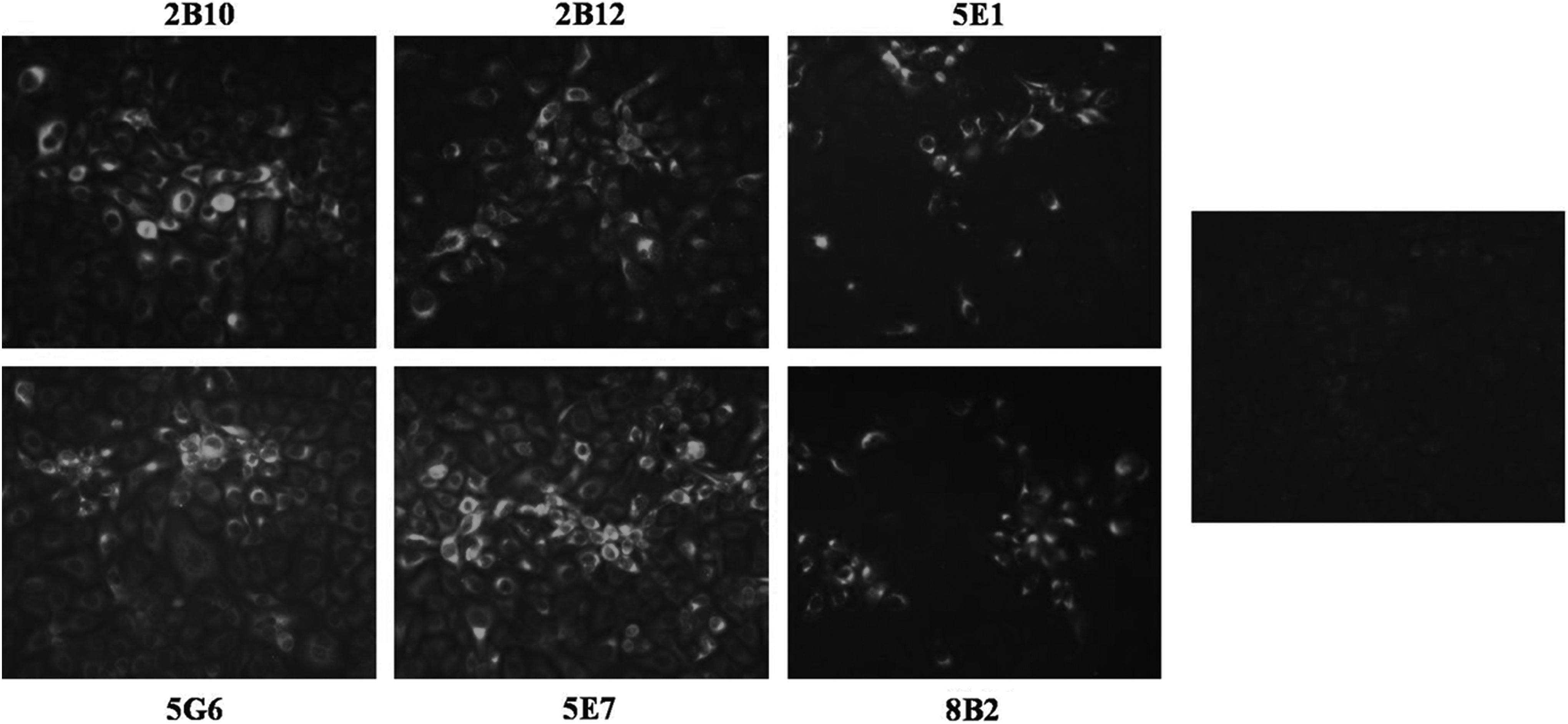
Indirect immunofluorescence assays. MARC-145 cells were infected with PRRSV vHuN4, and mAbs (2B10, 2B12, 5E1, 5G6, 5E7, and 8B2) were used as primary antibodies, respectively, followed by incubation of FITC-conjugated secondary antibody. MARC-145 cells were infected by vHuN4 (MOI = 0.1). MARC-145 cells were fixed at 36 hours postinfection and immunostained with the mAbs against the viral Nsp12 and FITC-conjugated goat antimouse IgG. mAb, monoclonal antibody.

Detection of antibody titers against rNsp12 in the ascetic fluid by indirect ELISA.
Western blot analysis.
Characterization and specificity of 5E7 against PRRSV of two genotypes
It is worth noting that 5E7 specifically recognized multiple type 2 PRRSV strains, including HP-PRRSV (vHuN4 and vHuN4-F112) and classical PRRSV (vAPRRS) strains, but not type 1 PRRSV vSHE strain, which could be used as a valuable differential diagnosis tool for two genotype PRRSVs (Fig. 5A). Results from the alignments (Fig. 5B) showed that the amino acid sequences of Nsp12 are highly conserved among type 2 strains, which now circulates in the world. However, two genotypes from two different continents shared very poor amino acid sequence identity, indicating the reason why 5E7 could not react with type 1 strain vSHE.
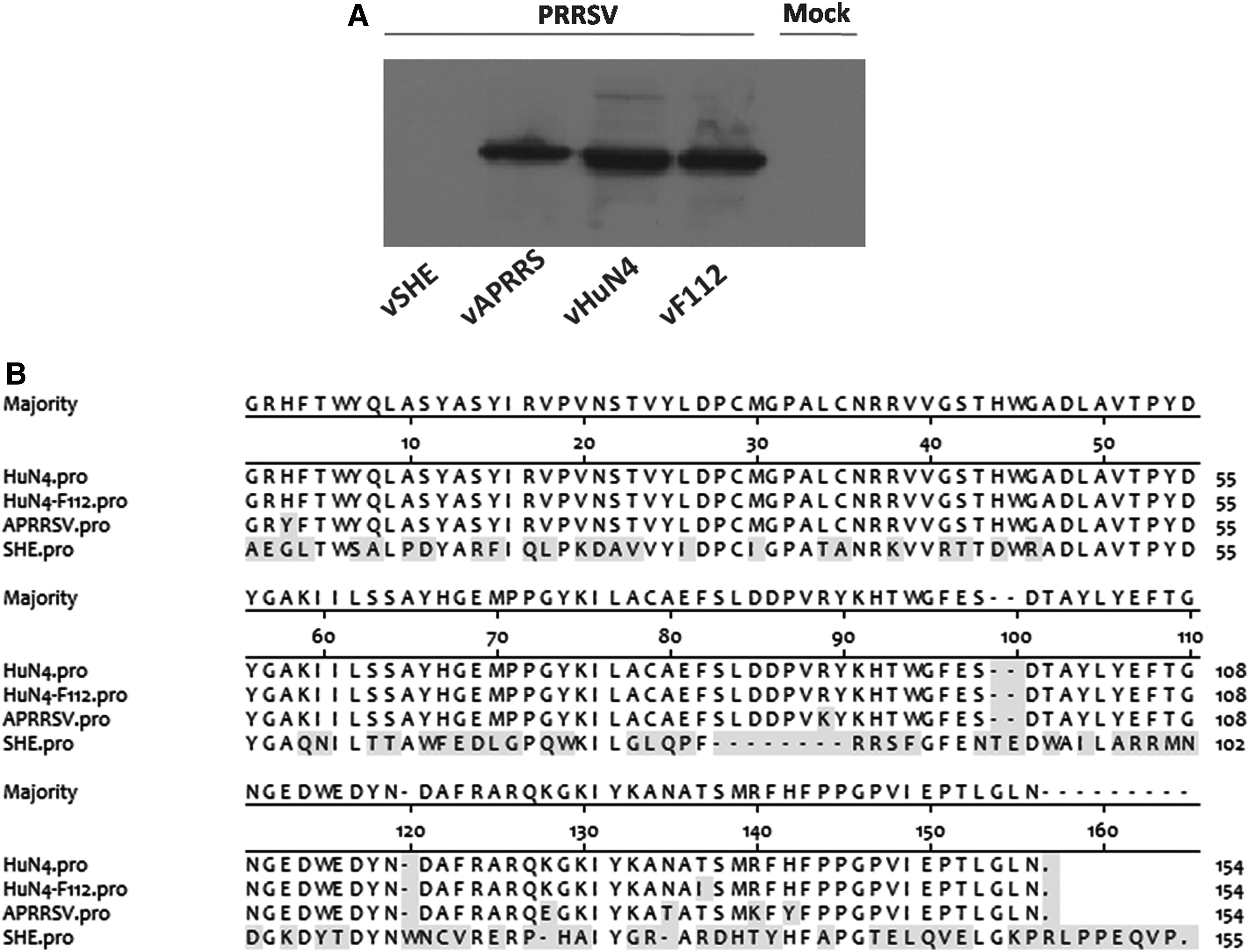
Specificity of 5E7 against PRRSV of two genotypes.
Discussion
PRRSV replicase-associated genes (ORF1a and ORF1b) encode the polyproteins pp1a and pp1ab, respectively. Proteolytic cleavage of pp1ab generates products nsp9 to nsp12.(16) The nsp9 to nsp12 are involved in virus genome transcription and replication.(9,17) Even though what we know thus far with regard to the function of PRRSV nonstructural proteins is exciting, few attempts had been done to Nsp12 that located in the 3′ end of ORF1b in viral genome. In this study, we prepared the 17.6 kDa recombinant Nsp12 purified protein (Fig. 1) and finally obtained six hybridoma cell strains named 2B10, 2B12, 5E1, 5G6, 5E7, and 8B2 secreting anti-Nsp12 mAbs. IFA and Western blot analysis showed that the mAbs specifically reacted with PRRSV Nsp12, which could be a useful tool to study the biological function of Nsp12 in the future (Figs. 3 and 4). The specific mAbs have no reaction with three other important swine viruses (CSFV, PEDV, and PRV). Notably, 5E7 specifically recognized type 2 strains including HP-PRRSV and classical PRRSV, but not type 1 PRRSV (Fig. 5), which can be used to establish a valuable tool to distinguish infections with type 2 PRRSV from type 1 PRRSV.
In summary, mAbs against PRRSV Nsp12 were successfully prepared. They are suitable for Western blot analysis, IFA, and ELISA, which may be a potential marker for developing differential diagnostic tests and for monitoring PRRSV of two genotypes in the future.
Footnotes
Acknowledgments
This study was supported by grants from the National Natural Science Foundation of China (Nos. 31602052 and 31670158), the National Key Research and Development Program of China (2016YFE0112500), the National Basic Research Program (973 Plan) (No. 2014CB542700), the National Sci-Tech Support Plan Program (2015BAD12B01-1), and the EU Horizon 2020 program project (Grant No.: SAPHIR-633184).
Author Disclosure Statement
No competing financial interests exist.