
Abstract
TRIM62 is a member of the tripartite interaction motif (TRIM) family and exerts crucial roles in innate immune response and cancer. To investigate the relationship between its distribution and avian retrovirus replication, in the present study, a monoclonal antibody (mAb) against chicken TRIM62 was generated. The open reading frame of chicken TRIM62 was amplified by polymerase chain reaction (PCR) and inserted into the expression vector pET-28a. The recombinant expression vectors were transformed into Escherichia coli BL21 (DE3). Then recombinant protein His-TRIM62 was expressed under isopropyl-β-D-thiogalactopyranoside (IPTG) induction and purified. BALB/c mice were immunized with the purified recombinant TRIM62 protein. The hybridomas were obtained by fusing mouse myeloma cell line SP2/0 with splenocytes of immunized mice. Western blot showed that the His-TRIM62 and endogenous TRIM62 were recognized by the mAb. The distribution of TRIM62 in no specific pathogen infection (SPF) chickens was detected by immunohistochemistry, and the positive signals of TRIM62 were mainly distributed in vascular endothelial. Our work indicated that the mAb against chicken TRIM62 would be a valuable tool for further study of the role of TRIM62 in avian retrovirus pathogenesis.
Introduction
T
.Otherwise, TRIM62 is a putative tumor suppressor gene. Low TRIM62 levels is an independent adverse prognostic factor in acute myeloid leukemia.(5) TRIM62 is a potential prognostic biomarker in cervical cancer and suppresses metastasis of cervical cancer via inhibiting c-Jun/Slug signaling-mediated EMT.(6) The expression of TRIM62 was downregulated in epithelial ovarian cancer.(7) Thus, the low expression level of TRIM62 is associated with the formation of tumor.
In our previous study, we found that expression of TRIM62 in DF-1 cells infected with avian reticuloendotheliosis virus (REV) at 48 h was significantly upregulated, and overexpression of chicken TRIM62 inhibited the replication of REV. In this study, we prepared the monoclonal antibody (mAb) against chicken TRIM62, and detected the distribution of TRIM62 in no specific pathogen infection (SPF) chicken using mAb. The prepared mAb would be a potential delivery tool for analysis of the distribution and expression of TRIM62 and further study of the pathogenesis of REV.
Materials and Methods
Cell line and animals
Myeloma (SP2/0) and DF-1 cell lines were purchased from the Shanghai Cell Biology Institutes, Chinese Academy of Sciences (Shanghai, China). BALB/C mice (6 weeks) were purchased from the Animal Center of Shandong University (Jinan, China). SPF chickens were purchased from Saisi Poultry Co., Ltd. (Jinan, China). All the animal protocols used in the study were approved by the Shandong Agriculture University Animal Care Committee.
Molecular cloning and expression vector construction
According to the mRNA sequence of chicken TRIM62 (GenBank ID no. 429807), specific primers (forward: 5′-CGGAATTCATGGCCTGCAGCCTGAAG-3′ and reverse: 5′-TTGCGGCCGCCTAGATGCGGACGGTGTTG-3′) were designed containing restriction enzyme sites EcoR I and Not I to amplify the coding sequence of TRIM62. Total RNA was extracted from the DF-1 cell line and cDNA synthesis was performed by using reverse transcription kit (Takara Bio, Dalian, China). The fragment of chicken TRIM62 gene was obtained by polymerase chain reaction (PCR) and ligated into expression vector pET32a (Takara, Dalian, China). PCR conditions were predegeneration for 5 minutes at 95°C, followed by 32 cycles of 30 seconds at 95°C, 30 seconds at 68°C, 1 minute 30 seconds at 72°C, and then a final extension step of 72°C for 10 minutes. The positive colonies were confirmed by DNA sequencing.
Expression and purification of recombinant chicken TRIM62
The ligated plasmid pET32a-TRIM62 was transformed into Escherichia coli BL21(DE3) (TianGen, Beijing, China). The recombinant protein expression was induced by isopropyl-β-D-thiogalactopyranoside (IPTG) at a final concentration of 0.5 mM for 4 hours at 37°C. The bacteria were harvested by centrifugation. Then, the pellet was resuspended in a lysis buffer RIPA, followed by sonication. The expression of recombinant protein was confirmed by sodium salt (SDS)-polyacrylamide gel electrophoresis and by western blot analysis with anti-His antibody (Sigma, St. Louis, MO). His-tagged TRIM62 proteins were purified using urea dialysis.
Immunization of mice
Six Balb/C mice were immunized with purified chicken TRIM62 (40 μg), emulsified with an equal amount of Freund's complete adjuvant by subcutaneous injection. At 2-week intervals, a second immunization was performed using the purified protein emulsified with an equal amount of Freund's incomplete adjuvant by intraperitoneal injection. Immunization was completed by one booster injection at a 2-week interval. Then, serum of the immunized mice was monitored for antibody titers against chicken TRIM62 by indirect enzyme-linked immunosorbent assay (ELISA).
Preparation of monoclonal antibody
Cell fusion was performed as previously described with modifications until the antibody titers reached a high level. Splenocytes obtained from the immunized mice were cultured in DMEM and mixed with SP2/0 at a ratio of 10:1. Then, HAT/HT selective medium was used to sort the fused cells until they became stable cell lines. The positive clone was screened for antibody production against chicken TRIM62 using an indirect ELISA. The serum from the immunized mouse was used as positive control and serum from unimmunized mice was used as negative control. By using the limiting dilution method, the positive hybridoma clones were subjected to single-cell cloning and subcloning. After subcloning three or four times, the stable hybridoma cell lines were expanded and cryopreserved in liquid nitrogen.
Indirect ELISA
The antibody titer determination was analyzed by indirect ELISA assay. The purified TRIM62 protein (1 μg/mL) was coated on plates at 100 μL per well in 96-well immunoplates at 4°C overnight. The plates were blocked with 5% skim milk for 1 hour at 37°C, and then incubated with 100 μL of anti-TRIM62 with serial 1:10 dilutions (normal serum was the negative control). After incubation for 2 hours at 37°C, the plates were washed and incubated with 100 μL of horseradish peroxidase (HRP)-conjugated goat anti-mice IgG (dilution, 1:500) for 1 h at 37°C. The enzyme reaction was performed with one component TMB ELISA. The absorbance was measured at 450 nm using a microplate reader.
Western blot analysis for mAb specificity
Equal amounts of DF-1 cell lysates, 293T cell lysates, and recombinant chicken TRIM62 were separated by 10% SDS-PAGE and then transferred to nitrocellulose membrane. The membrane was blocked with 5% skim milk and incubated with prepared mAb or the supernatant from SP2/0 culture. After washing three times in phosphate-buffered saline with Tween 20 (PBST), the membrane was incubated with HRP-conjugated goat anti-mouse IgG. The blot was scanned and visualized using an enhanced chemiluminescence detection system (Thermo Fisher Scientific, Waltham, MA).
Immunohistochemistry
To detect distribution of chicken TRIM62, SPF chicken tissue were fixed with formalin, embedded in paraffin, and cut at 4 μm. Endogenous peroxidase activity was quenched by treating sections in 3% hydrogen peroxide in methanol. Sections were treated with 0.1% trypsin in 0.1% calcium (pH 7.8) for 10 minutes to remove cross-linking matrix and reveal antigen. Slides were then incubated with prepared mAb at a concentration of 1: 500, washed with PBS, and incubated with biotinylated secondary anti-mouse antibody (Santa Cruz) at a concentration of 1:5000. The supernatant from SP2/0 culture was used as control. The immunoreaction was visualized when brown precipitates were incubated by 3, 3′- diaminobenzidine-hydrogen peroxide substrate solution. Subsequently, the reaction was stopped by water. The slides were then counterstained with hematoxylin. Positive signal distribution was observed under light microscopy (Olympus, Japan).
Tissue sections were scored for presence or absence of TRIM62 and the location/amount of protein within a tissue.
Results
Expression of recombinant chicken TRIM62 protein
Total RNA was extracted from DF-1 and was used for cDNA synthesis. The TRIM62 gene was obtained by PCR amplification with chicken TRIM62-specfic primers, using cDNA as template. The results showed that a gene fragment about 1428 bp was generated (Fig. 1A). The resulting fragments encoding TRIM62 were cloned into pMD18T vector, resulting in a recombinant plasmid. The recombinant plasmid was identified by restriction enzyme digestion (Fig. 1B) and sequencing.
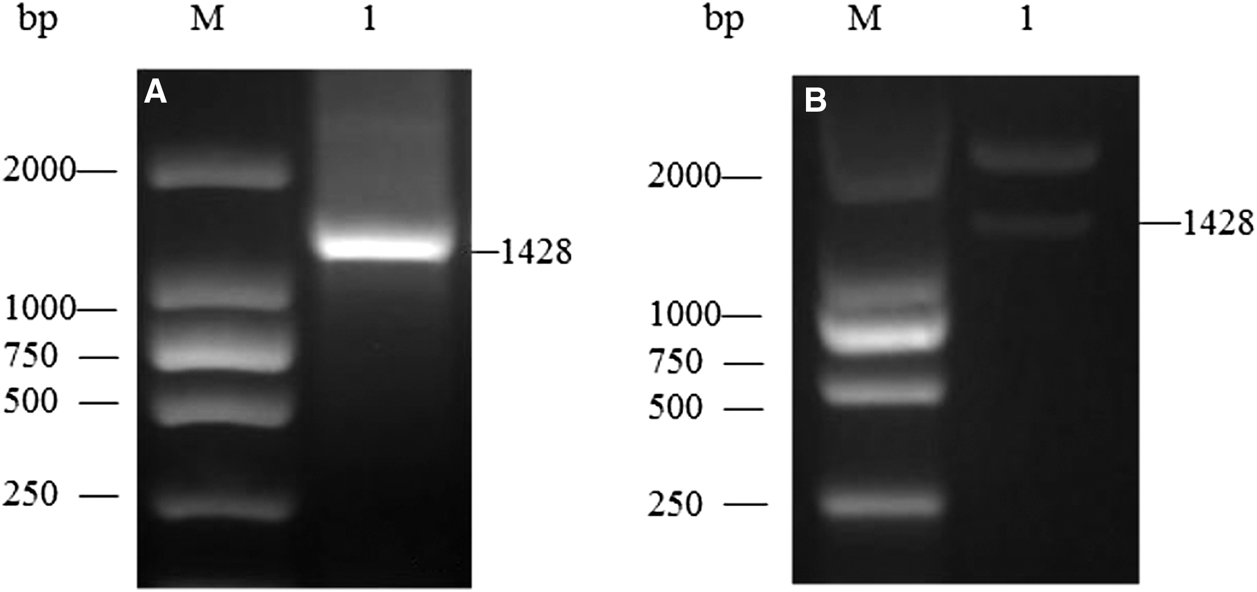
Amplification of coding sequence of chicken TRIM62 and prokaryotic expression vector construction.
The form of expression of TRIM62 protein was confirmed by SDS-PAGE. The results showed that the expression efficiencies of TRIM62 protein were presented in the cells (Fig. 2A) and reached the highest expression for 2 hours induced by 0.5 mM IPTG (Fig. 2B). The TRIM62 protein was purified by gradient urea dialysis. As shown in Figure 2C, the purified chicken TRIM62 protein was of high purity. Western blot was performed to confirm the purified protein using anti-His tag antibody, the empty vector-transformed bacteria served as control. The results demonstrated that purified protein was recognized in the predicted size (Fig. 2D).
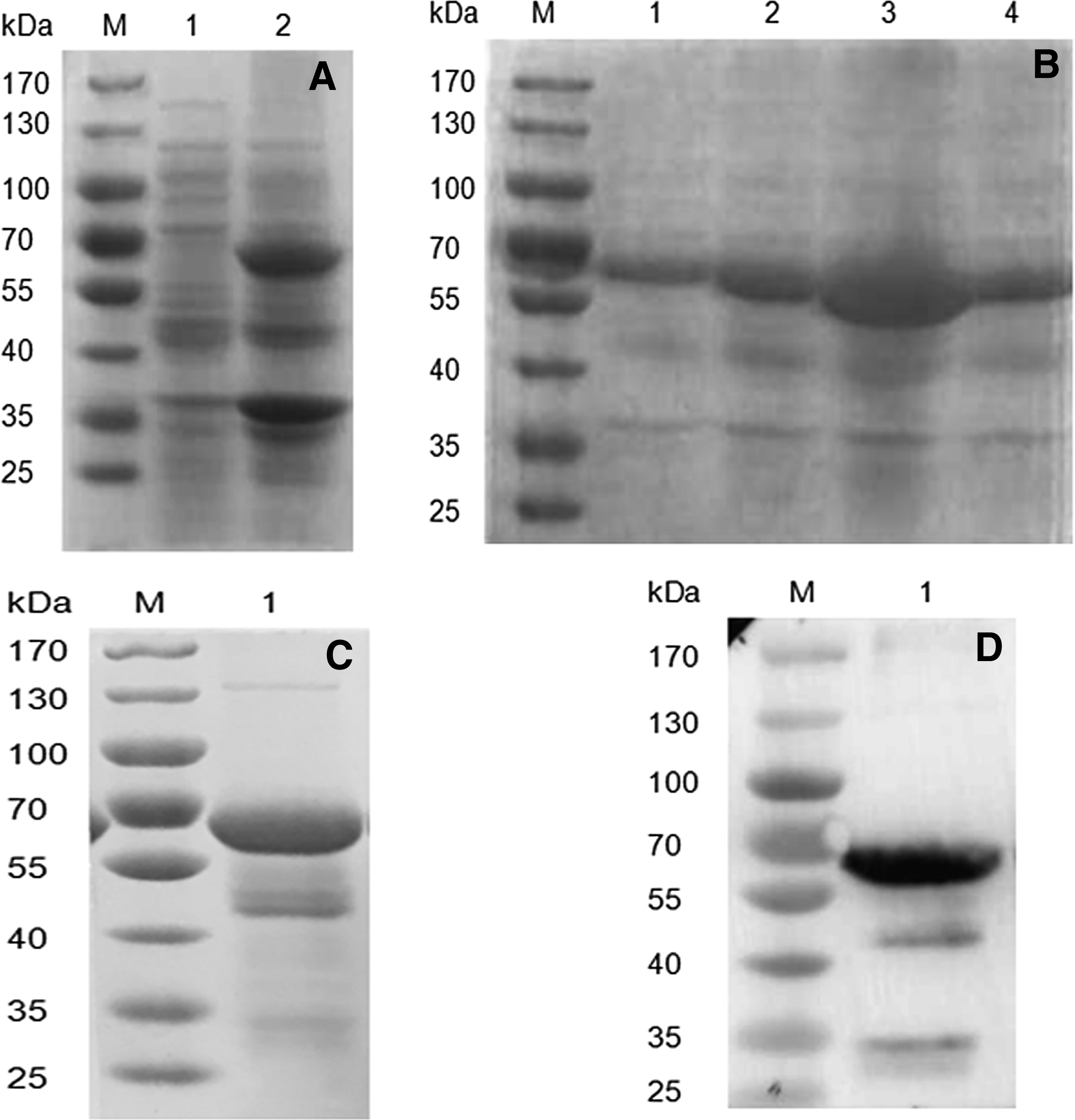
The expression, purification, and identification of recombinant chicken TRIM62.
Affinity and specificity of chicken TRIM62 monoclonal antibodies
After subcloning four times by limiting dilution and screening, we then utilized the chicken TRIM62 as immunogen, and three clones of hybridoma cell lines were acquired and named 2D3, 4B2, and 5D1. Antibody titers of the supernatant were determined by ELISA (Table 1). The antibodies were assayed at different dilutions ranging from 1:10 to 1:109. The titers were determined to be 1:105– to 1:106 (Table 2).
The screened cell with S/P>2.0 was considered to be hybridoma. The serum from unimmunized mice was used as negative control.
The supernatant gradient was diluted, the ratio of absorbance at 450 nm ≥2, the dilution gradient is the antibody titer.
To explore the specificity of mAb, recombinant and endogenous chicken TRIM62 were confirmed with mAb by western blot. The results proved that the mAb had the specific binding capacity to the recombinant and natural chicken TRIM62 (CEF and DF-1 cells). However, the endogenous TRIM62 from human cells (293T cells) and mouse cells (myeloma cells) were not detected (Fig. 3). The results demonstrated that the mAb could be used for further analysis.

Western blot analysis of the specificity of prepared mAb. Lysates of different cell lines were prepared for analysis using mAb 2D3. M: protein marker; TRIM62: purified TRIM62 expression construct, CEF: CEF cells lysates; DF-1: DF-1 cells lysates; 293T: 293T cells lysates; SP2/0: mice myeloma cells lysates. mAb, monoclonal antibody.
The distribution of chicken TRIM62 in SPF chicken tissues
To further detect the distribution of chicken TRIM62 in different organs, immunohistochemistry was performed with the prepared mAb to detect the presence of TRIM62. Microscopic observation showed that the TRIM62 positive signals were mainly distributed in the epithelial cells of proventriculus glands duct, pulmonary bronchioles, duodenum glands duct, and renal tubules. Otherwise, positive signals were detected in vascular endothelium of brain and spleen (Fig. 4).

Immunohistochemistry analysis of the distribution of chicken TRIM62. The prepared mAb was used as primary antibody to detect the presence of TRIM62. The supernatant from SP2/0 culture was used as control. There was no signal observed in control. Significant positive signals were mainly presented in the epithelial cells of proventriculus glands duct, pulmonary bronchioles, duodenum glands duct and renal tubules, and vascular endothelium of brain and spleen.
Discussion
TRIM62 regulated inflammatory and innate immune signaling, and the regulation of TRIM62 was associated with its antiretroviral activity.(4) Also, TRIM62 has been found to act as a tumor suppressor of several cancer.(6) However, the precise biological role of chicken TRIM62 and related mechanism remains unknown in avian diseases. In our previous study, we found that the level of chicken TRIM62 was associated with the replication of REV (data not shown) in vitro. REV is an oncogenic and immunosuppressive retrovirus.(8) In this study, our purpose was to prepare an antibody against the chicken TRIM62, which can be used as a tool to analyze the role of chicken TRIM62 in avian retrovirus disease.
In our present work, high-level expression of the chicken TRIM62 protein was used as antigen to prepare mAb. The titer determination and western blot detection indicated that the mAb was successfully produced. The antibody is a high-affinity mAb against chicken TRIM62 protein, which suggests that the newly prepared antibody can be used in further research on the distribution and biological function of chicken TRIM62.
TRIM62 is expressed in all normal tissues. The expression is localized only in the duct and glandular epithelium, and its subcellular localization in the cytoplasm [1, 2].(1,9) In this study, results of immunohistochemistry demonstrated that the chicken TRIM62 is mainly distributed in proventriculus, pulmonary, duodenum, kidney, brain, and spleen. Positive signals were presented in both the glandular epithelium and vascular endothelium. The localization and distribution of chicken TRIM62 may be associated with the function of chicken TRIM62. We would determine the speculation in further studies.
Footnotes
Acknowledgments
This work was supported by the National Natural Science Foundation of China (31772703), Shandong Provincial Natural Science Foundation (ZR2017MC011) and Funds of Shandong “Double Tops” Program.
Author Disclosure Statement
No competing financial interests exist.