




Abstract
Esophageal cancer is one of the highly malignant cancers. It comprises two of the most common histological tumor types: squamous cell carcinoma (SCC) and adenocarcinoma. SCC accounts for about 90% of esophageal cancers. Despite developments in treatment strategies, the prognosis and survival rate remain poor. Podocalyxin (PODXL) is a highly glycosylated type-I transmembrane protein. It is expressed in normal tissues such as kidney, heart, breast, and pancreas. Upregulation of PODXL correlates with tumor progression, invasion, and metastasis. Therefore, this glycoprotein could be a potential biomarker for predicting the prognosis of some cancers, for instance, brain, colorectal, oral, lung, bladder, prostate, and ovarian cancers. We previously developed a specific and sensitive anti-PODXL monoclonal antibody (mAb), PcMab-47 (mouse IgG1, kappa) and its mouse IgG2a-type (47-mG2a). We showed their utility in immunohistochemical analysis of oral cancers. Herein, we demonstrate that PcMab-47 and 47-mG2a can also be used to detect esophageal squamous cell carcinoma (ESCC) with this technique. These two antibodies, respectively, stained 123/130 (94.6%) and 127/130 (97.7%) ESCC cases, indicating that they can detect PODXL with high sensitivity in this carcinoma. Of more than 3+ cases, 47-mG2a was more effective than PcMab-47, respectively, staining 56/127 (44.1%) and 41/123 (33.3%). Therefore, 47-mG2a can be used for the detection of PODXL in ESCC using immunohistochemical analysis.
Introduction
E
Podocalyxin (PODXL) is a type-I transmembrane protein highly glycosylated with N-glycan or O-glycan(12) belonging to the CD34 family of sialomucins. It is expressed in normal tissues, such as kidney, heart, breast, and pancreas. PODXL upregulation has been correlated with tumor progression, invasion, and metastasis.(13) It is used as a diagnostic marker in numerous cancers, including brain,(14) colorectal,(15) oral,(16) lung,(17,18) bladder,(19) prostate,(20) and ovarian cancers.(21) Indeed, Borg et al. reported that PODXL expression in esophageal AC is an independent prognostic biomarker for poor overall survival.(22) Therefore, specific and sensitive anti-PODXL mAbs are required.
We previously established anti-PODXL mAbs PcMab-47 (mouse IgG1, kappa)(23) and mouse IgG2a-type (47-mG2a).(24) In this study, we investigated their utility for immunohistochemical analysis of esophageal squamous cell carcinoma (ESCC).
Materials and Methods
Tissues
Cancer tissue microarrays of esophageal cancers were purchased from Cybrdi, Inc. (Frederick, MD; Cat. No. CC02-01-001): Case-1–62; and, US Biomax, Inc. (Rockville, MD; Cat. No. BC02011): Case-63–130.
Antibodies
PcMab-47, a mouse anti-PODXL mAb (IgG1, kappa), was developed as previously described.(23) Appropriate VH and VL complementary DNAs (cDNAs) of mouse PcMab-47 and CH and CL of mouse IgG2a were subcloned into pCAG-Ble and pCAG-Neo vectors (FUJIFILM Wako Pure Chemical Industries Ltd., Osaka, Japan), respectively, to generate 47-mG2a.(24) Antibody expression vectors were transfected into ExpiCHO-S cells using the ExpiCHO Expression System (Thermo Fisher Scientific, Inc., Waltham, MA). 47-mG2a was purified using Protein G-Sepharose (GE Healthcare Bio-Sciences, Pittsburgh, PA).
Immunohistochemical analyses
Histologic sections (4 μm thick) were deparaffinized in xylene and then rehydrated and autoclaved in citrate buffer (pH 6.0; Agilent Technologies, Inc., Santa Clara, CA) for 20 minutes. Sections were then incubated with 0.5 or 5 μg/mL primary mAbs for 1 hour at room temperature and treated using an Envision+ kit (Agilent Technologies, Inc.) for 30 minutes. Color was developed using 3,3-diaminobenzidine tetrahydrochloride (Agilent Technologies, Inc.) for 2 minutes, and sections were counterstained with hematoxylin (FUJIFILM Wako Pure Chemical Industries Ltd.). The intensity of staining was evaluated as −, 1+, 2+, or 3+.
Results and Discussion
Previously, we immunized mice with recombinant PODXL and produced PcMab-47 (mouse IgG1, kappa).(23) This antibody was then engineered into a mouse IgG2a-type mAb (47-mG2a).(24) 47-mG2a exhibited a much higher PODXL-binding affinity compared with PcMab-47. Immunohistochemical analysis of oral cancer tissues revealed that those mAbs sensitively stain cells in a cytoplasmic pattern. In this study, we performed immunohistochemical analysis using PcMab-47 and 47-mG2a against ESCC.
Representative immunohistochemistry results of PcMab-47 and 47-mG2a against ESCC are shown in Figure 1. Both antibodies stained cancer cells in a cytoplasmic pattern (Fig. 1). As shown in Table 1 and Supplementary Table S1, PcMab-47 stained 123/130 (94.6%) and 47-mG2a 127/130 (97.7%) of ESCCs, indicating that both antibodies can be used to detect PODXL in ESCC using this method. Among more than 3+ cases, 47-mG2a was more sensitive than PcMab-47 with 56/127 (44.1%) and 41/123 (33.3%) occurrences, respectively (Table 1). In conclusion, 47-mG2a can be used to detect PODXL in ESCC by immunohistochemical analysis.
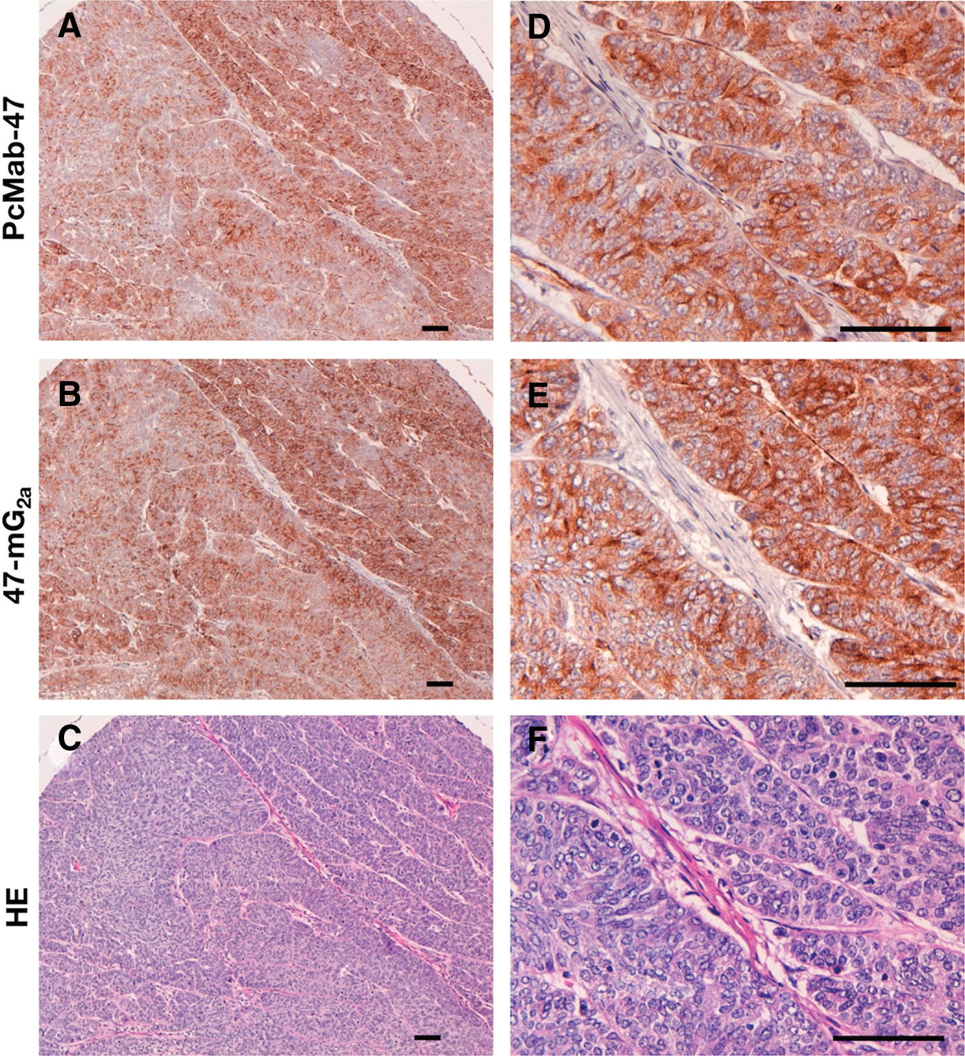
Immunohistochemical analysis by anti-PODXL antibodies against esophageal squamous cell carcinoma (Case-26). Sections were incubated with 5 μg/mL of PcMab-47
The intensity of staining was evaluated as −, 1+, 2+, 3+.
Footnotes
Acknowledgments
We thank Takuro Nakamura, Miyuki Yanaka, Noriko Saidoh, Saori Handa, and Yoshimi Nakamura for their excellent technical assistance. This research was supported by AMED under Grant Numbers: JP17am0301010 (Y.K.), JP17am0101078 (Y.K.), and JP17ae0101028 (Y.K.).
Author Disclosure Statement
The authors have no conflict of interest.
References
Supplementary Material
Please find the following supplemental material available below.
For Open Access articles published under a Creative Commons License, all supplemental material carries the same license as the article it is associated with.
For non-Open Access articles published, all supplemental material carries a non-exclusive license, and permission requests for re-use of supplemental material or any part of supplemental material shall be sent directly to the copyright owner as specified in the copyright notice associated with the article.
