
Abstract
Podoplanin (PDPN) is expressed in lymphatic endothelial cells, where it induces platelet aggregation through C-type lectin-like receptor-2 (CLEC-2). This protein has been characterized for a number of animal species using specific anti-PDPN monoclonal antibodies (mAbs). We recently established the mAb against horse PDPN (horPDPN) named PMab-219. Therefore, in this study, we investigated whether PMab-219 can detect lymphatic endothelial cells in horse tissues. Immunohistochemical analysis demonstrated that PMab-219 strongly stained lymphatic endothelial cells in horse colon tissues, indicating that it will be useful for investigating the function of horPDPN in these cells.
Introduction
H
Monoclonal antibodies (mAbs) against human,(18) mouse,(18) rat,(19) rabbit,(20) bovine,(21) dog,(22) cat,(23) and horse(24,25) PDPNs have been developed, and an anti-cat PDPN mAb (PMab-52) cross-reacted with tiger PDPN.(26) The anti-horse PDPN (anti-horPDPN) mAb PMab-219 has been shown to be useful in flow cytometry, Western blot, and immunohistochemical analyses.(25) Therefore, in this study, we investigated whether PMab-219 can detect lymphatic endothelial cells in horse tissues.
Materials and Methods
Immunohistochemical analyses
Normal horse tissues were collected after autopsy at Hokkaido University (Hokkaido, Japan) fixed in 10% neutral-buffered formalin, and processed routinely to prepare paraffin-embedded tissue sections. These sections were directly autoclaved in citrate buffer (pH 6.0; Nichirei Biosciences, Inc., Tokyo, Japan) for 20 minutes, blocked using SuperBlock T20 (PBS) Blocking Buffer (Thermo Fisher Scientific, Inc., Waltham, MA) for 15 minutes, incubated with PMab-219 (10 μg/mL) for 1 hour at room temperature, and then treated using the Envision+ Kit (Agilent Technologies, Inc., Santa Clara, CA) for 30 minutes. Color was developed with 3,3′-diaminobenzidine tetrahydrochloride (Agilent Technologies, Inc.) for 2 minutes, and counterstaining was performed with hematoxylin (FUJIFILM Wako Pure Chemical Corporation, Osaka, Japan).
Results and Discussion
We recently developed a Cell-Based Immunization and Screening (CBIS) method to produce sensitive and specific mAbs against membrane proteins, such as CD44,(27) CD133,(28) and PD-L1,(29) which we used to design anti-horPDPN mAbs.(25) Two mice were immunized with Chinese hamster ovary (CHO)/horPDPN cells using an immunization and screening procedure, and the developed hybridomas were seeded into 96-well plates and cultivated for 10 days. Wells that were positive for CHO/horPDPN and negative for CHO-K1 were then selected using flow cytometry. Nine hybridomas were developed: one clone of IgG1, one clone of IgG2a, five clones of IgG3, and two clones of IgM. Among these, PMab-219 (IgG2a, kappa) successfully recognized the horse kidney cell line FHK-Tcl3.1 in flow cytometry and detected horPDPN as a 40-kDa band in CHO/horPDPN cells using Western blot analysis.(25) Furthermore, immunohistochemical analysis revealed that PMab-219 strongly stained CHO/horPDPN cells.(25) However, it remained unclear whether PMab-219 was useful for detecting lymphatic endothelial cells in horse tissues.
In this study, we found that PMab-219 strongly stained lymphatic endothelial cells in equine colon tissues, whereas leaving vascular endothelial cells unstained (Fig. 1). Furthermore, it weakly stained podocytes in equine kidney tissues (Fig. 2). Although it has been reported that type I alveolar cells in the lung also express PDPN,(9) PMab-219 did not stain these cells in equine lung tissues (data not shown), possibly due to the varying expression levels of horPDPN in different tissues. These results indicate that PMab-219 will be useful for elucidating the pathophysiological functions of horPDPN in equine lymphatic endothelial cells in future studies.
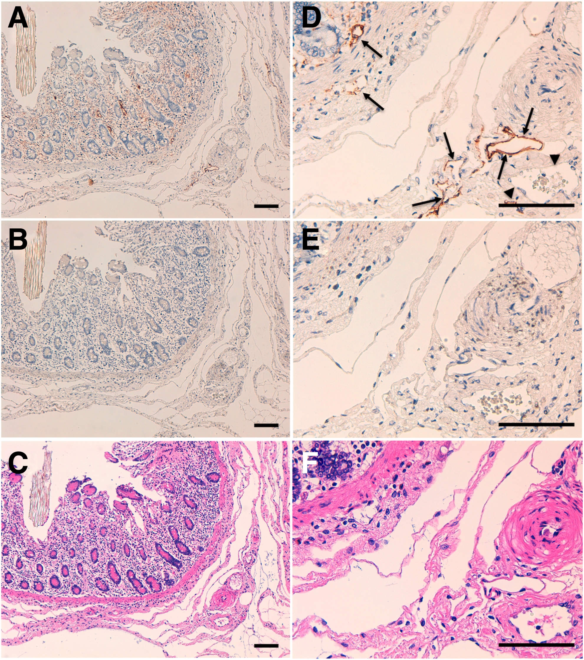
Immunohistochemical analysis of equine colon tissues. Equine colon tissue sections were incubated with 10 μg/mL of PMab-219

Immunohistochemical analysis of equine kidney tissues. Equine kidney tissue sections were incubated with 10 μg/mL of PMab-219
Footnotes
Acknowledgments
This research was supported in part by AMED under Grant Numbers JP18am0101078 (Y.K.), JP18am0301010 (Y.K.), and JP18ae0101028 (Y.K.), and by JSPS KAKENHI Grant Number 17K07299 (M.K.K.) and Grant Number 16K10748 (Y.K.).
Author Disclosure Statement
No competing financial interests exist.