
Abstract
Horse podoplanin (horPDPN), a type I transmembrane sialoglycoprotein, is expressed on the podocytes of the kidneys, alveolar type I cells of the lungs, and lymphatic endothelial cells. PDPN is a platelet aggregation-inducing factor, and it primarily possesses three platelet aggregation-stimulating (PLAG) domains, that is, PLAG1, PLAG2, and PLAG3, at the N-terminus and several PLAG-like domains. In a previous study, we reported on a mouse anti-horPDPN monoclonal antibody (mAb) clone, PMab-202. Although the effectiveness of PMab-202 in flow cytometry and Western blotting is known, its exact binding epitope remains unknown to date. In this study, enzyme-linked immunosorbent assay and flow cytometry were used to identify the epitope of PMab-202. We found that the critical epitopes of PMab-202 include Lys64, Thr66, and Phe70 of horPDPN. We believe that our findings can be applied in the production of more functional anti-horPDPN mAbs.
Introduction
Podoplanin (PDPN) induces platelet aggregation by binding to C-type lectin-like receptor-2 (CLEC-2).(1–8) PDPN is a type I transmembrane sialoglycoprotein expressed in normal tissues, including renal corpuscles, alveolar type I cells of the lungs, and lymphatic endothelial cells.(3,9) The interaction between the PDPN of lymphatic endothelial cells and CLEC-2 of platelets facilitates embryonic blood/lymphatic vessel separation.(10) Expression of human PDPN, a protein associated with malignant progression and cancer metastasis,(5,11,12) has been reported in several malignant tumors, such as brain tumors,(11,13–15) mesotheliomas,(16,17) oral squamous cell carcinomas,(18) esophageal cancers,(19) lung cancers,(20) osteosarcomas,(21–23) and testicular tumors.(24)
In a previous study, we developed monoclonal antibodies (mAbs) not only against human,(25) mouse,(25) and rat(26) PDPNs but also against rabbit,(27) dog,(28) cat,(29) bovine,(30) pig,(31) and horse(32–34) PDPNs (horPDPNs). PDPN primarily possesses three platelet aggregation-stimulating (PLAG) domains: PLAG1, PLAG2, and PLAG3; these domains are present at the N-terminus of PDPN.(2) One PLAG-like domain (PLD), important for PDPN–CLEC-2 interaction, has been reported to be present in the middle of PDPN.(35) Almost all mAbs against PDPNs have been reported to react with the PLAG domains or PLDs.(35,36)
In this study, we determined the epitope responsible for the binding of PMab-202 to horPDPN using enzyme-linked immunosorbent assay (ELISA) and flow cytometry.
Materials and Methods
Cell line
Chinese hamster ovary (CHO)-K1 was obtained from American Type Culture Collection (ATCC, Manassas, VA). The horse kidney cell line FHK-Tcl3.1 was prepared at Yamaguchi University.(37) horPDPN bearing an N-terminal PA16 tag (PA16-horPDPN) was inserted into a pCAG-Ble vector (FUJIFILM Wako Pure Chemical Corporation, Osaka, Japan).(33) The PA16 tag comprised 16 amino acids (GLEGGVAMPGAEDDVV).(38) Furthermore, the CHO-K1 cells were transfected with pCAG-Ble/PA16-horPDPN using Lipofectamine LTX with Plus Reagent (Thermo Fisher Scientific, Inc., Waltham, MA). Stable transfectants were selected by limiting dilution and cultivating in a medium containing 0.5 mg/mL Zeocin (InvivoGen, San Diego, CA). The CHO/horPDPN cells were cultured in Roswell Park Memorial Institute (RPMI) 1640 medium (Nacalai Tesque, Inc., Kyoto, Japan), whereas FHK-Tcl3.1 was cultured in Dulbecco's modified Eagle's medium (DMEM; Nacalai Tesque, Inc.).(33) RPMI 1640 and DMEM were supplemented with 10% heat-inactivated fetal bovine serum (Thermo Fisher Scientific, Inc.), 100 units/mL penicillin, 100 μg/mL streptomycin, and 25 μg/mL amphotericin B (Nacalai Tesque, Inc.). The cells were grown at 37°C in a humidified environment under a 5% CO2 atmosphere.
Enzyme-linked immunosorbent assay
The horPDPN peptides synthesized using PEPScreen (Sigma-Aldrich Corp., St. Louis, MO) were immobilized on Nunc Maxisorp 96-well immunoplates (Thermo Fisher Scientific, Inc.) at concentrations of 1 μg/mL for 30 minutes at 37°C. After blocking with SuperBlock T20 (PBS) Blocking Buffer (Thermo Fisher Scientific, Inc.), the plates were incubated with 1 μg/mL purified PMab-202, followed by 1:2000 dilution of peroxidase-conjugated antimouse IgG (Agilent Technologies, Inc., Santa Clara, CA). The enzymatic reaction was performed using 1-Step Ultra TMB-ELISA (Thermo Fisher Scientific, Inc.). Optical density was measured at 655 nm using an iMark microplate reader (Bio-Rad Laboratories, Inc., Berkeley, CA). These reactions were performed at 37°C using a total sample volume of 50–100 μL.
Flow cytometry
CHO/horPDPN or FHK-Tcl3.1 cells were harvested after a brief exposure to 0.25% trypsin/1 mM EDTA (Nacalai Tesque, Inc.), and were washed with 0.1% bovine serum albumin/phosphate-buffered saline. CHO/horPDPN cells were treated with 0.1 μg/mL PMab-202 or 0.1 μg/mL PMab-202 plus 50 μg/mL peptides for 30 minutes at 4°C. FHK-Tcl3.1 cells were treated with 1 μg/mL PMab-202 or 1 μg/mL PMab-202 plus 10 μg/mL peptides for 30 minutes at 4°C. Thereafter, the cells were treated with Alexa Fluor 488-conjugated antimouse IgG (1:2000; Cell Signaling Technology, Inc., Danvers, MA). Fluorescence data were collected using SA3800 Cell Analyzers (Sony Corp., Tokyo, Japan).
Results and Discussion
In a previous study, we immunized mice with a synthesized peptide (horPP6279) corresponding to the amino acids 62–79 of horPDPN.(33) ELISA screening indicated positive signals in 21 of 960 (2.2%) wells. Among these 21 wells, 6 (29%) tested positive against CHO/horPDPN in flow cytometry. One clone, that is, PMab-202 (IgG1, kappa), among the six positive clones was established by limiting dilution. PMab-202 could detect endogenous horPDPN expressed in FHK-Tcl3.1, a horse kidney cell line, in flow cytometry and Western blotting. However, PMab-202 was not advantageous for immunohistochemical analysis.
We first synthesized a series of peptides of horPDPN (Table 1). Using ELISA, PMab-202 detected 53–72 and 63–82 corresponding to the amino acids 53–72 and 63–82 of horPDPN, respectively. Next, we synthesized the point mutants of 53–72 peptides (Table 2). Using ELISA, PMab-202 detected the following antigens: T53A, G54A, S55A, P56A, A57G, L58A, V59A, P60A, R61A, S62A, T63A, S65A, G67A, G68A, D69A, E71A, and D72A. However, F70A was not recognized, and it weakly reacted with K64A and T66A, indicating that Lys64, Thr66, and Phe70 are the critical epitopes of PMab-202.
Determination of PMab-202 Epitope by Enzyme-Linked Immunosorbent Assay
+++, OD655 ≧ 0.6; −, OD655 < 0.1.
Determination of PMab-202 Epitope by Enzyme-Linked Immunosorbent Assay
+++, OD655 ≧ 0.6; +, 0.1 ≦ OD655 < 0.4; −, OD655 < 0.1.
Next, we performed a blocking assay using flow cytometry. We found that PMab-202 reacted with the CHO/horPDPN cell line (Fig. 1). This reaction was partially neutralized by T53A. However, K64A, T66A, and F70A did not block the reaction of PMab-202 with CHO/horPDPN. Similarly, PMab-202 reacted with the FHK-Tcl3.1 cell line (Fig. 1). Notably, this reaction was completely neutralized by T53A. In contrast, K64A, T66A, and F70A did not block the reaction of PMab-202 with FHK-Tcl3.1, thereby confirming that Lys64, Thr66, and Phe70 of horPDPN are critical for PMab-202 detection. As shown in Figure 2, Lys64, Thr66, and Phe70 are included between PLAG3 and PLD.

Flow cytometry using PMab-202 and point mutants of horPDPN. CHO/horPDPN cells were treated with 0.1 μg/mL PMab-202 or 0.1 μg/mL PMab-202 plus 50 μg/mL peptides for 30 minutes at 4°C. FHK-Tcl3.1 cells were treated with 1 μg/mL PMab-202 or 1 μg/mL PMab-202 plus 10 μg/mL peptides for 30 minutes at 4°C. Thereafter, the cells were treated with Alexa Fluor 488-conjugated antimouse IgG. Solid line with gray shade, control (second Ab only); dotted line, PMab-202 or PMab-202 plus peptides. CHO, Chinese hamster ovary; horPDPN, horse podoplanin.
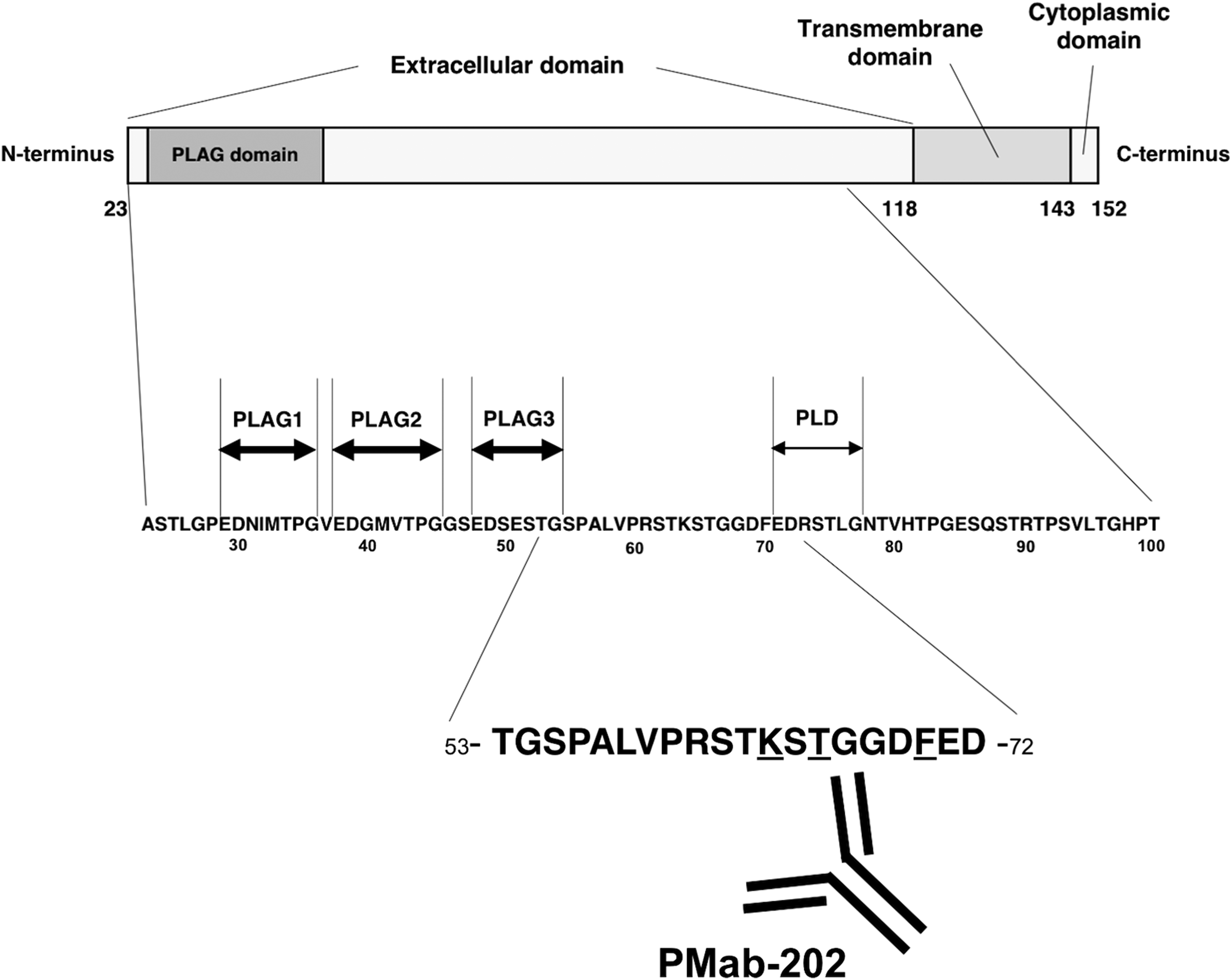
Schematic illustration of the epitope recognized by PMab-202. Underlined amino acids are the critical epitope PMab-202. PLAG, platelet aggregation-stimulating; PLD, PLAG-like domain.
Taken together, the study findings indicate that the critical epitopes of PMab-202 are Lys64, Thr66, and Phe70 of horPDPN. We believe that these findings will be beneficial in the production of more functional anti-horPDPN mAbs.
Footnotes
Acknowledgments
We thank Takuro Nakamura, Miyuki Yanaka, Saori Handa, Kayo Hisamatsu, and Yoshimi Nakamura for excellent technical assistance. FHK-Tcl3.1 was kindly provided by Ken Maeda (Yamaguchi University). This research was supported, in part, by AMED under Grant Nos.: JP18am0101078 (Y.K.), JP18am0301010 (Y.K.), and JP18ae0101028 (Y.K.), and by JSPS KAKENHI Grant No. 17K07299 (M.K.K.) and Grant No. 16K10748 (Y.K.).
Author Disclosure Statement
Y.K. received research funding from ZENOAQ Resource Co., Ltd. The other authors have no conflict of interest.