
Abstract
Cancer stem cells contribute to tumorigenesis, metastasis, recurrence, and chemoresistance. CD133/prominin-1—a pentaspan membrane glycoprotein—has been used as a stem cell biomarker for the isolation of stem-like cells from a variety of normal and pathological tissues. In our previous studies, we developed several anti-CD133 monoclonal antibodies using Cell-Based Immunization and Screening (CBIS) methods, followed by characterization of their efficacy by flow cytometry, western blotting, and immunohistochemical analyses. One of the 100 clones, CMab-43 (IgG2a, kappa), demonstrated a sensitive and specific reaction against colon cancer cells. This study aimed to investigate the antitumor activity of CMab-43. Caco-2 cells (human colon cancer cell line) were subcutaneously implanted into the flanks of nude mice. CMab-43 and control mouse IgG were injected three times into the peritoneal cavity of mice. Tumor formation was observed in the control and CMab-43-treated mice of Caco-2 xenograft models. CMab-43 significantly reduced tumor development of Caco-2 xenograft in comparison with the control mouse IgG on days 12, 14, and 17. Our results cumulatively suggest that CMab-43 is useful for antibody therapy against CD133-expressing colon cancers.
Introduction
Cancer stem cells (CSCs) or tumor-initiating cells possess several properties of non-neoplastic stem cells. CSCs are also characterized by extensive proliferation, self-renewal, invasion, metastasis, and chemoresistance.(1–3) Several protein markers, such as CD133 and CD44 as well as side-population cells, have been utilized to isolate CSCs from cancerous tissues as well as to investigate the CSC properties in cancer tissues.(1,4–12) CD133, also known as prominin-1, was first described as a cell surface marker on hematopoietic stem cells.(13) It is a pentaspan membrane glycoprotein, composed of an N-terminal extracellular tail, two small cytoplasmic loops, two large extracellular loops containing several potential glycosylation sites, and a short C-terminal intracellular tail.(14) Its expression also serves as a prognostic marker of gliomas.(15)
In our previous studies, we developed novel anti-CD133 monoclonal antibodies (mAbs)(16) using Cell-Based Immunization and Screening (CBIS) method.(17–20) We first expressed the full length of CD133 in LN229 glioblastoma cells, immunized mice with LN229/CD133 cells, and then performed the first screening by flow cytometry. After limiting dilution, we established 100 anti-CD133 mAbs, reacting with LN229/CD133 cells but not with LN229 cells. Subsequently, we performed the second and third screening, with western blotting and immunohistochemical analyses, respectively. Among the 100 mAbs, 11 strongly reacted with CD133 in western blotting analysis. One of the 11 clones, CMab-43 (IgG2a, kappa), demonstrated a sensitive and specific reaction against colon cancer cells. Among the mouse IgG subclasses, IgG2a(21) and IgG2b(22) are known to possess antibody-dependent cellular cytotoxicity (ADCC) and complement-dependent cytotoxicity (CDC). In this study, we investigated the antitumor activity of CMab-43 against mouse xenograft models of human colon cancer.
Materials and Methods
Cell lines
Caco-2 was obtained from the American Type Culture Collection (ATCC, Manassas, VA) and was cultured in the Dulbecco's modified Eagle's medium (DMEM) medium (Nacalai Tesque, Inc., Kyoto, Japan) supplemented with 10% heat-inactivated fetal bovine serum (FBS;Thermo Fisher Scientific, Inc., Waltham, MA), 100 U/mL of penicillin, 100 μg/mL of streptomycin, and 25 μg/mL of amphotericin B (Nacalai Tesque, Inc.) at 37°C in a humidified atmosphere containing 5% CO2 and 95% air.
Antibodies
The mouse anti-CD133 mAb CMab-43 was developed as described elsewhere.(16) CMab-43 hybridoma was cultured in Hybridoma-SFM medium (Thermo Fisher Scientific, Inc.), supplemented with 100 U/mL of penicillin, 100 μg/mL of streptomycin, and 25 μg/mL of amphotericin B (Nacalai Tesque, Inc.) at 37°C under a humidified 5% CO2 and 95% air atmosphere. CMab-43 was purified using Protein G-Sepharose (GE Healthcare Bio-Sciences, Pittsburgh, PA). Control mouse IgG was purchased from Sigma-Aldrich Corp. (St. Louis, MO).
Antitumor activity of CMab-43
Female BALB/c nude mice (6-week old) were purchased from Charles River (Kanagawa, Japan) and used in experiments when they were 10 weeks old. Caco-2 (0.3 mL of 1.33 × 10 8 /mL in DMEM) were mixed with 0.5 mL of BD Matrigel Matrix Growth Factor Reduced (BD Biosciences, San Jose, CA). A 100-μL suspension (containing 5 × 10 6 cells) was injected subcutaneously into the left flanks of nude mice. After day 1, 50 μg of CMab-43 and control mouse IgG in 100 μL PBS were injected into the peritoneal cavity of each mouse. Additional antibodies were then injected on days 7 and 14. The tumor diameter and volume were determined as previously described.(23) The mice were euthanized 19 days after cell implantation. All data were expressed as mean ± SEM. Statistical analysis was performed using the Tukey–Kramer's test. p < 0.05 was considered to be statistically significant.
Results and Discussion
We had previously reported the development of the mouse mAb CMab-43 against CD133.(16) Flow cytometry demonstrated that CMab-43 reacted with LN229/CD133 cells but not with LN229 brain tumor cells, which indicates that CMab-43 is specific for CD133. CMab-43 also recognized as an endogenous CD133 in Caco-2 colon cancer cells. Western blotting against LN229, LN229/CD133, CHO-K1, CHO/CD133, and Caco-2 cells revealed that CMab-43 detected a strong signal at ∼100 kDa in LN229/CD133 and CHO/CD133 but detected a moderate signal in Caco-2 cells, which indicates the usefulness of CMab-43 in western blotting. Furthermore, the calculated KD values for CMab-43 against LN229/CD133 and Caco-2 cells were 4.4 × 10⁻⁹ and 2.6 × 10⁻⁹ M, respectively, indicating a high affinity for CD133-expressing cell lines. The immunohistochemical analysis demonstrated that CMab-43 stained 83.3% of colon adenocarcinomas, which indicated the usefulness of CMab-43 for immunohistochemical analysis.(16) As CMab-43 was determined to be an IgG2a subclass of mouse IgG, it demonstrates the potential of ADCC and CDC activities.(21)
To study the antitumor activity of CMab-43 on cell growth in vivo, Caco-2 cells were subcutaneously implanted into the flanks of nude mice. CMab-43 and control mouse IgG were injected three times (on days 1, 7, and 14 after cell injections) into the peritoneal cavity of mice. Tumor formation was observed in mice from the control and CMab-43-treated groups in Caco-2 xenograft models. CMab-43 significantly reduced the tumor development of Caco-2 xenograft in comparison with that in control mouse IgG on days 12, 14, and 19 (Fig. 1). Caco-2 xenograft mice models on day 19 are shown in Figure 2A. The resected tumors of Caco-2 xenografts are depicted in Figure 2B. The tumor weight of mice in CMab-43 treated was significantly lower than that in the control mouse IgG group in Caco-2 xenograft models (Fig. 2C). However, body weight was not significantly different among the two groups in the Caco-2 xenograft models (Fig. 2D).
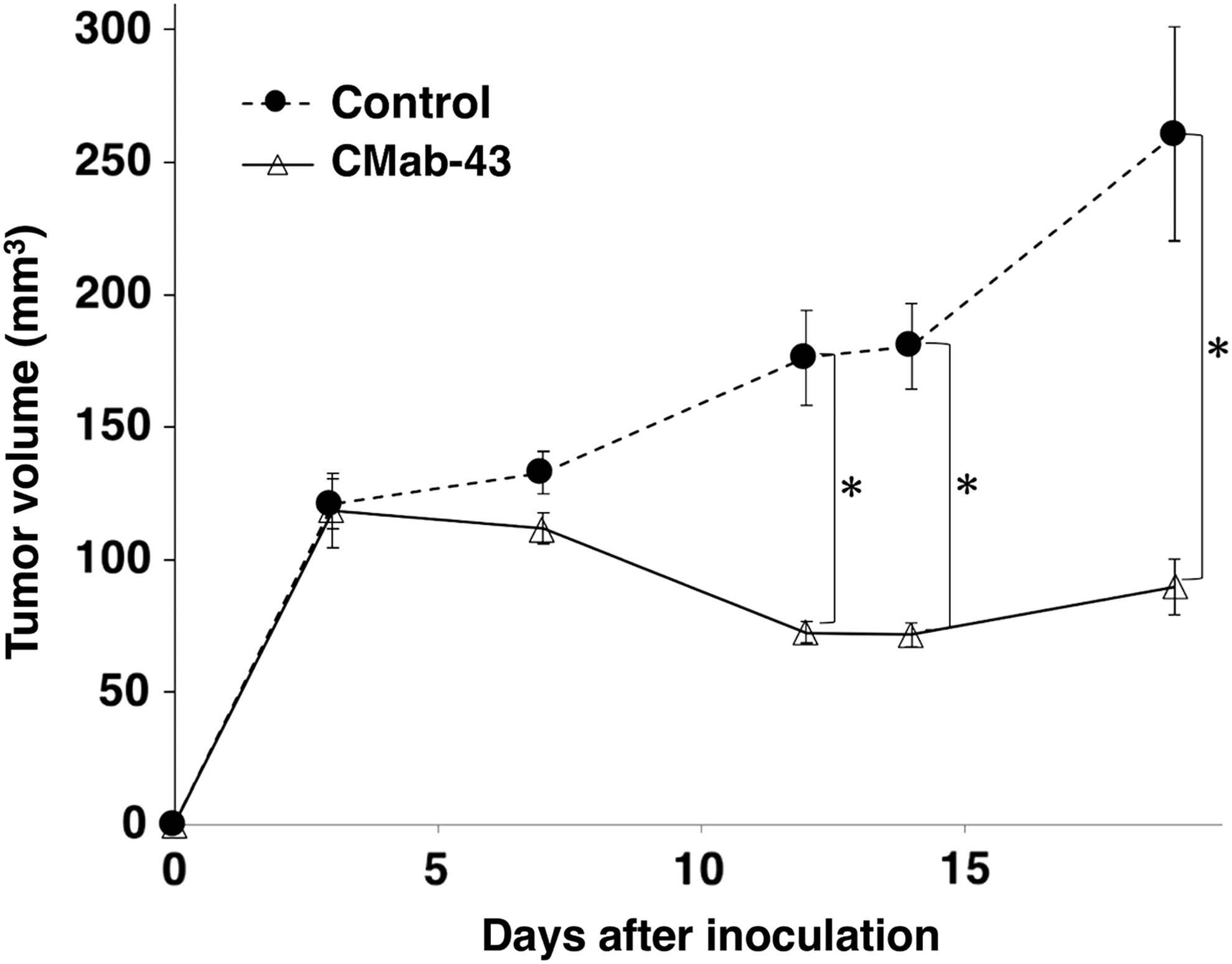
Antitumor activity of CMab-43 against Caco-2 xenograft model. Tumor volume of Caco-2 xenografts. Caco-2 cells were injected subcutaneously into female nude mice. The indicated antibodies (50 μg/day; 2.5 mg/kg) were administered intraperitoneally on days 1, 7, and 14 after cell inoculation. The tumor volume was measured at the indicated time points. The values are presented as mean ± SEM. *p < 0.01, Tukey–Kramer's test; SEM, standard error of the mean.

Evaluation of antitumor activity of CMab-43 against Caco-2 xenograft model.
In conclusion, CMab-43 is applicable for antibody therapy against human colon cancers expressing CD133. Further studies on antitumor activities against CD133-expressing xenografts are, therefore, necessary to obtain a more detailed understanding of antibody therapy against CD133.
Footnotes
Acknowledgments
We thank Akiko Harakawa, Shun-ichi Ohba, Miyuki Yanaka, Kayo Hisamatsu, Saori Handa, and Yoshimi Nakamura for their excellent technical assistance. This research was supported in part by AMED under grant nos. JP18am0101078 (Y.K.), JP18am0301010 (Y.K.), and JP18ae0101028 (Y.K.), and by JSPS KAKENHI grant no. 17K07299 (M.K.K.) and grant no. 16K10748 (Y.K.).
Author Disclosure Statement
No competing financial interests exist.